MYH7 disorders
|
Childhood Gowers-Laing: Adult |
Gowers-Laing: Adult
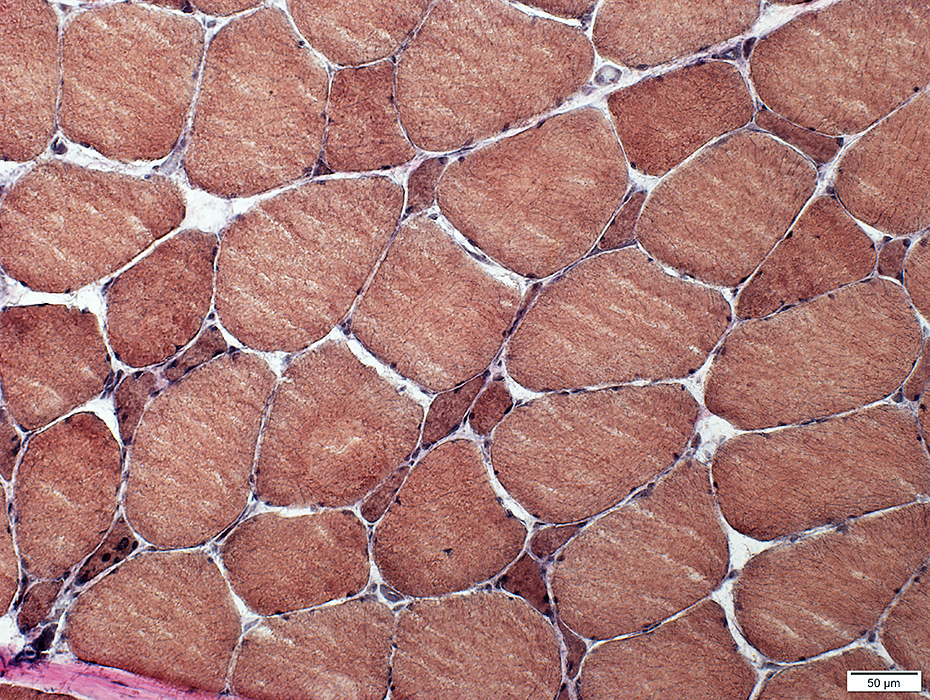
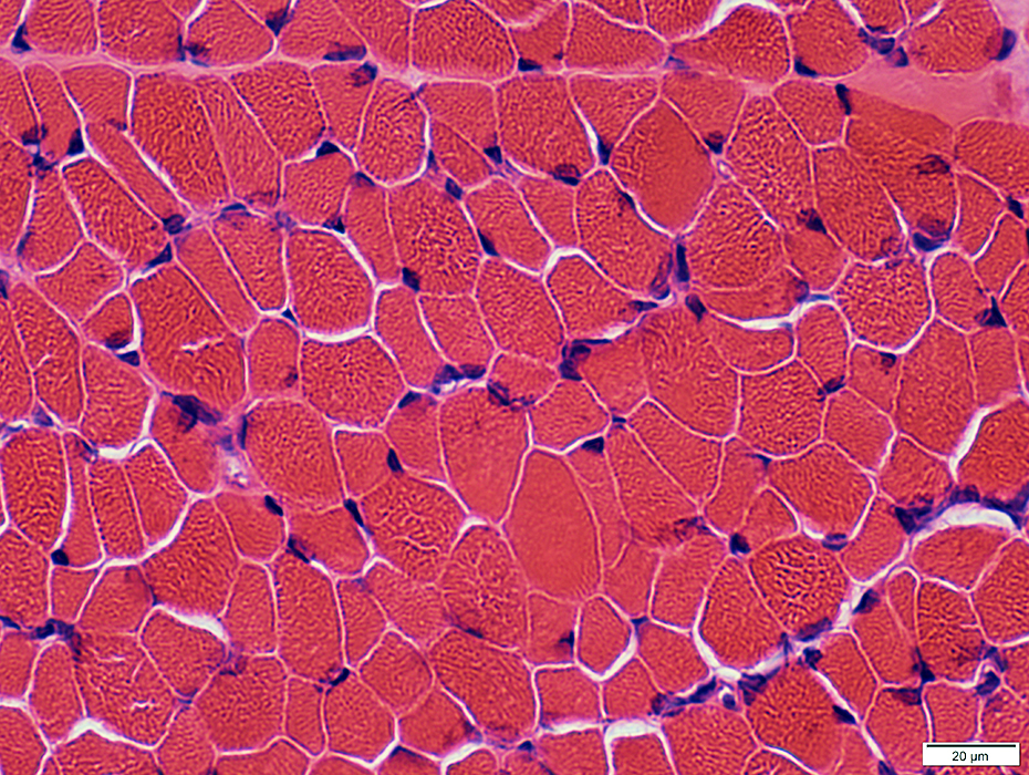
Muscle fiber sizesVaried: Small to Hypertrophied
Bimodal distribution
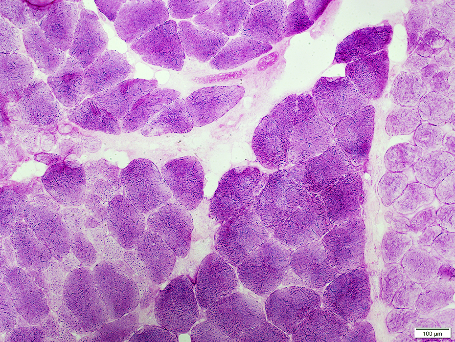
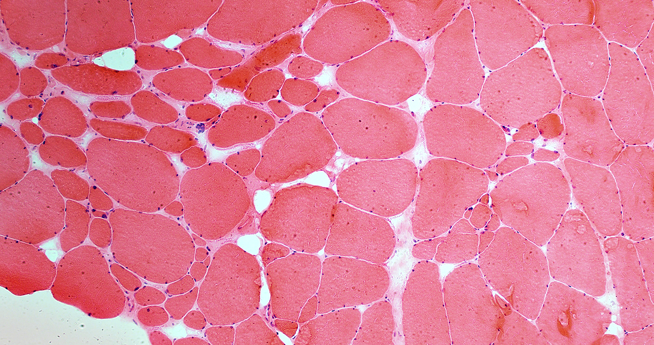 H & E stain |
 H & E stain |
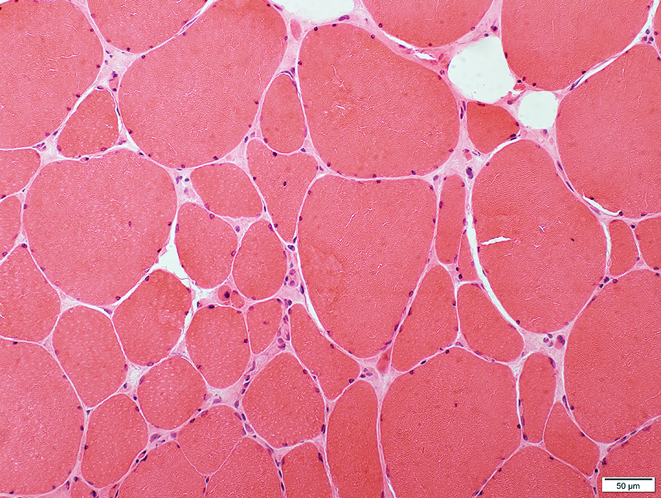
Often present in clusters
May be intermediate sized or very small
May be round, polygonal or angular
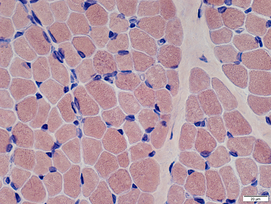
 H & E stain |
 H & E stain |
Varied shapes & sizes
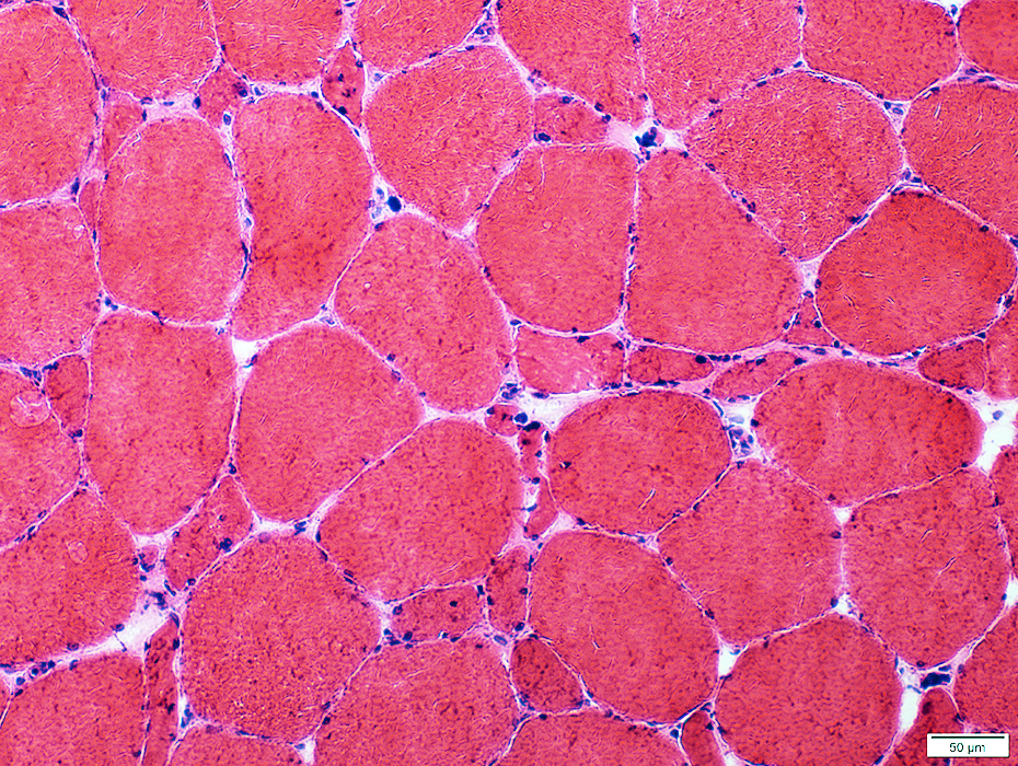
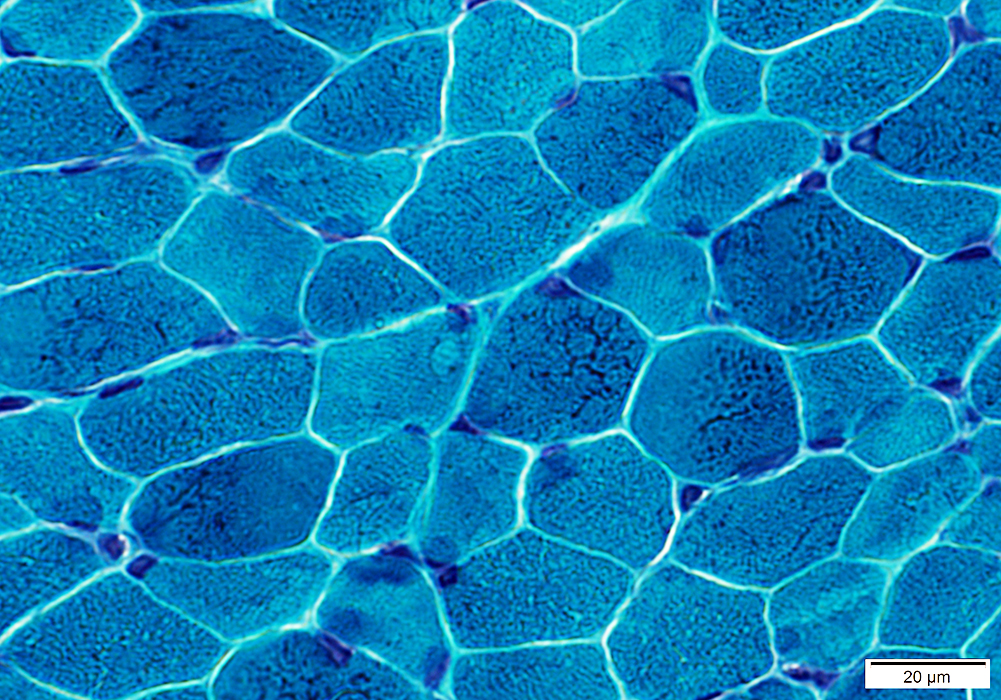
 Gomori trichrome stain |
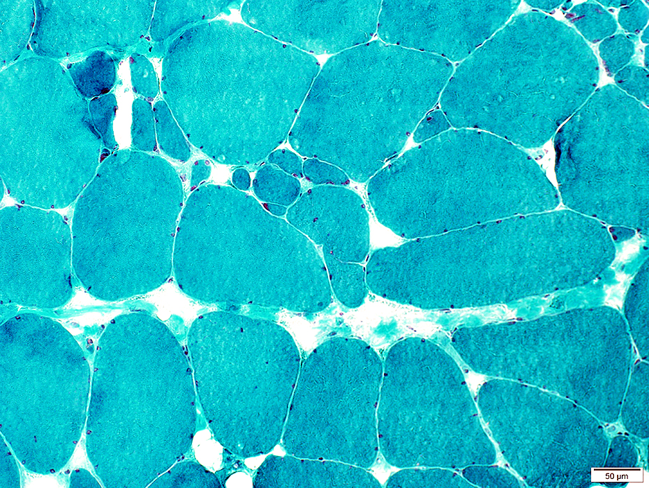 Gomori trichrome stain |
Varied
Bimodal distribution
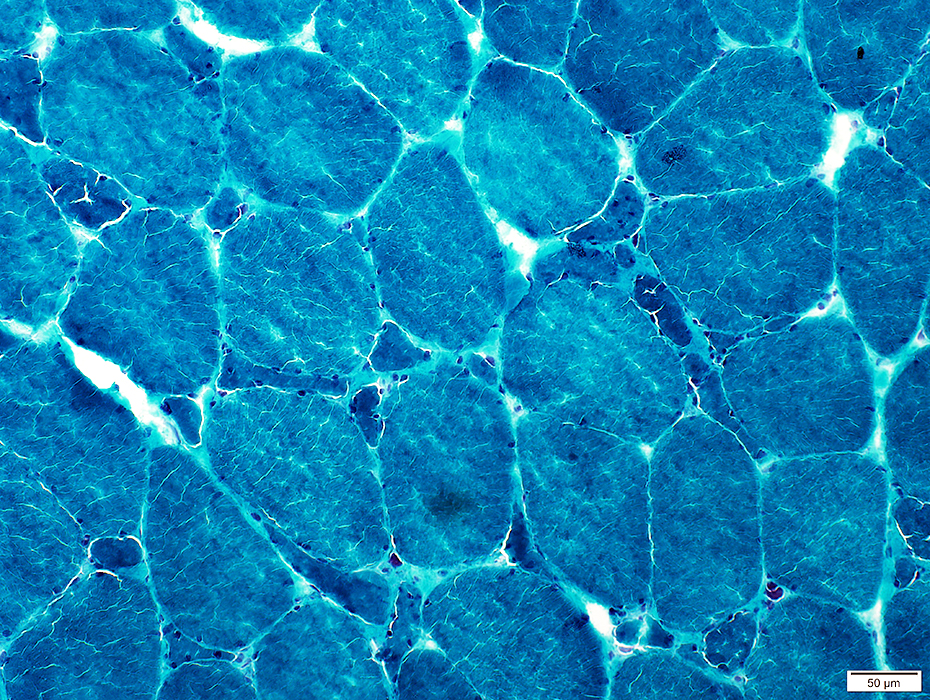
 VvG stain |
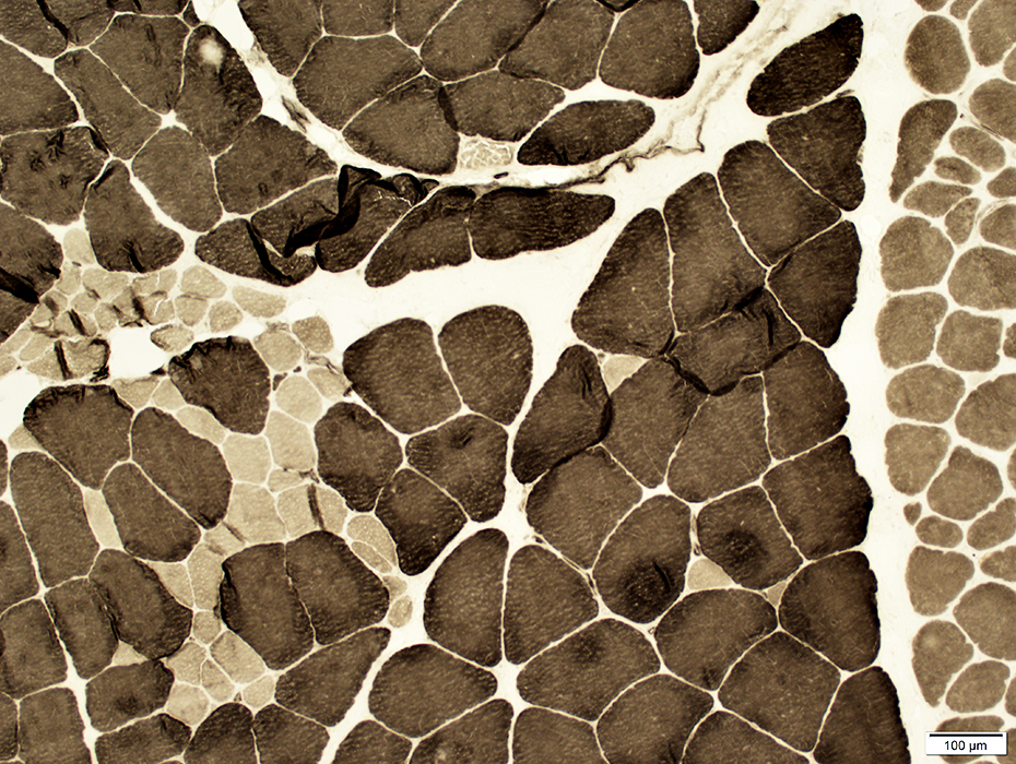
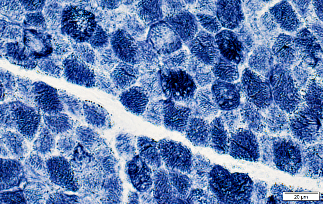
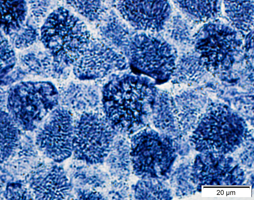
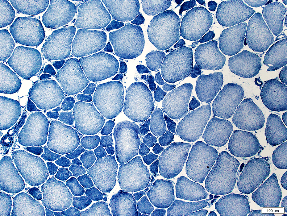 NADH stain |
Stain dark with NADH: Likely type I
 NADH stain |
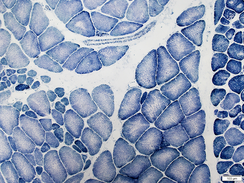 NADH stain |
Cores
In some muscle fibers within a group of type I fibers
Other fibers have irregular internal architecture
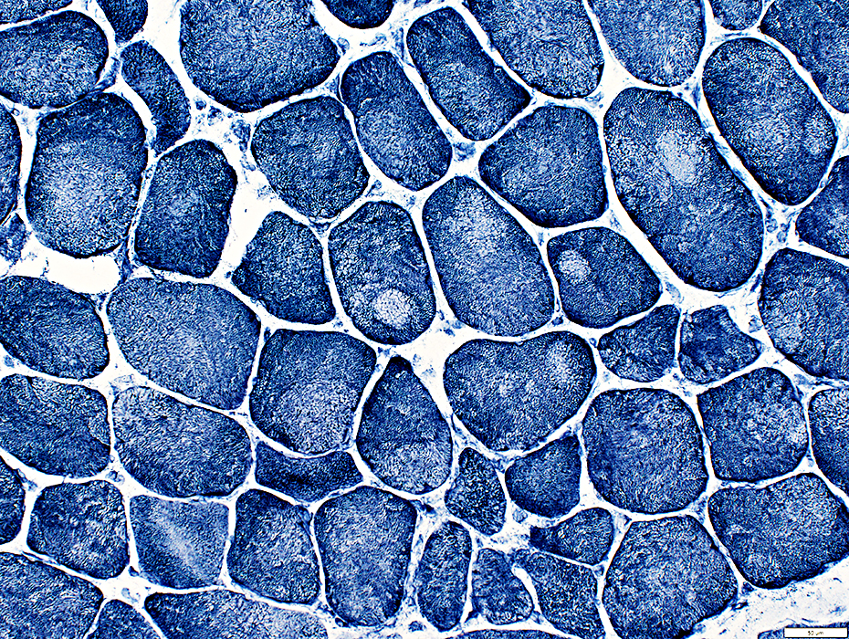 NADH stain |
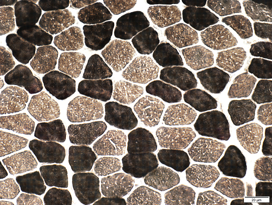
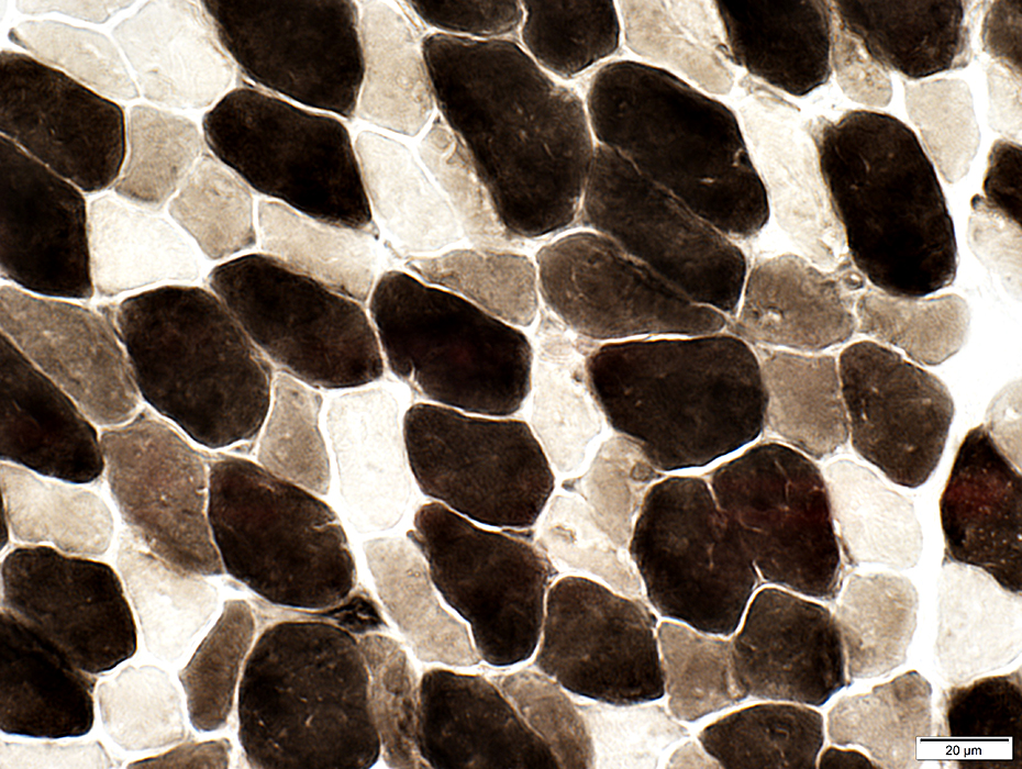
 ATPase pH 4.3 stain |
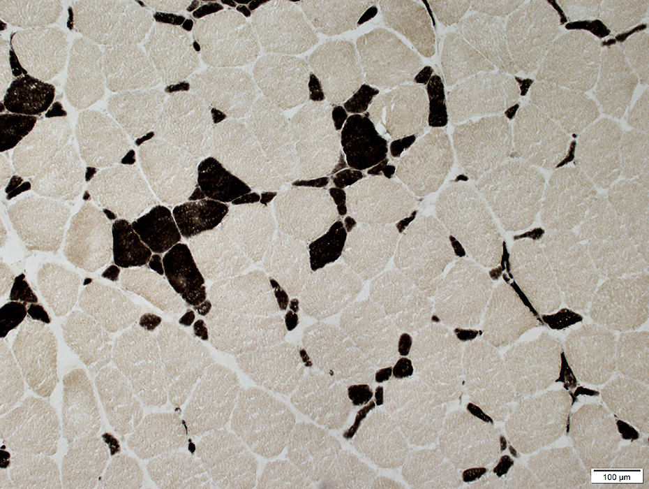 ATPase pH 4.3 stain |
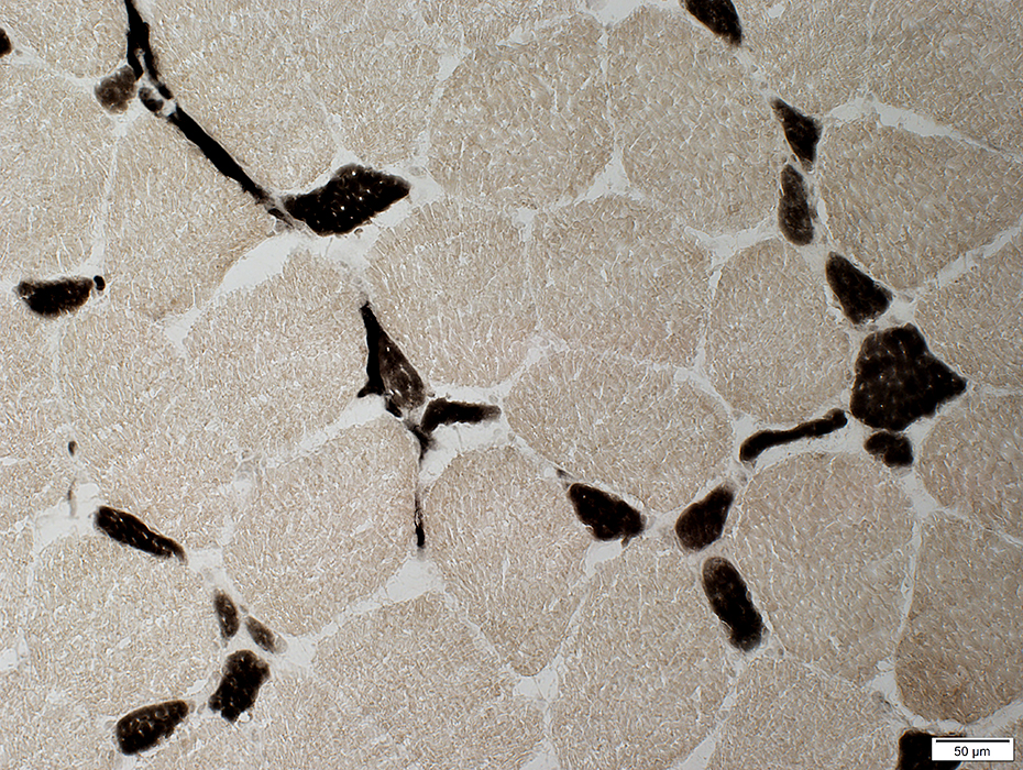 ATPase pH 4.3 stain |
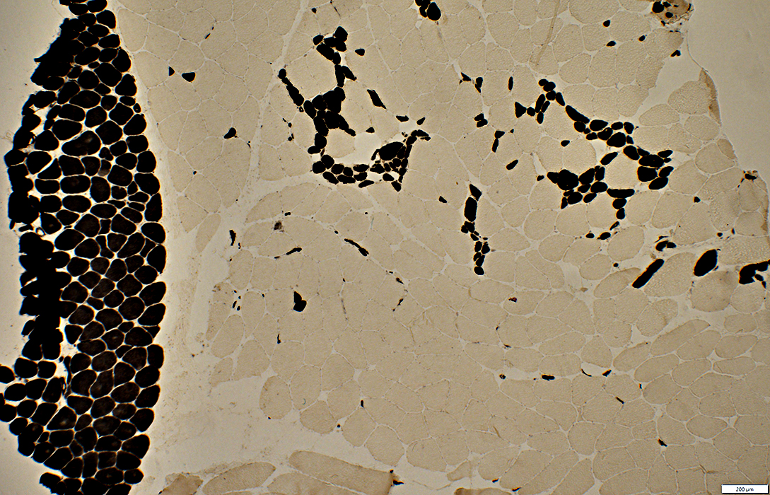
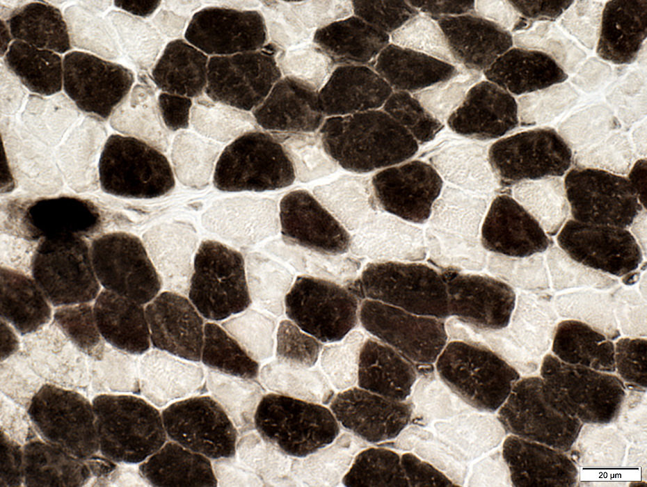
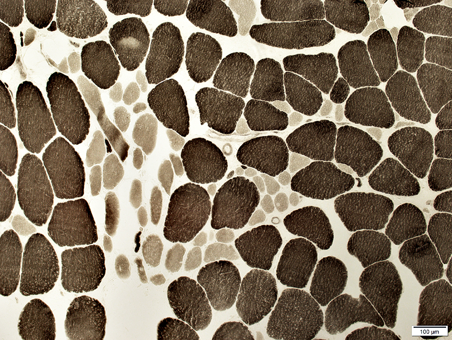 ATPase pH 9.4 stain |
May be large or small
May be present in groups or clusters
 ATPase pH 9.4 stain |
Serial sections with different stains
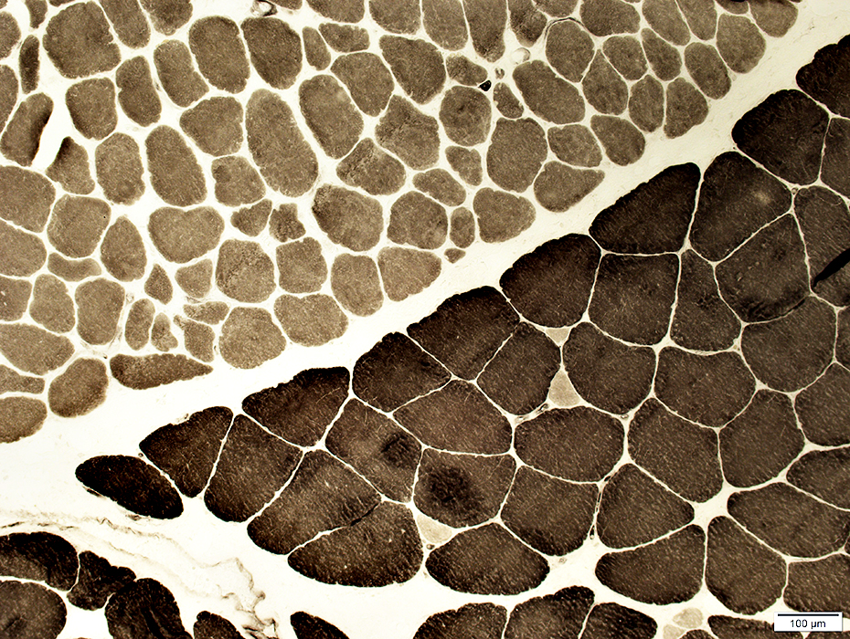
 ATPase pH 4.3 stain |
 ATPase pH 9.4 stain |
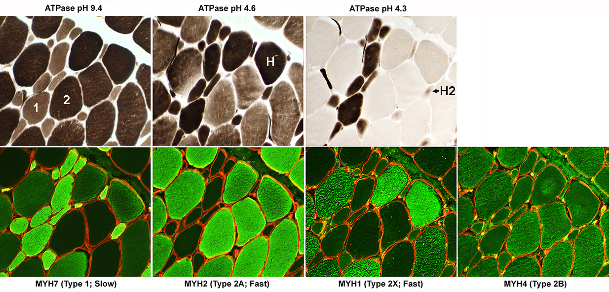
Abnormal Fiber Types
1: Contain both MYH7 & MYH42A (No staining at ATPase pH 4.6): Not present
2: Express MYH2 & MYH4
H: Dark at both ATPase pH 9.4 & 4.6; Express MYH7, MYH2 & MYH1
H2: Express both MYH7 & MYH1
 |
 PAS stain |
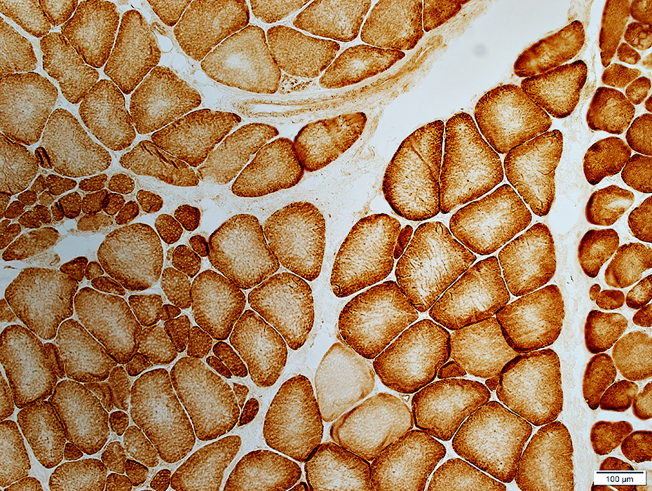 Cytochrome oxidase stain |
MYH7 mutation: Childhood
 H&E stain |
Varied
Bimodal distribution
 Congo red stain |
Muscle fiber internal architecture
Irregular
"Smudged" regions with reduced staining
 Gomori trichrome stain |
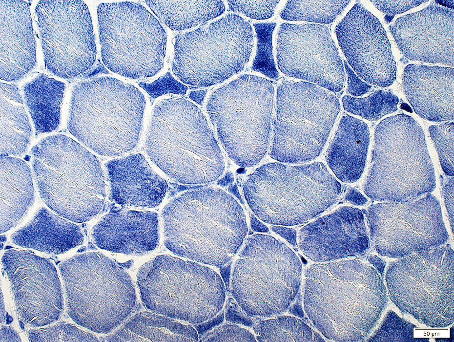
 NADH stain |
May be coarse with variable dark & light-stained areas
Core-like pathology: Minicores; Eccentric cores
 NADH stain |
Muscle fiber internal architecture
Reduced mitochondrial staining in some areas
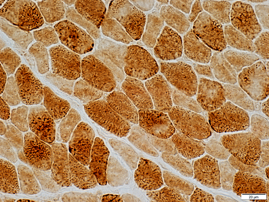 Cytochrome oxidase stain |
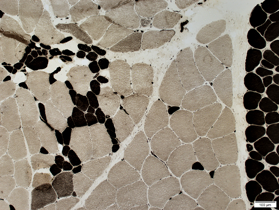
 ATPase pH 9.4 stain |
Type 2, especially 2B, fibers tens to be smaller than type 1
 ATPase pH 4.3 stain |
 ATPase pH 4.6 stain |
Neuromuscular junctions
Large & Dark stained for esterase
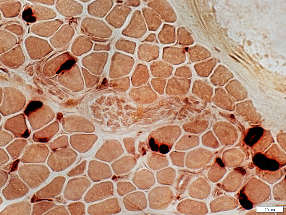 Esterase stain |
Return to MYH7
Return to Neuromuscular home page
11/14/2021