MTTL1 Encephalomyopathy (A3251G heteroplasmic mutation) 1
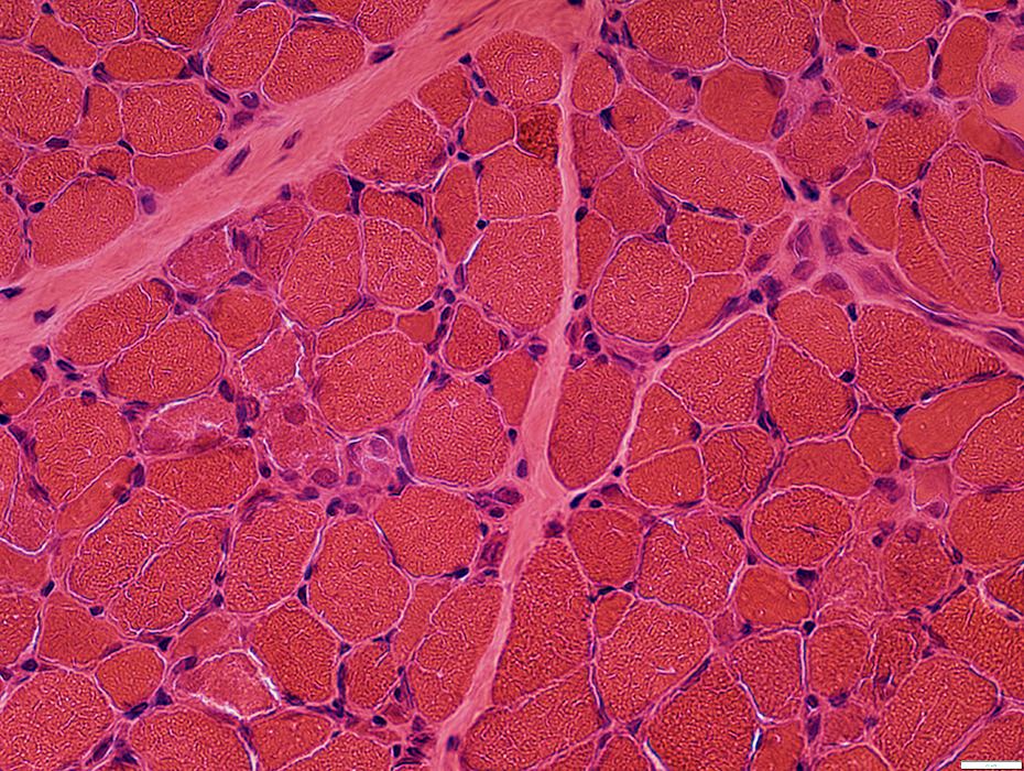
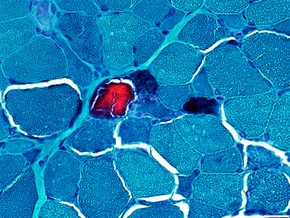
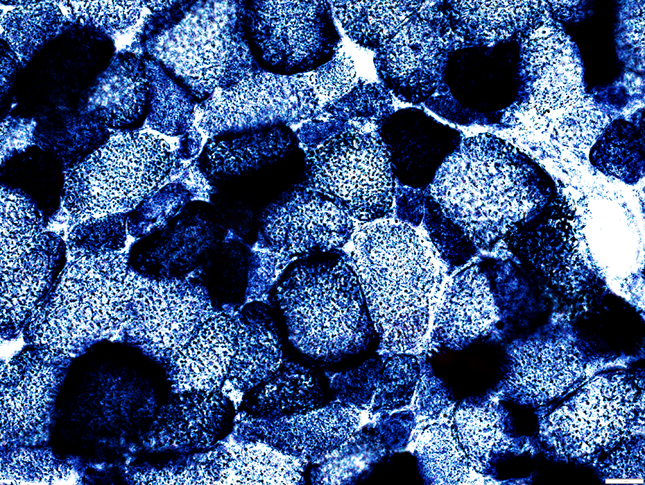
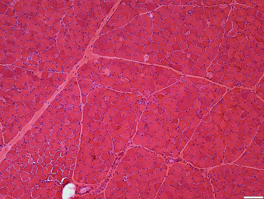 H&E stain |
 Congo red stain |
Muscle fibers: Lipid storage with round, clear droplets
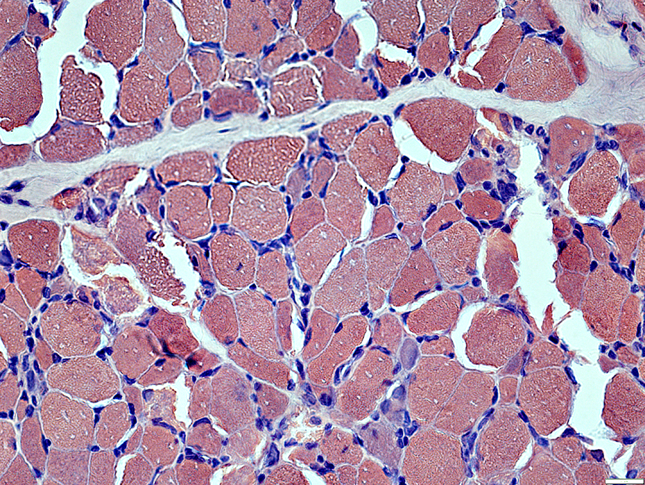
 H&E stain |
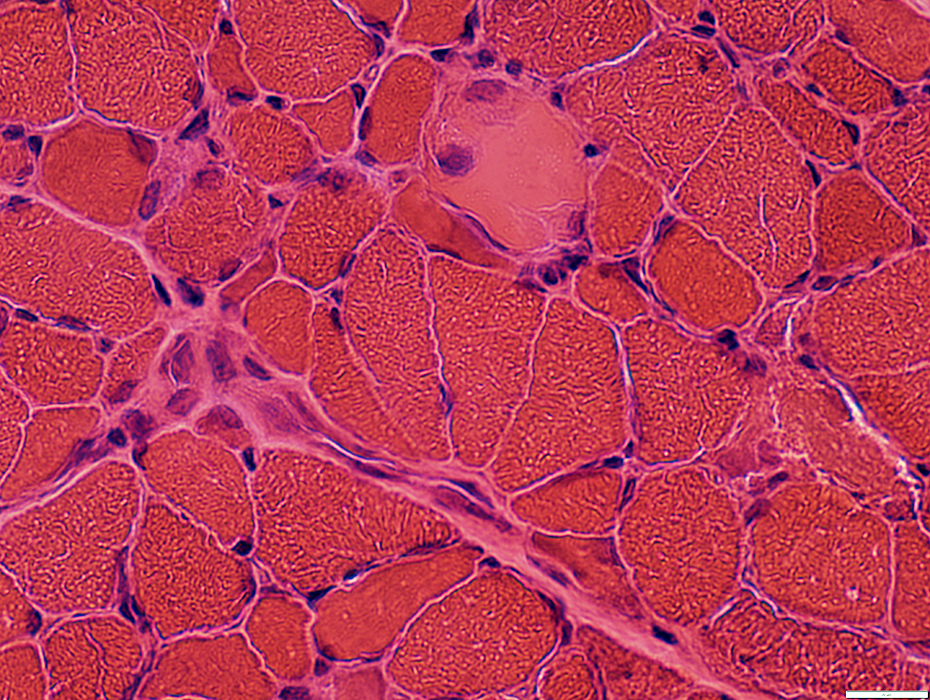
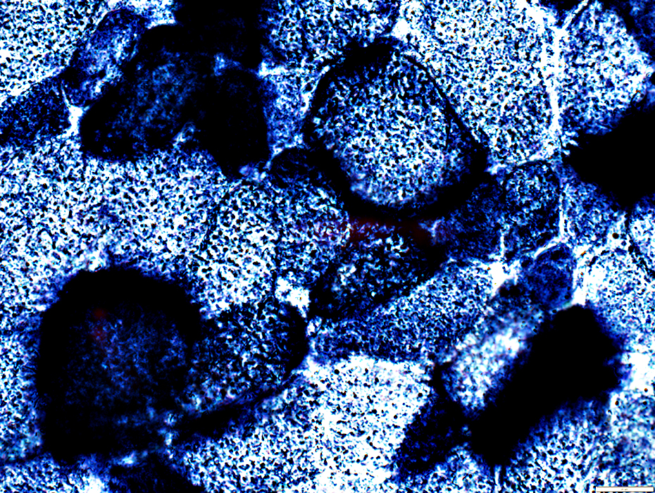
 H&E stain |
Anatomic Pattern: Scattered
Stages: Varied
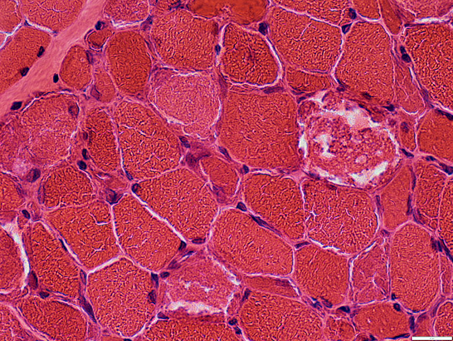
 H&E stain |
 H&E stain |
Anatomic Pattern: Scattered
Stages: Varied
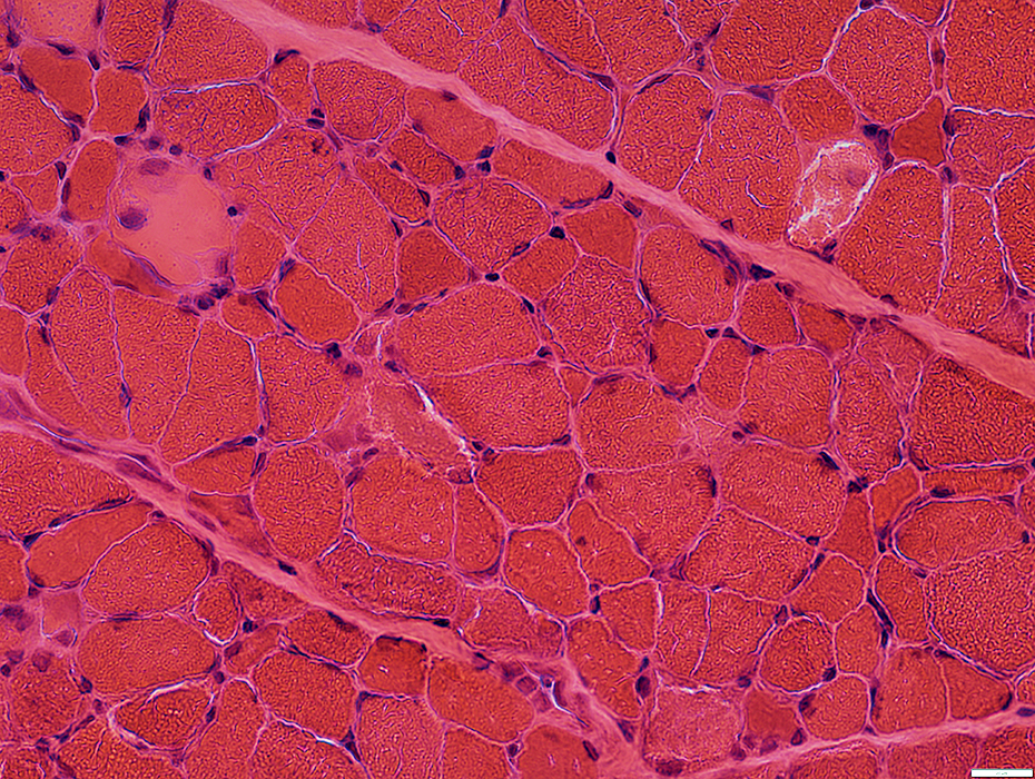
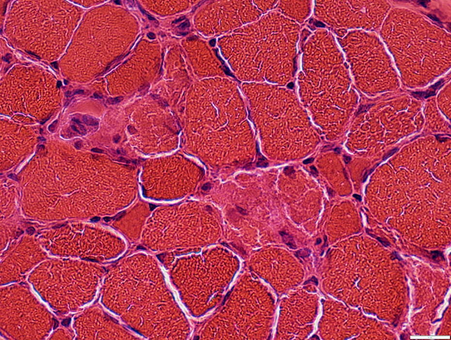 H&E stain |
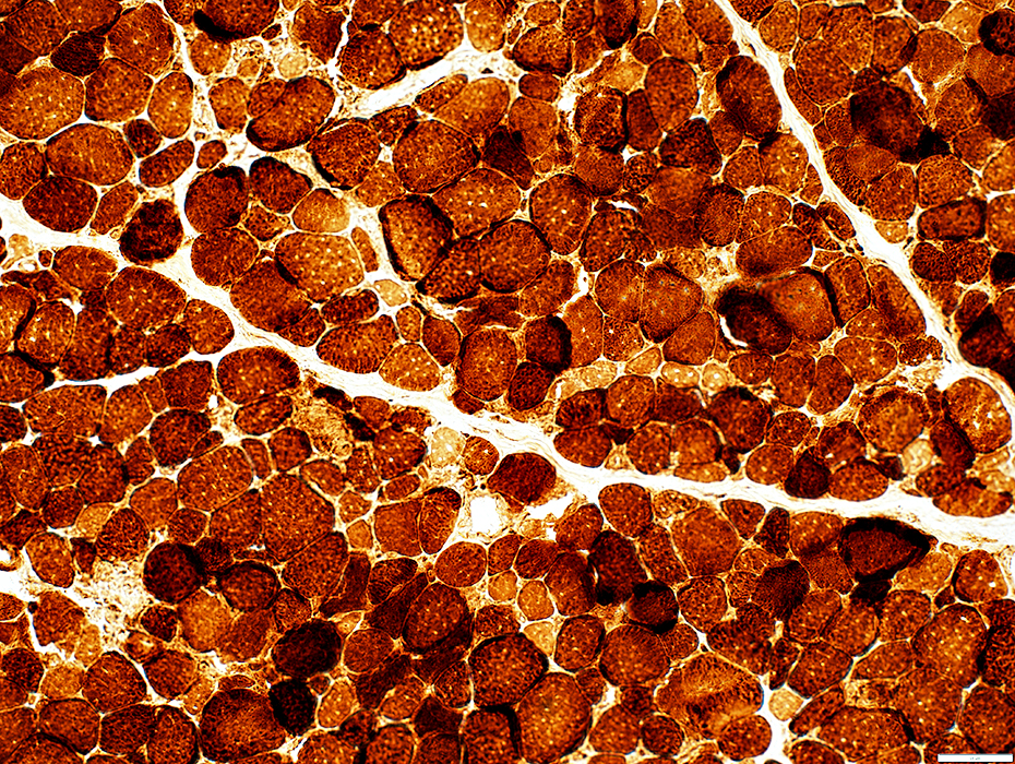
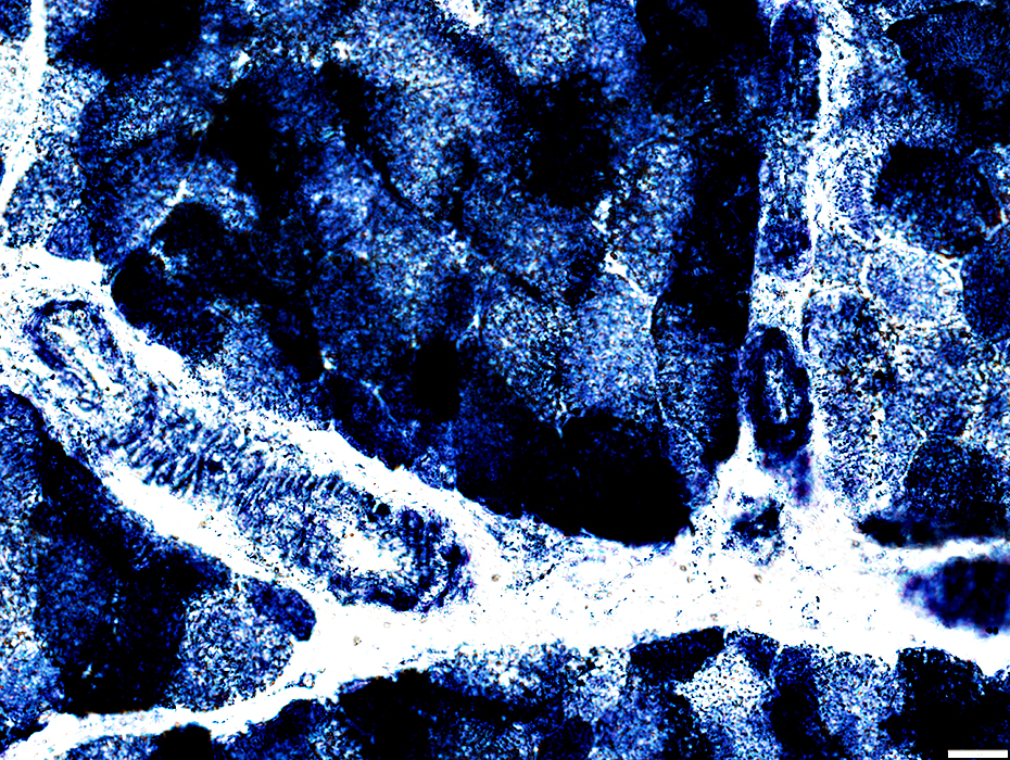
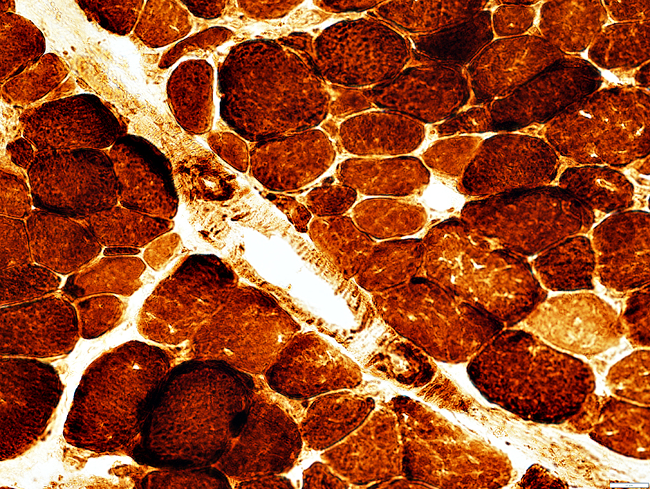
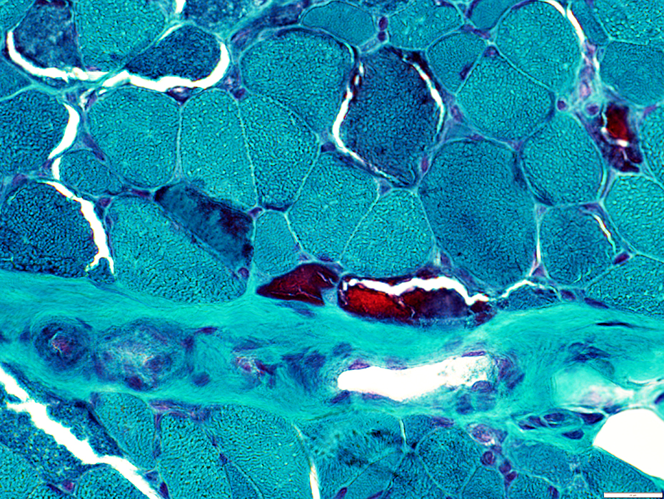 Gomori trichrome stain |
Fibers with prominent mitochondrial proliferation are stained dark red.
 Gomori trichrome stain |
Mitochondrial Pathology: Mitochondrial oxidative enzymes in muscle fibers
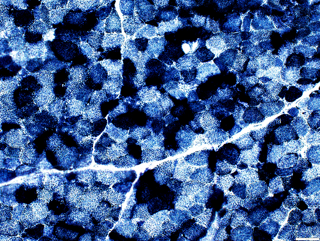
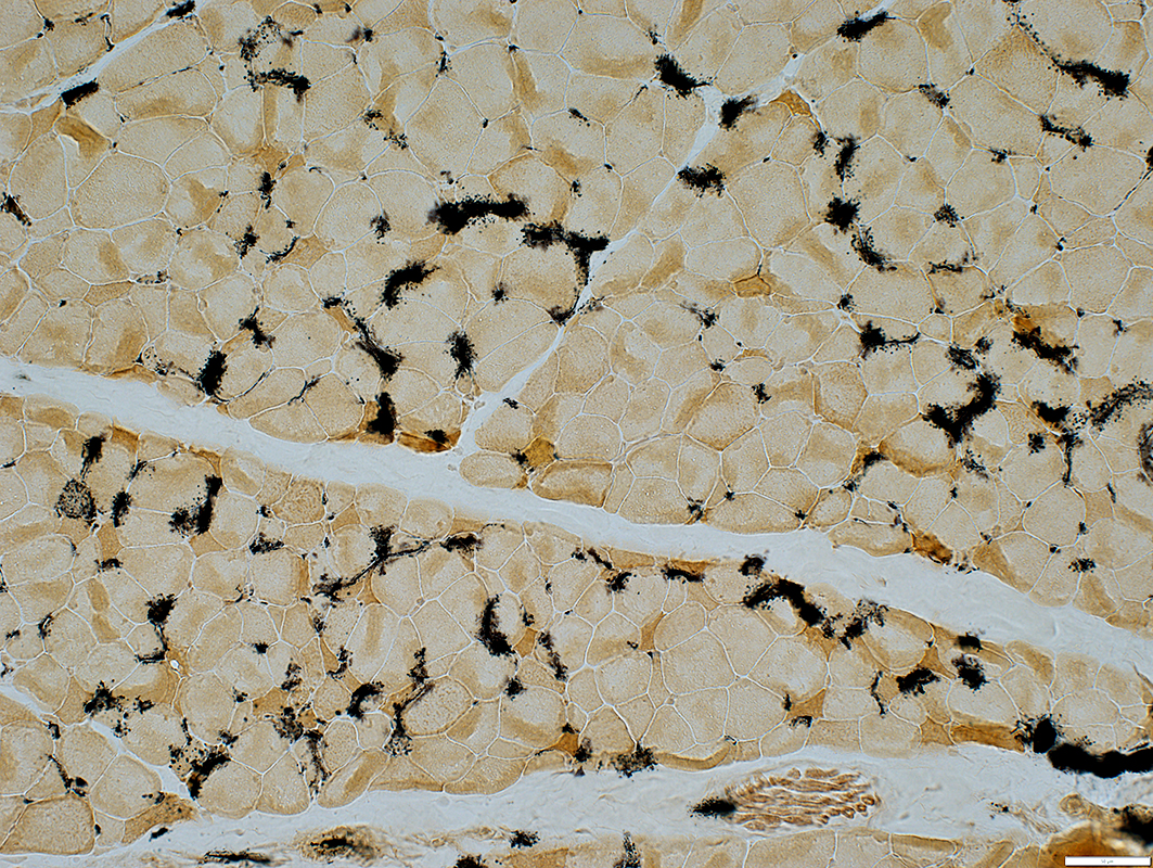
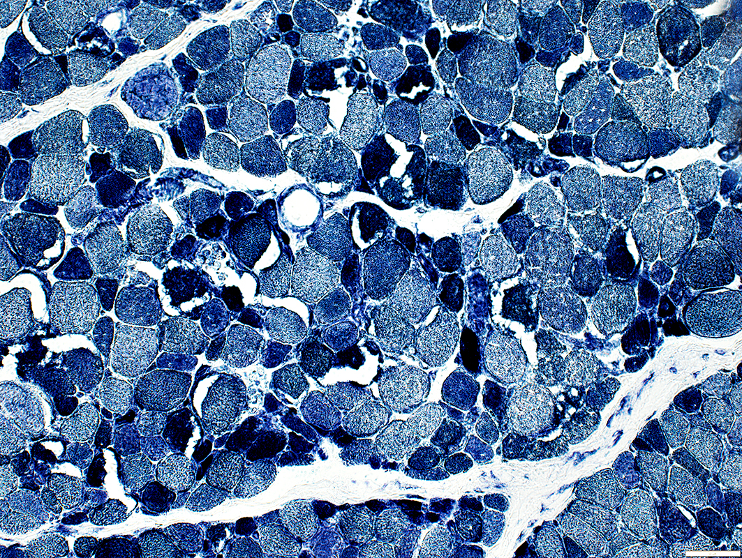
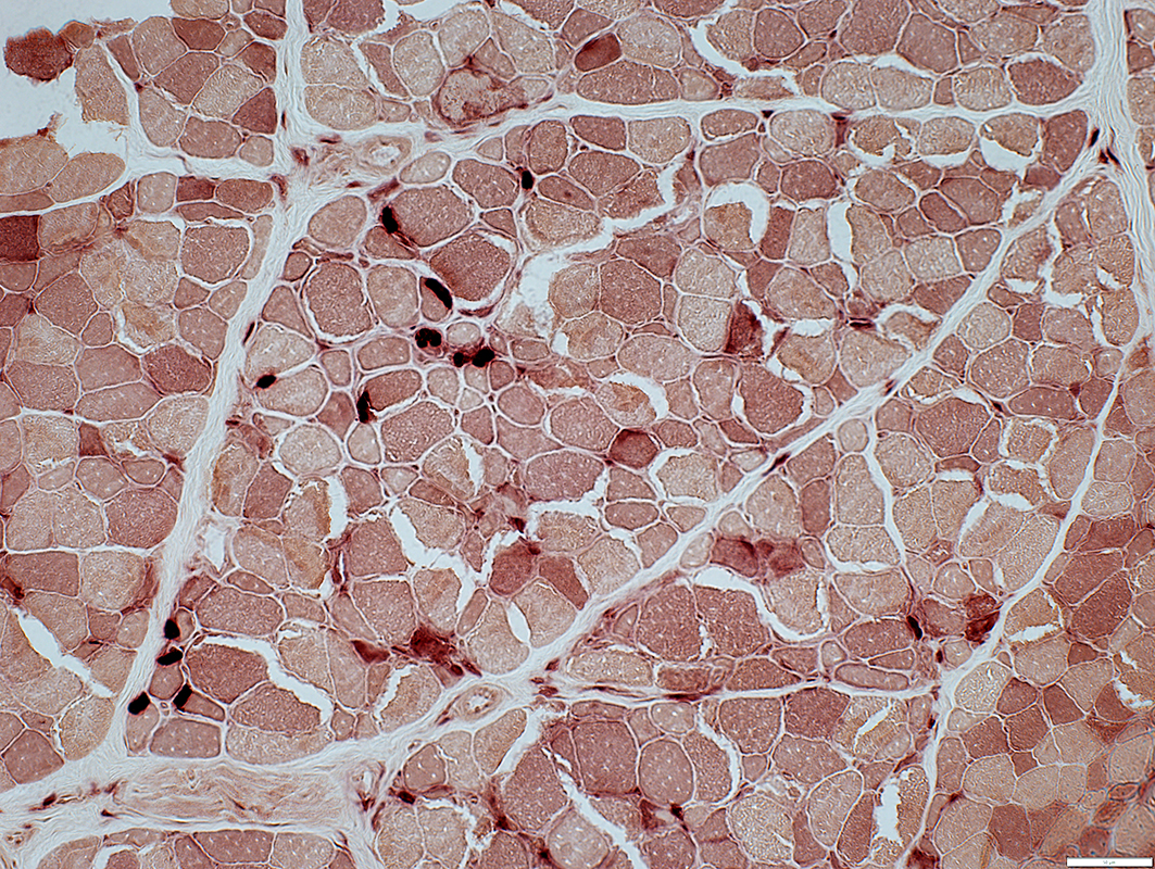
 Cytochrome oxidase (COX) stain |
Scattered muscle fibers with increased staining
No COX- fibers
 Cytochrome oxidase (COX) stain |
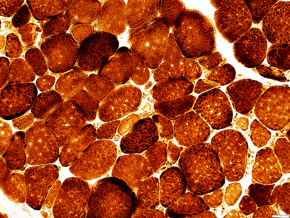
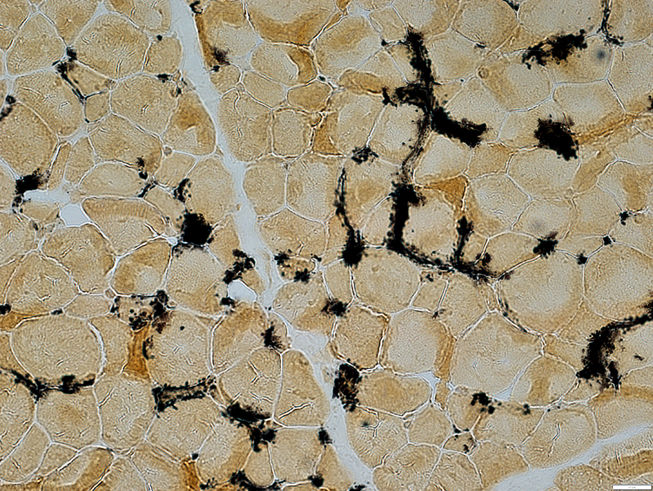
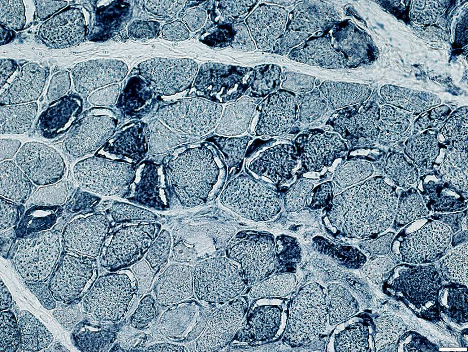
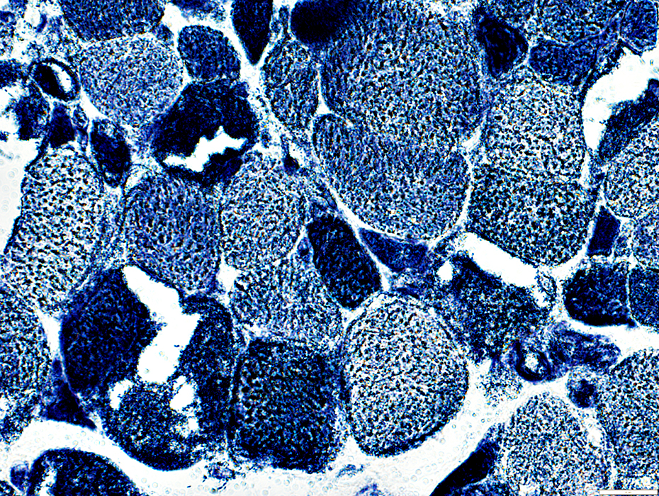
 Succinate Dehydrogenase (SDH) stain |
 Succinate Dehydrogenase (SDH) stain |
Succinate Dehydrogenase (SDH)
 Succinate Dehydrogenase (SDH) stain |
Scattered muscle fibers with increased staining
 Succinate Dehydrogenase (SDH) stain |
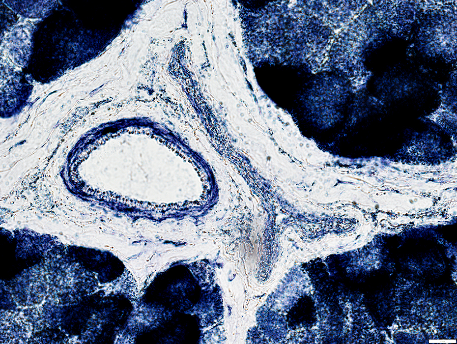
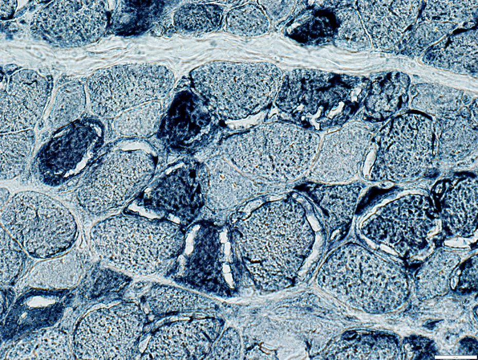
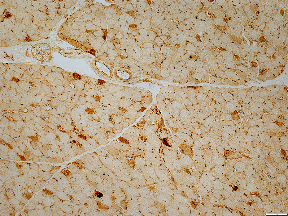
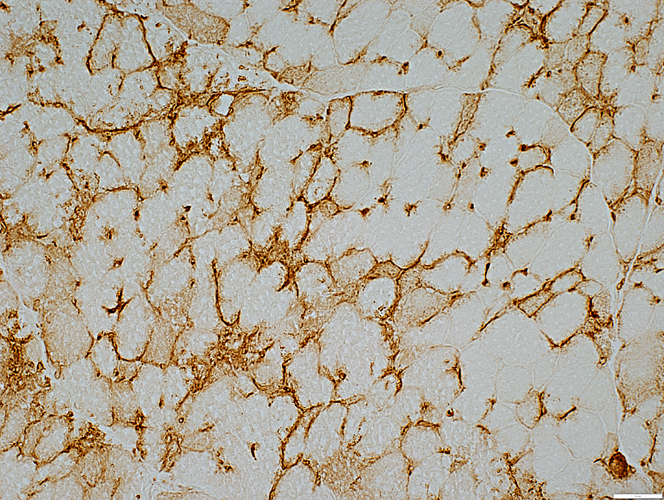
Mitochondrial Pathology: Mitochondrial oxidative enzymes in vessel walls
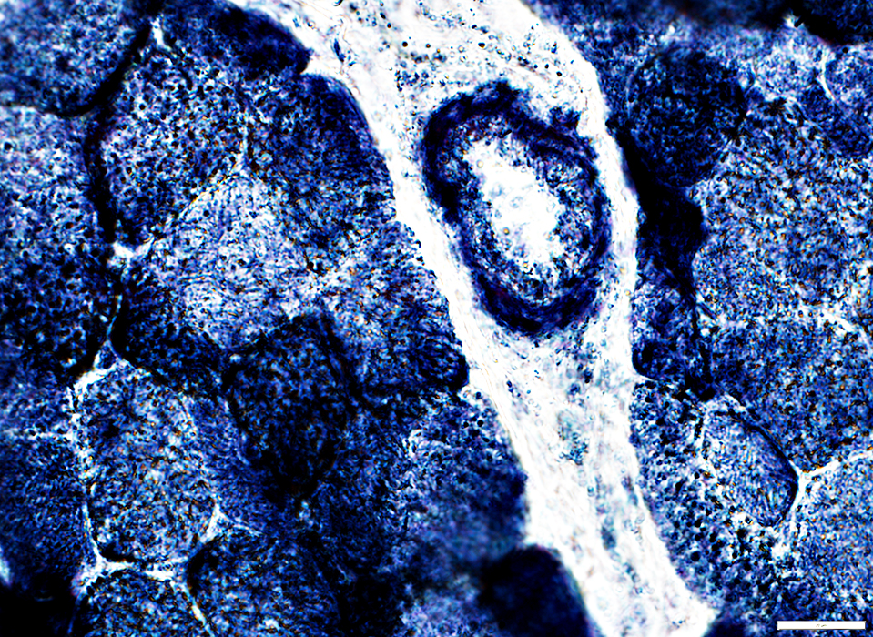 Succinate Dehydrogenase (SDH) stain |
Increased staining of wall of intermediate sized vessels in permysium
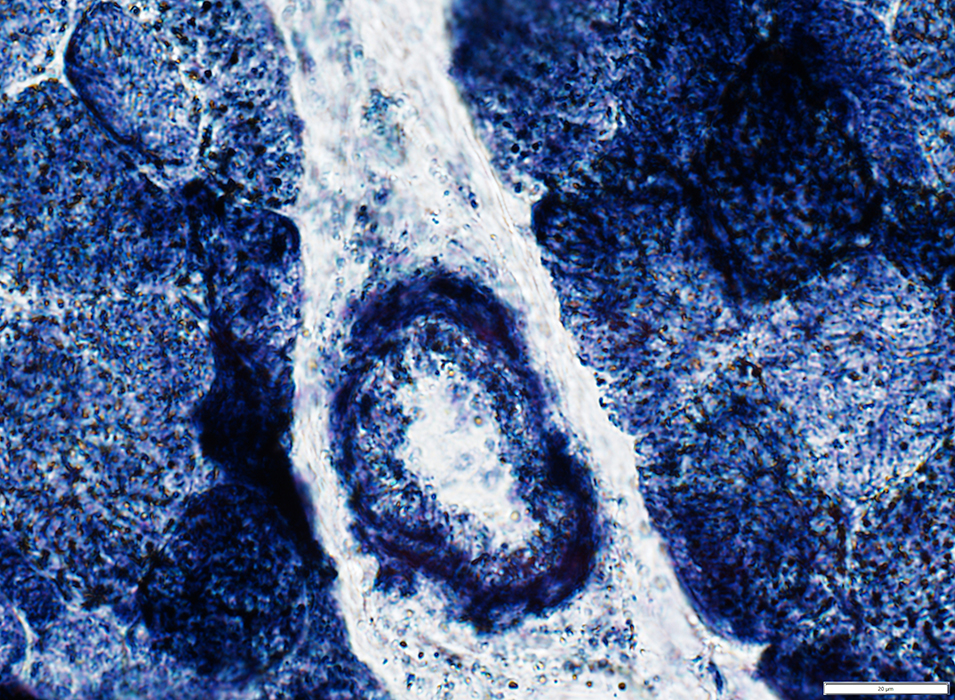 Succinate Dehydrogenase (SDH) stain |
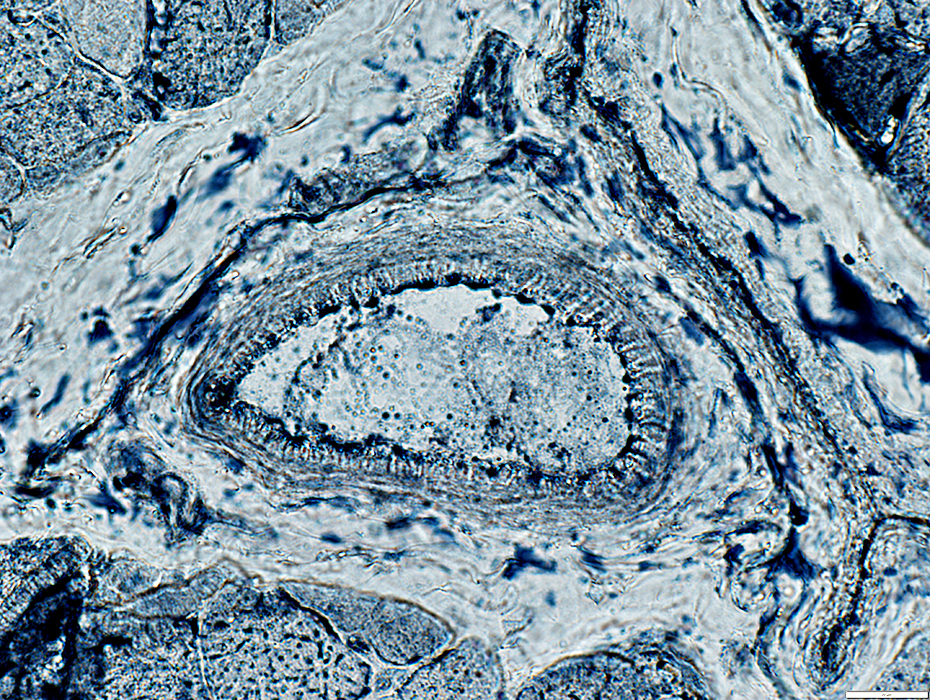
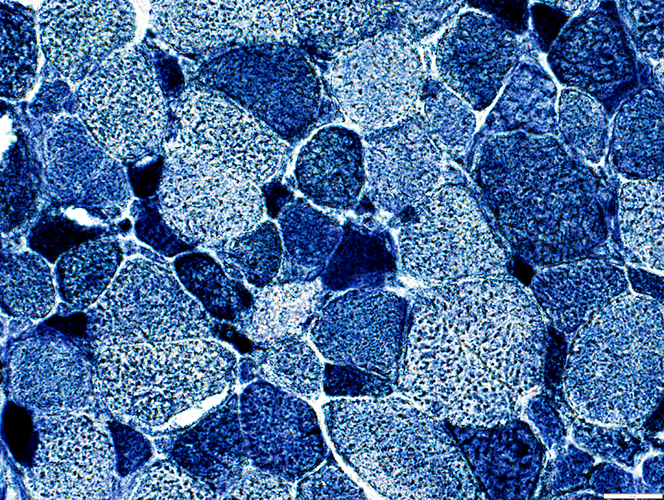
Cytochrome oxidase (COX)
Increased staining of walls of intermediate sized vessels in permysium
 Cytochrome oxidase (COX) stain |
Vessels
Capillaries
 Alkaline phosphatase stain |
 Alkaline phosphatase stain |
 Alkaline phosphatase stain |
Perimysial Vessels
 Congo red stain |
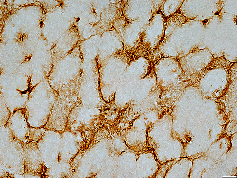
 Succinate Dehydrogenase (SDH) stain |
Increased SDH in vessel wall & endothelial cells
Increased lipid in vascular endothelial cells
 Sudan Black stain |
Lipid Storage in muscle fibers
Many smaller, and some larger, muscle fibers have dark stained cytoplasm & small to moderate sized lipid droplets
 Sudan stain |
 Sudan stain |
Many smaller, and some larger, muscle fibers have dark stained cytoplasm & small to moderate sized lipid droplets
Clear rims, possibly lipid droplets, surround parts of some muscle fibers
 Sudan Black stain |
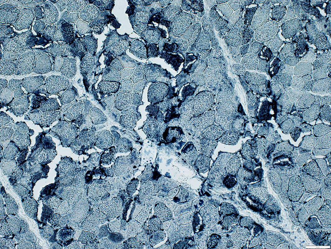
 NADH stain |
Many small muscle fibers are dark stained
 NADH stain |
 NADH stain |
Neuromuscular Junctions
Many appear large
Esterase stains cytoplasm of scattered large and small muscle fibers dark
 Esterase stain |
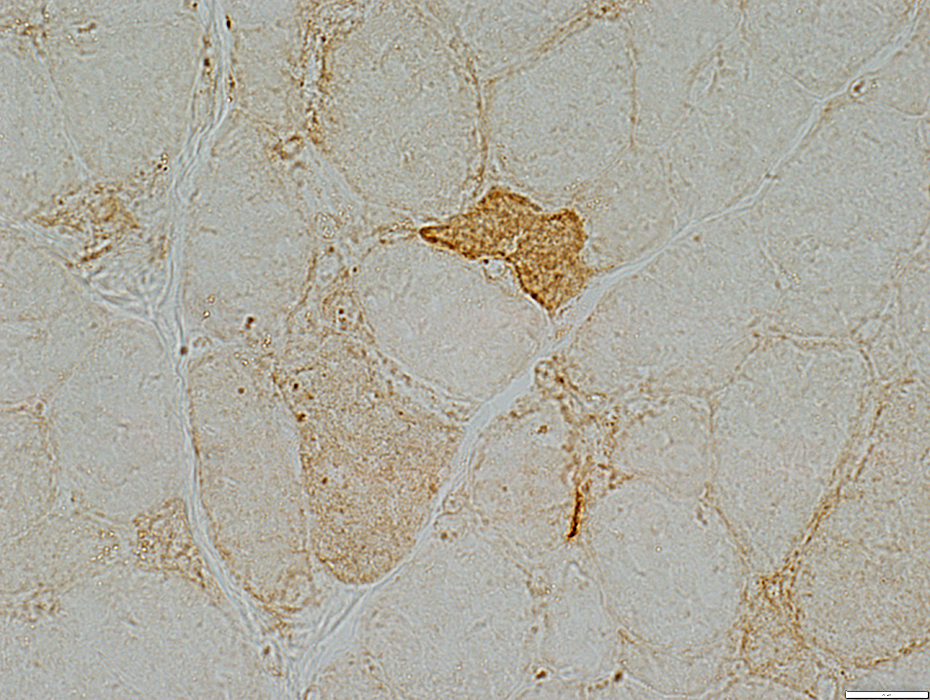
LC3 stain |
LC3 stains cytoplasmic aggregates, clustered or scatterd, in many muscle fibers
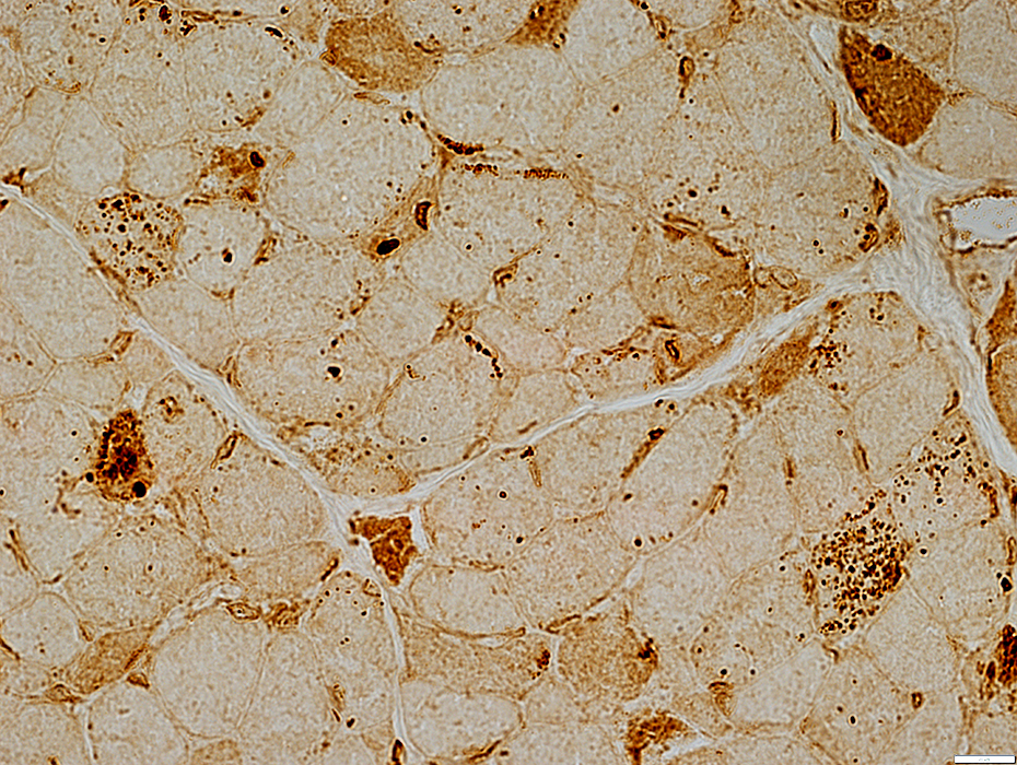 LC3 stain |
 LC3 stain |
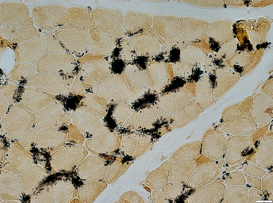
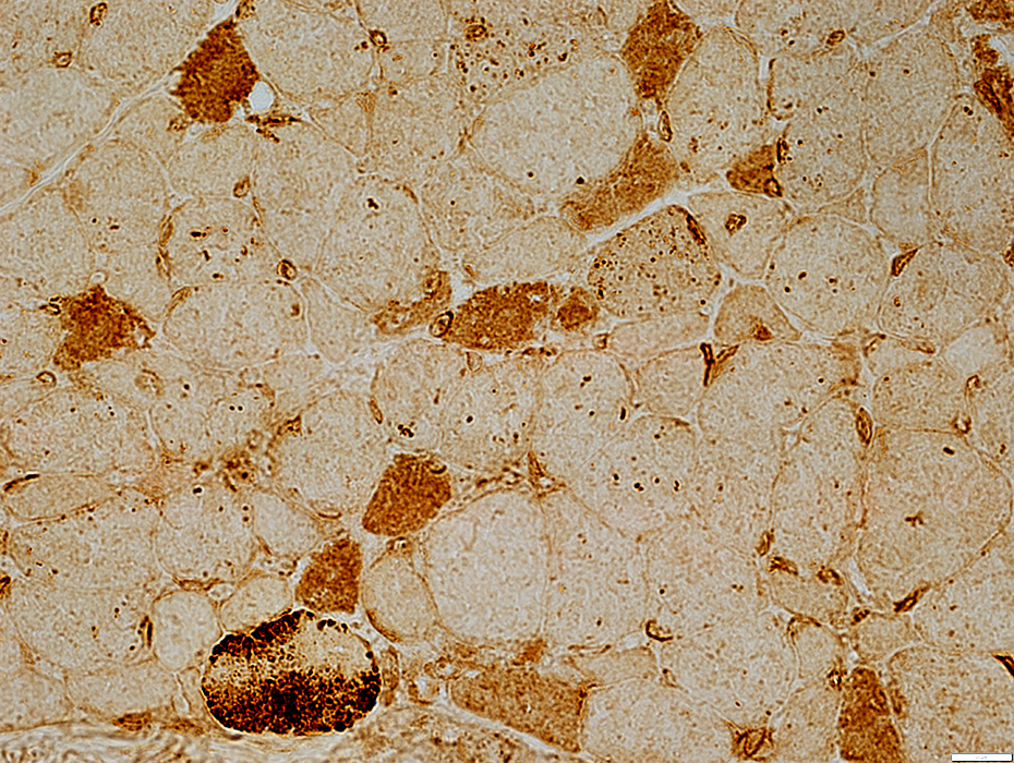
 MxA stain |
MxA stains the cytoplasm of scattered smaller muscle fibers
 MxA stain |
 MxA stain |
Control muscle: No MxA staining in muscle fibers
 MxA stain |
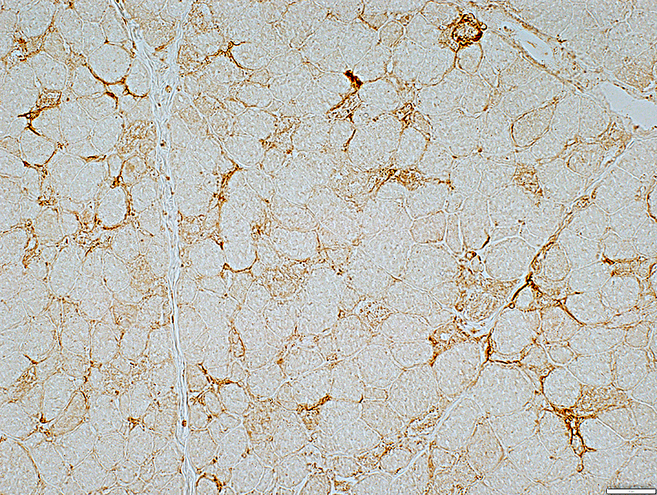
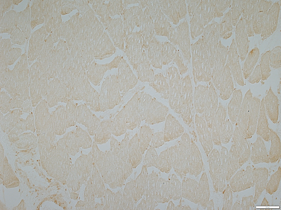
 MHC Class I stain |
Cytoplasm of scattered intermediate sized muscle fibers
Irregular cells associated with endomysial capillaries
 MHC Class I stain |
Return to: MELAS
Return to: Neuromuscular Home Page
1. J Med Case Rep 2019;13:63
9/14/2022