Mi-2 antibodies: α & β
|
Case 1 Case 2 |
Case 1
85 yo female with 1 year of progressive dysphagia, & symmetric weakness, proximal legs & arms + distal arms with 50 lb weight loss
CK = 2,800.
Inflammatory Myopathy
Muscle fibers
Morphology: Varied sizes; Many small
Internal architecture
Cytoplasm
Immune
MHC1: Upregulated on muscle fibers
IgG present in myonuclei
SCRT1 expression: Increased
Inflammation
Types
Lymphocytes: Foci & Scattered in Endomysium, Types
Histiocytes: Scattered in Endomysium; Few in Perimysium
Distribution: Endomysial; Foci & Scattered
Cell Foci contain: Small vessels with endothelium & thin wall
Connective tissue
Perimysium: Fragmented in some regions
Endomysium: Increased; C5b9 deposition
Vessels
Endomysial capillaries: Large; Reduced numbers; Alkaline phosphatase +
Nailfold capillaroscopy
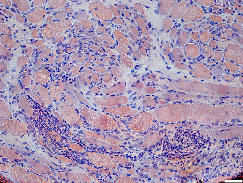
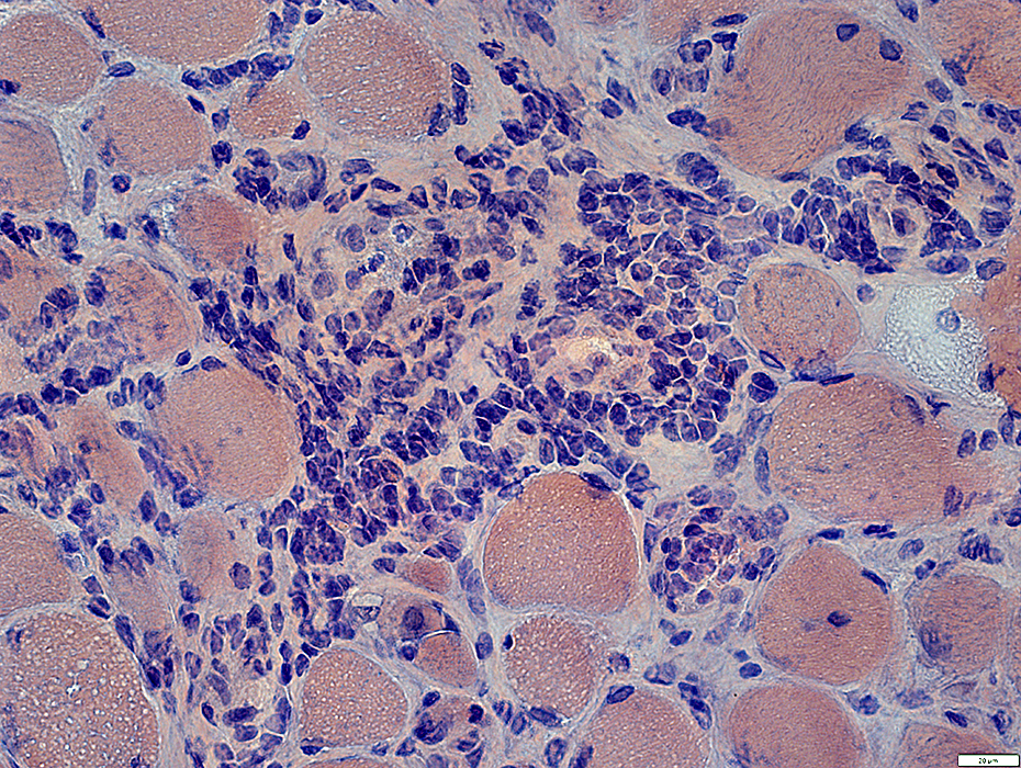
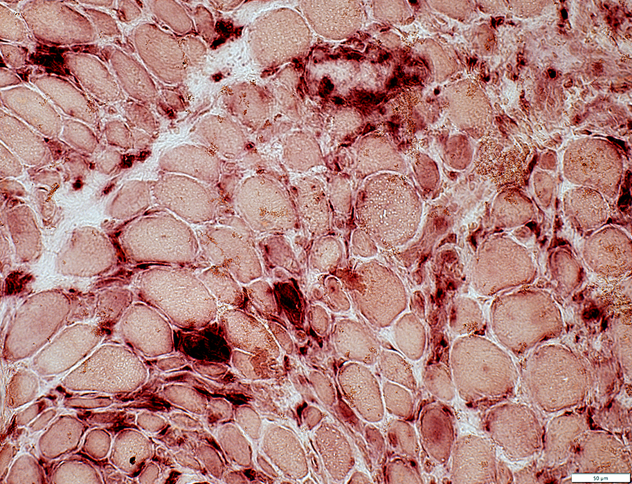
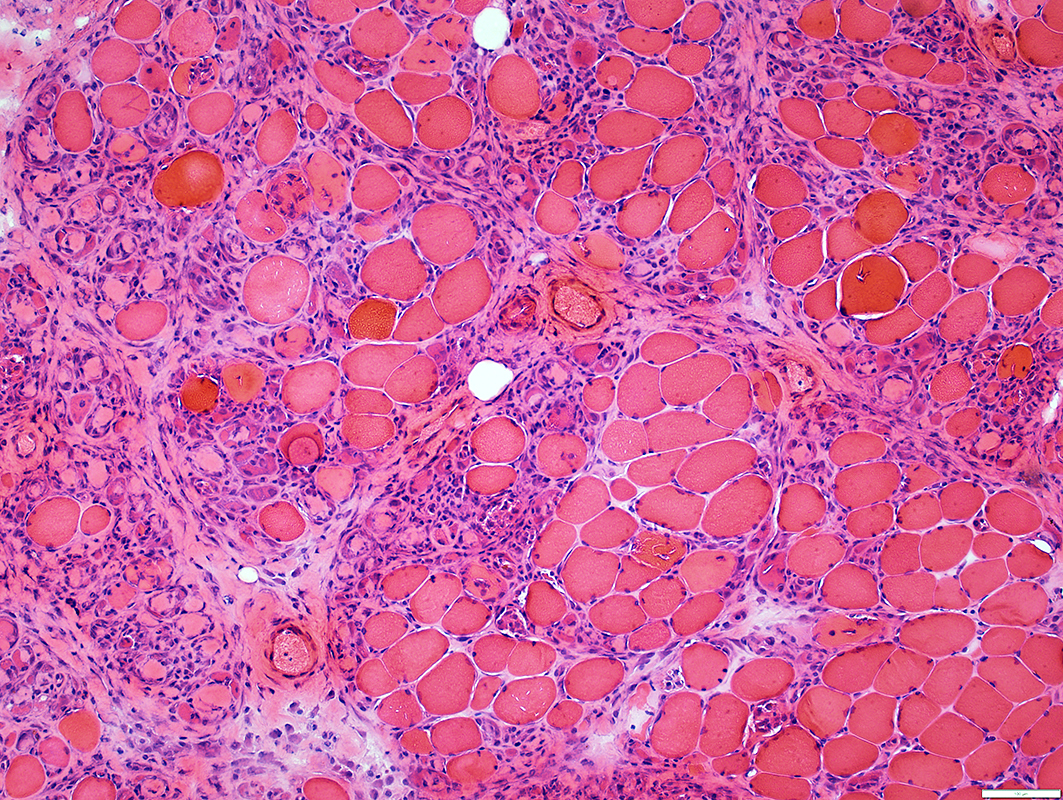
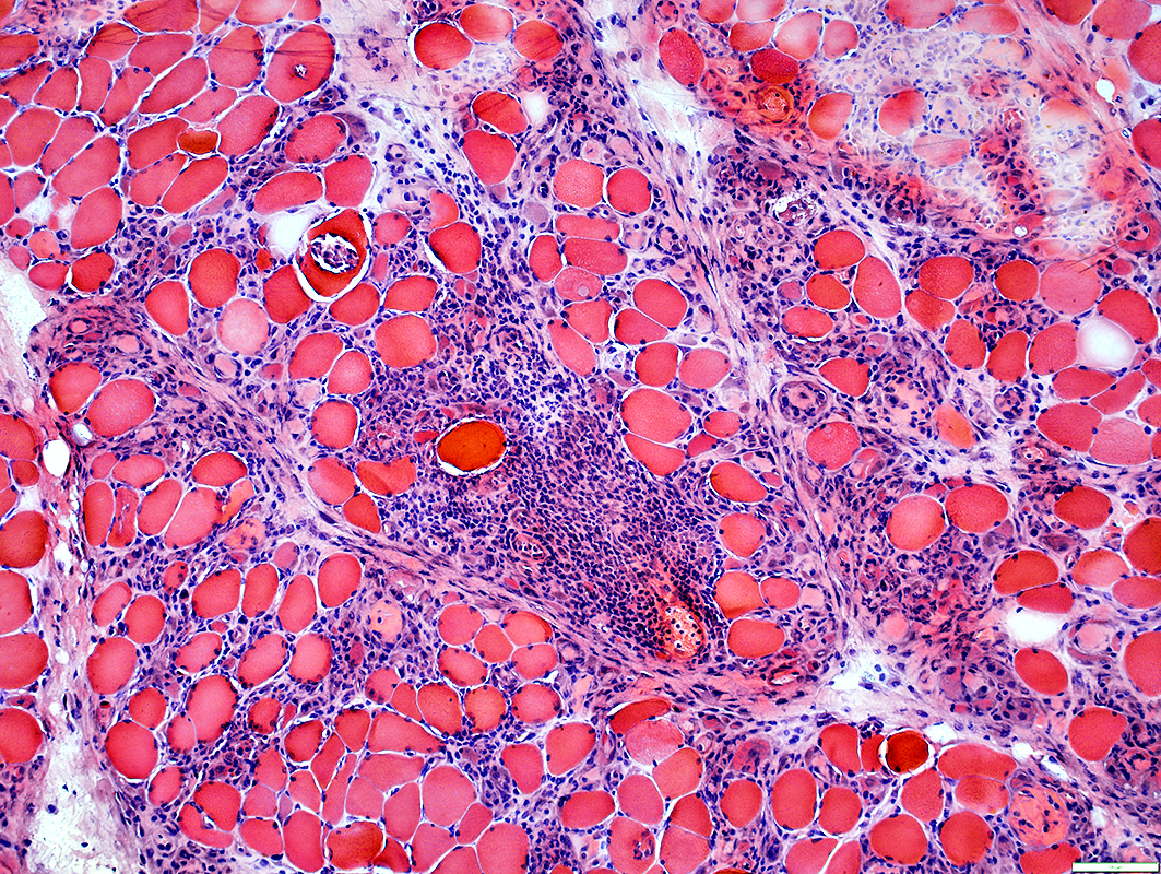
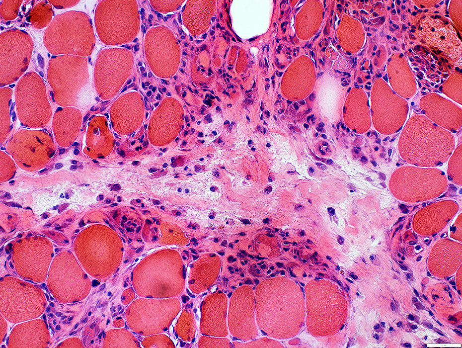
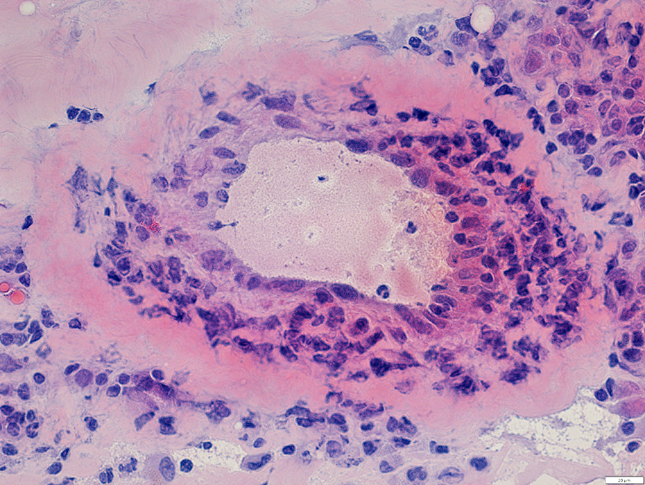
 H&E stain |
Sizes: Varied; General atrophy
Inflammation
Scattered in endomysium & perimysium
Lymphocyte Foci
Endomysial connective tissue
Increased between muscle fibers
 Congo red stain |
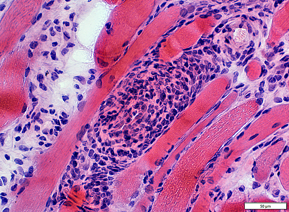
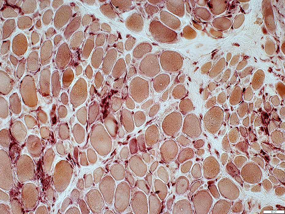
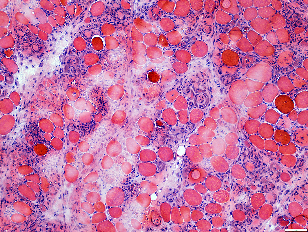
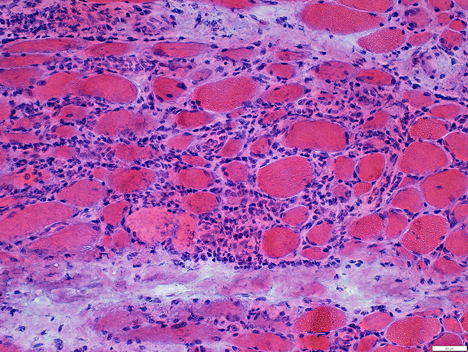 H&E stain |
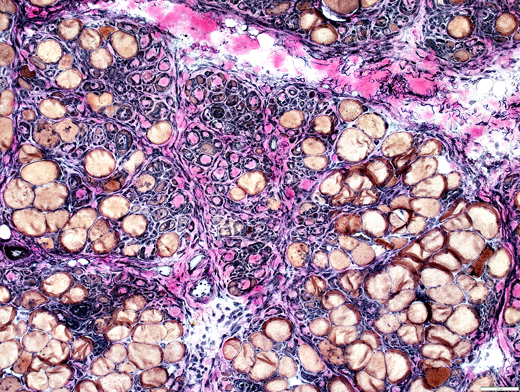
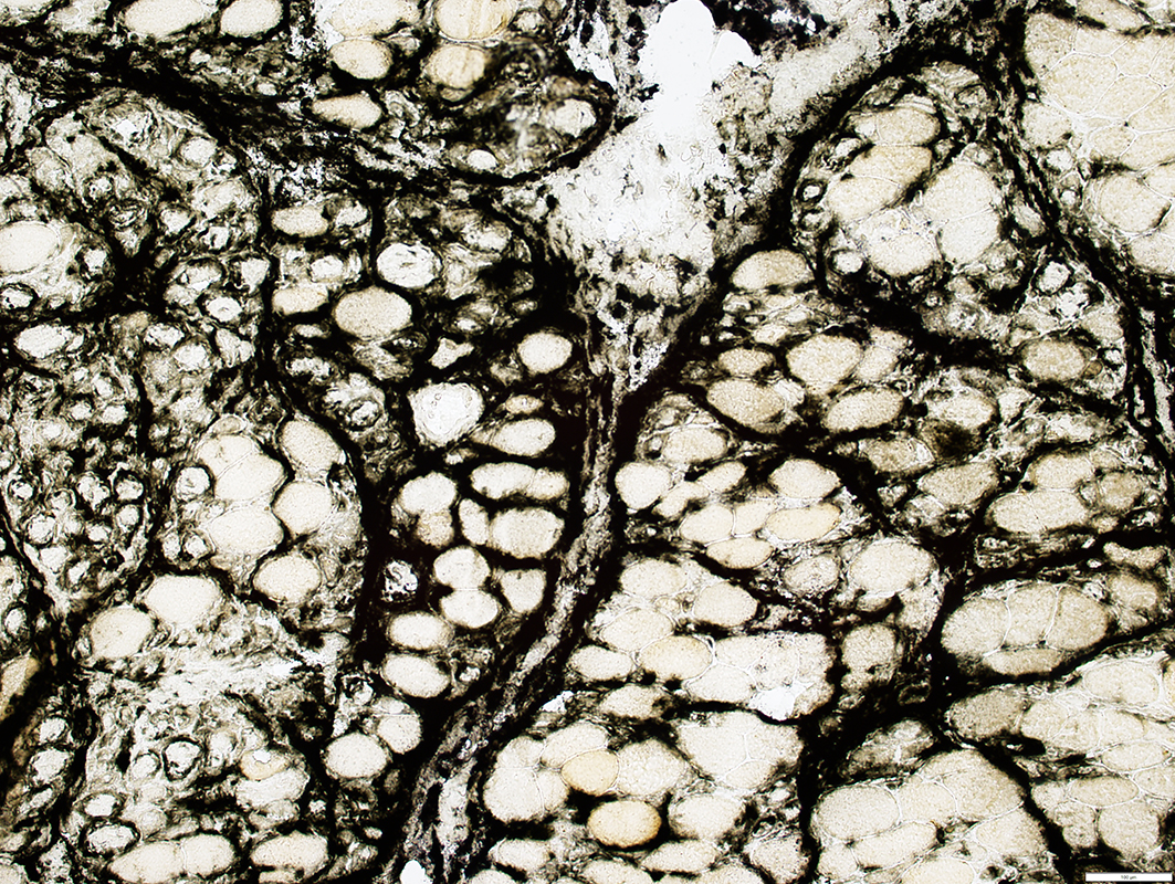
Scattered in endomysium betweem muscle fibers
Perimysial connective tissue: Fragmented
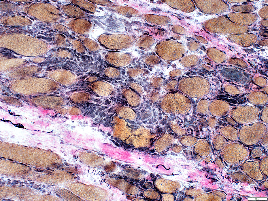 VvG stain |
 Congo red stain |
Often contain smaller vessel with thin wall
 Congo red stain |
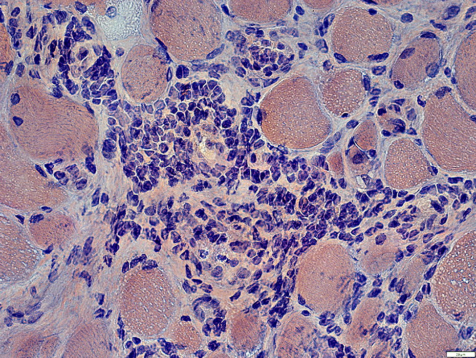
 H&E stain |
 CD4 stain |
Often contain CD4 cells
 CD4 stain |
 CD8 stain |
|
Lymphocyte cell foci CD8 cells Scattered within foci and in endomysium Form small clusters 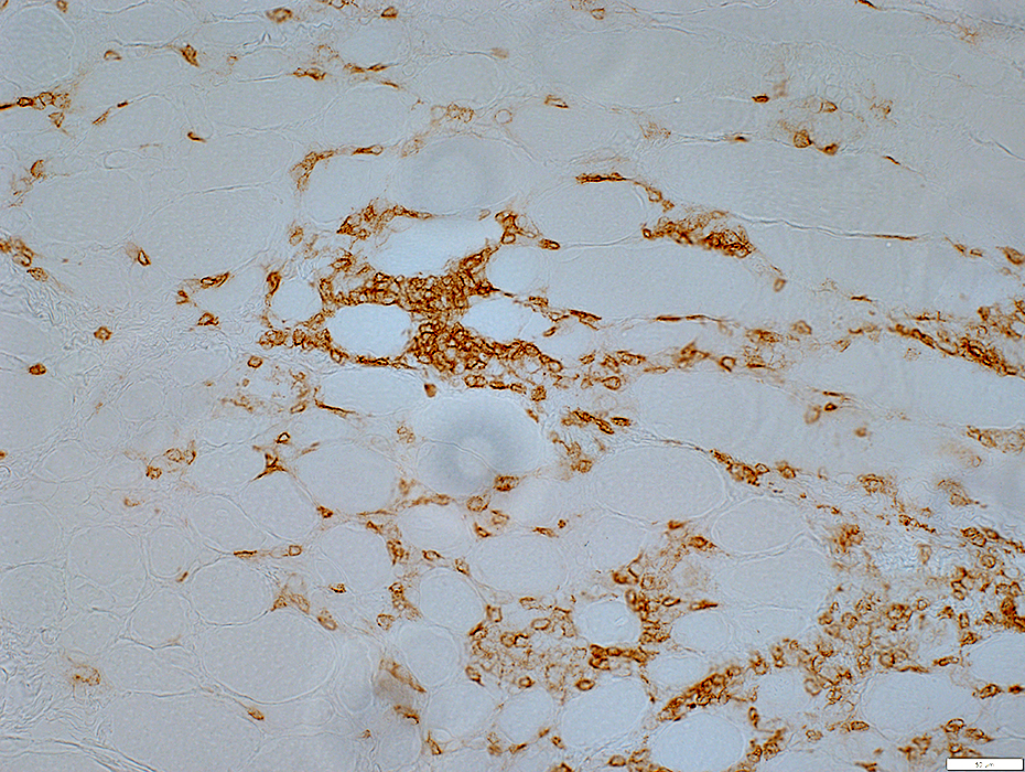 CD8 stain |
 CD8 stain |
 CD20 stain |
CD20 cells
Scattered in endomysium
Form small clusters in perimysium
 CD20 stain |
Histiocytes
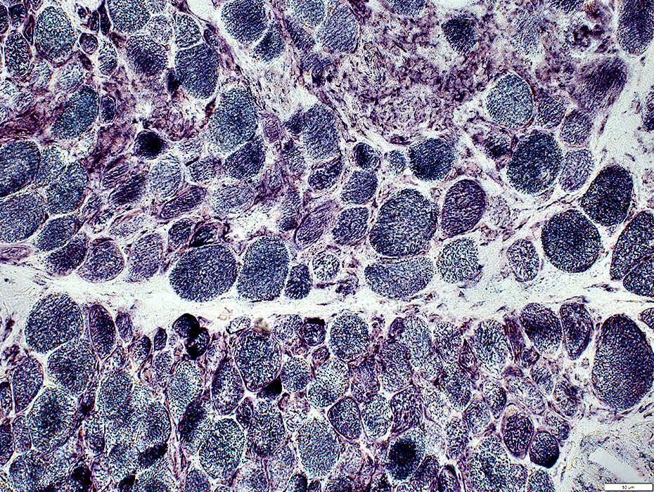
 Acid phosphatase stain |
Scattered in: Endomysium & Perimysium
Associated with damaged muscle fibers
 Acid phosphatase stain |
Esterase+ histiocytes are
Scattered in: Endomysium & Perimysium
 Esterase stain |
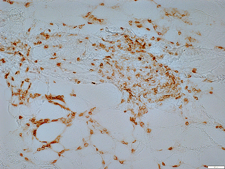
 CD163 stain |
Scattered in: Endomysium & Perimysium
 CD163 stain |
 HAM56 stain |
Scattered in: Endomysium & Perimysium
 HAM56 stain |
 HAM56 stain |
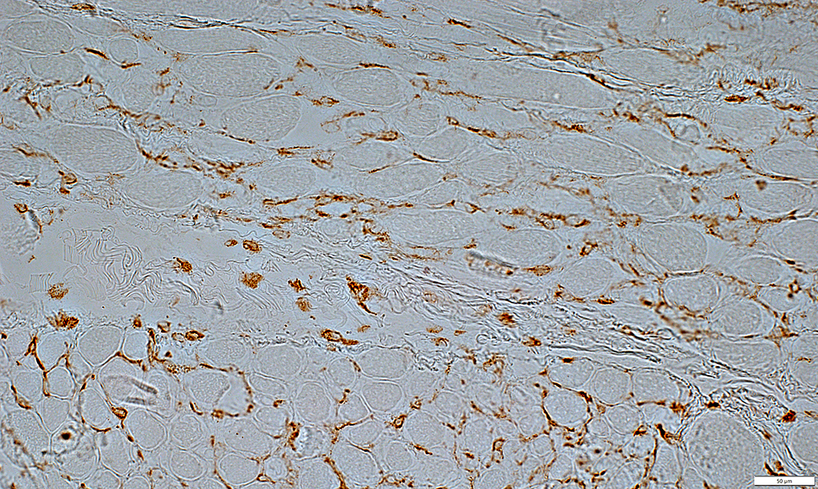
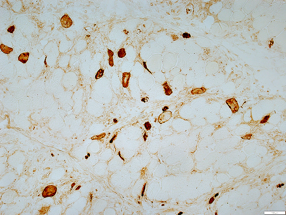
 MxA stain |
Endomysial cells
Muscle fiber cytoplasm: Scattered very small fibers
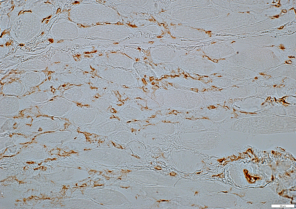
 MxA stain |
 MxA stain |
Capillary pathology
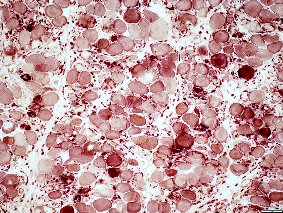
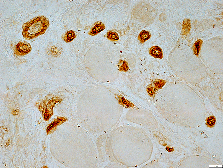
 Alkaline phosphatase stain |
Increased staining by alkaline phosphatase
Muscle fibers
Cytoplasm of scattered very small, immature muscle fibers is stained by alkaline phosphatase
 Alkaline phosphatase stain |
Endomysial Capillaries
Size: Large
Number: Reduced; Scattered muscle fibers with no adjacent capillary
 UEA1 stain |
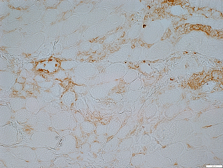
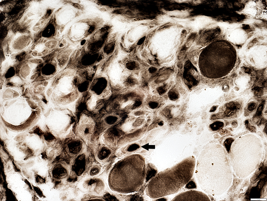
 C5b-9 stain |
Endomysial connective tissue
Cytoplasm of rare necrotic muscle fiber (Below; Arrow)
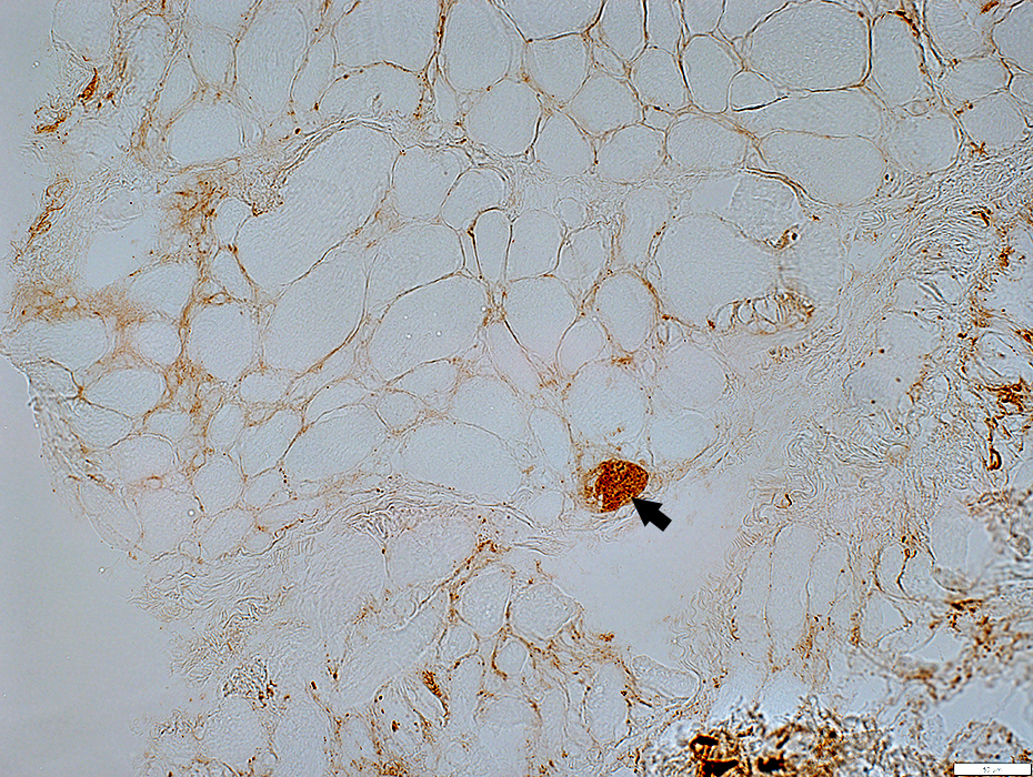 C5b-9 stain |
 C5b-9 stain |
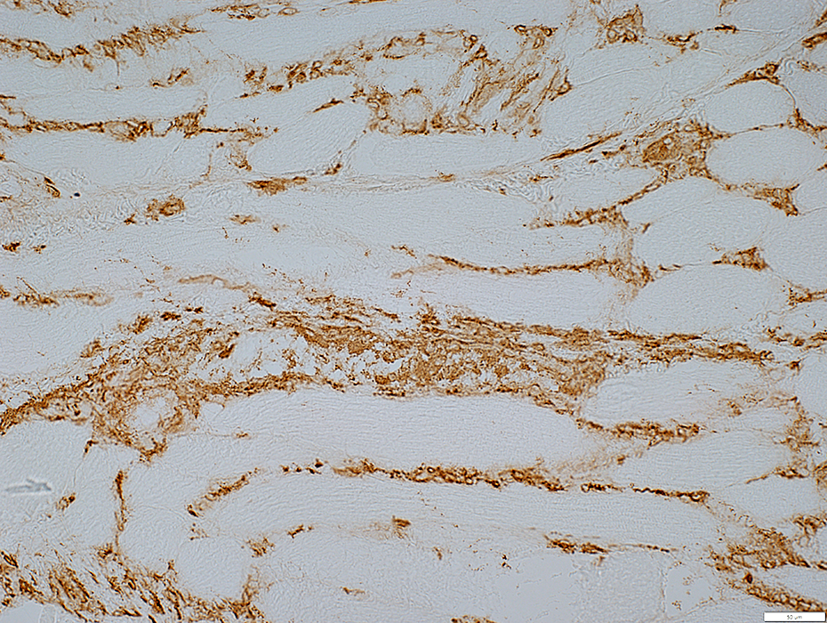
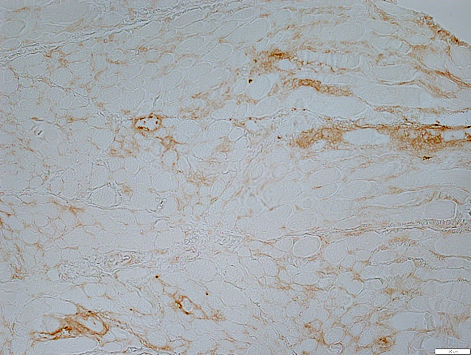
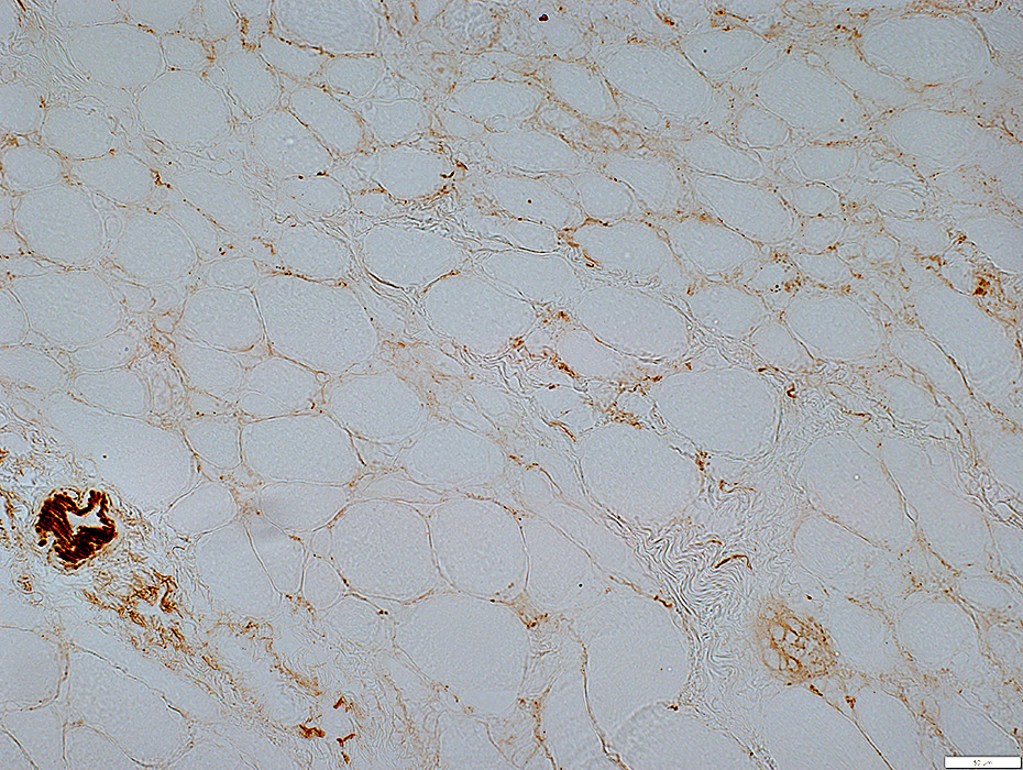
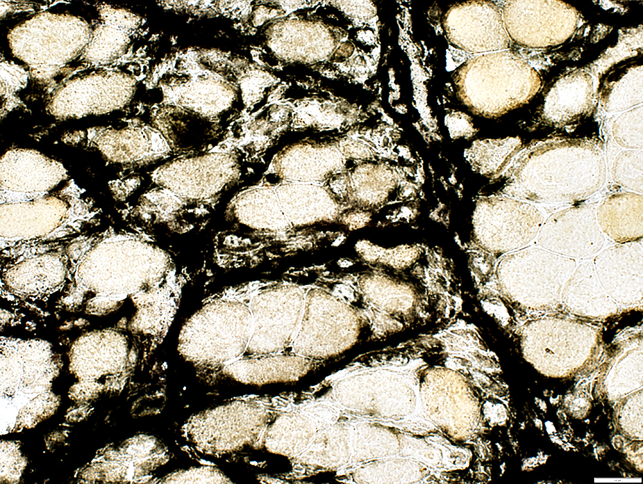
Endomysial Connective Tissue
Increased between muscle fibers
Stains for decorin
 Decorin stain |
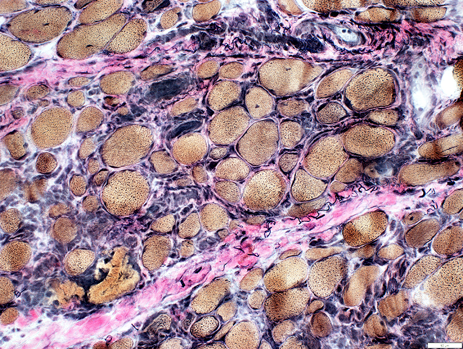 VvG stain |
Sizes: Varied
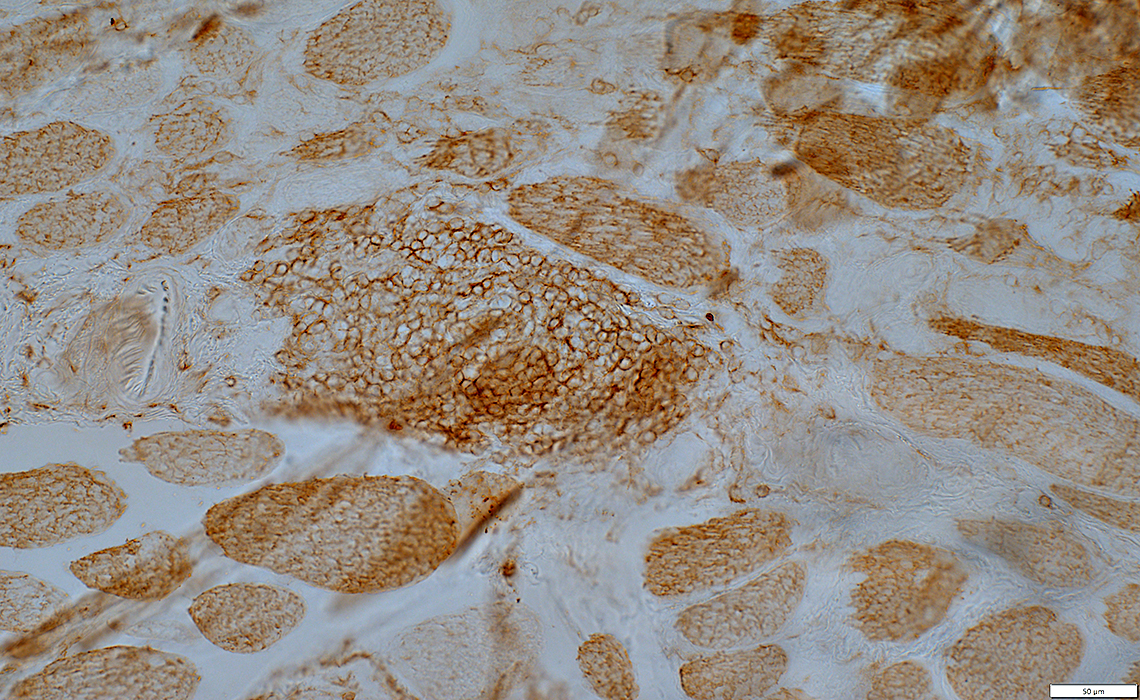
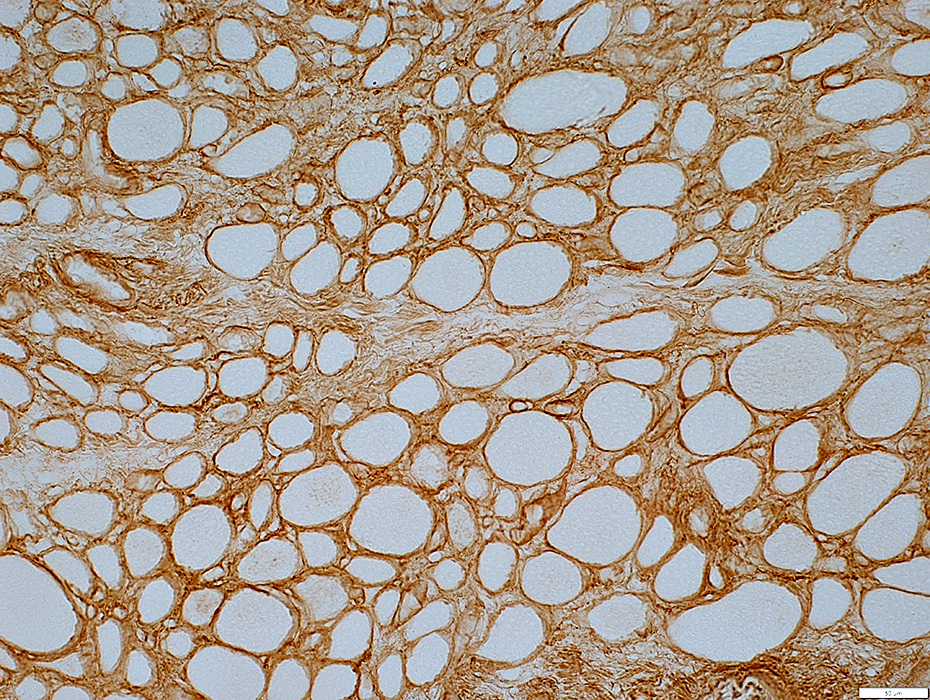
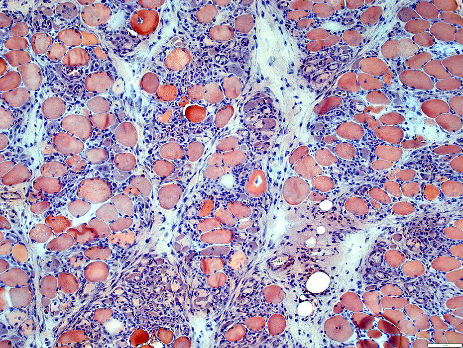
MHC1: Difffusely upregulated by muscle fibers (Below)
 MHC1 stain |
 MHC1 stain |
Sizes: Varied
MHC1: Difffusely upregulated by muscle fibers (Below)
Inflammation
Many MHC1+ cells in endomysium & perimysium
 MHC1 stain |
Muscle Fibers
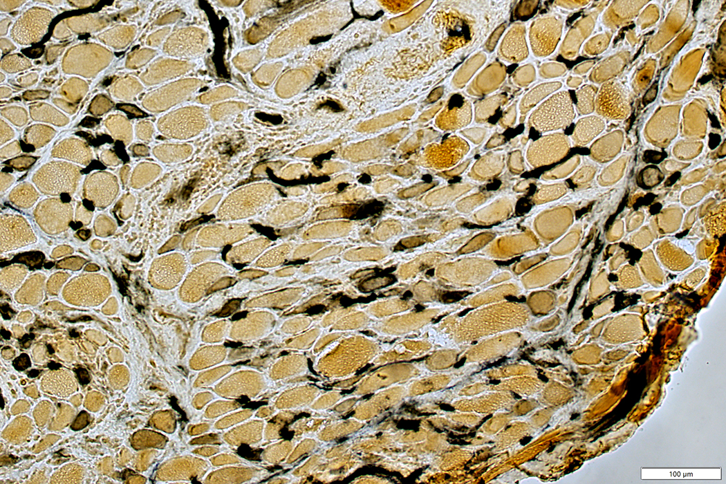
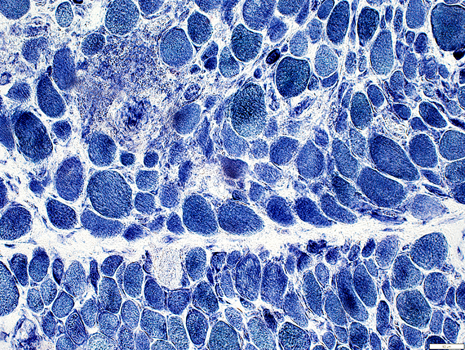
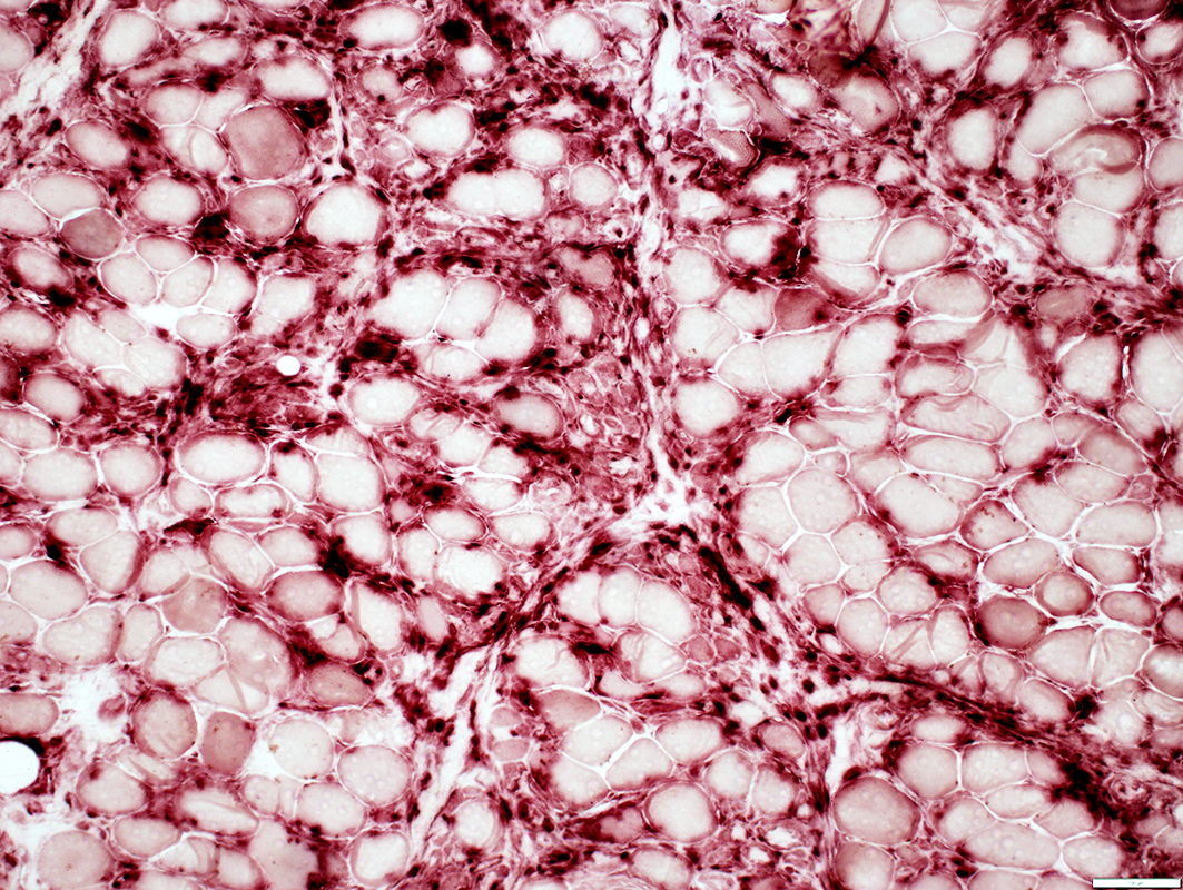
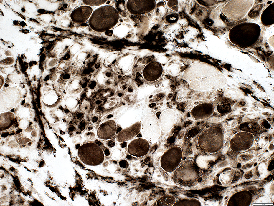
 ATPase pH 9.4 stain |
No grouping
Many immature, intermediate-stained 2C fibers (Below)
Few pale type 2A Fibers (Below)
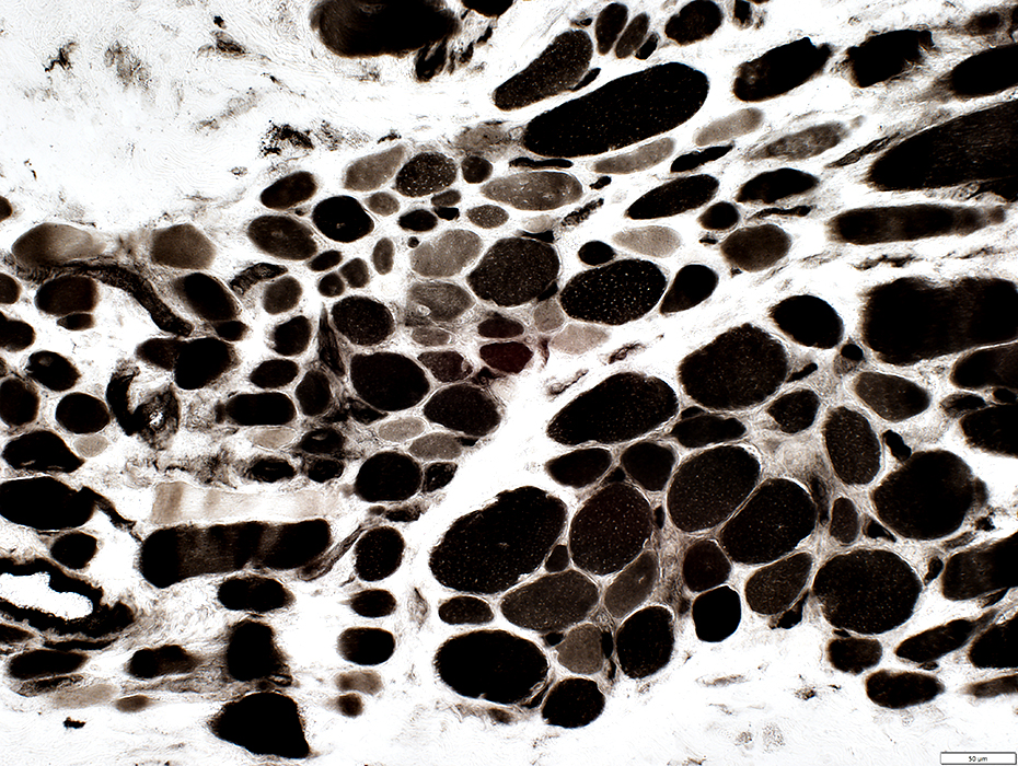 ATPase pH 4.3 stain |
 LC3 stain |
Present in muscle fibers in some regins of muscle
LC3 in muscle fibers: Multiple punctate aggregates scattered through cytoplasm, or clustered in some regions
 LC3 stain |
 LC3 stain |
Muscle Fiber Internal Architecture
Coarse & Immature
 NADH stain |
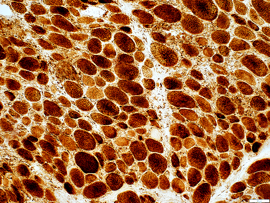 COX stain |
No definite mitochondrial pathology
COX stain is present in all muscle fibers
SDH stain is increased in some small fibers, possibly related to immaturity
 SDH stain |
Nailfold Capillaroscopy
 From: B Hopfinger |
 From: B Hopfinger |
Mi-2 antibodies: α & β
Case 283 yo female with 6 months of progressive dysphagia, & symmetric weakness, proximal legs & arms + distal arms
Inflammatory Myopathy with Perimysial Pathology
|
Myopathy Necrosis MHC-I LC3 aggregates MxA Inflammation Histiocytes Connective tissue Endomysium Perimysium Vessels Capillaries Perimysial |
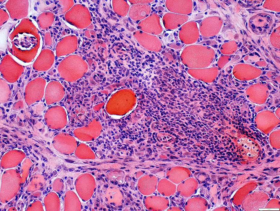
Myopathic Features
 H&E stain |
Muscle fibers
Varied sizes
Necrosis & Regeneration: Scattered
Patchy involvement (Most on bottom left of image)
Perimysial connective tissue: Cellular
Pale round structures: Surrounded by cells (Left)
 VvG stain |
 H&E stain |
Muscle fibers
Varied sizes
Necrosis & Regeneration: Scattered
Multifocal
Perimysial connective tissue damage: Cellular; Pale
Muscle Fiber Necrosis: Scattered
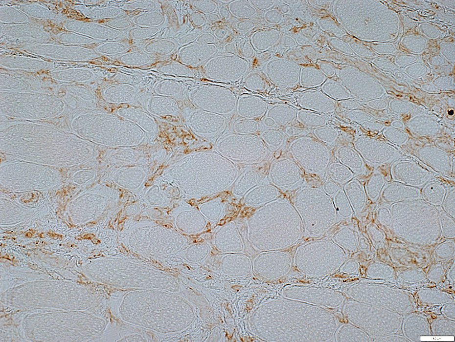
 C5b-9 stain |
Cytoplasm of scattered muscle fibers
Connective tissue: Endomysium & Perimysium
Endomysial cells
 C5b-9 stain |
 C5b-9 stain |
Cytoplasm of scattered muscle fibers
Connective tissue: Endomysium & Perimysium
Endomysial cells
 C5b-9 stain |
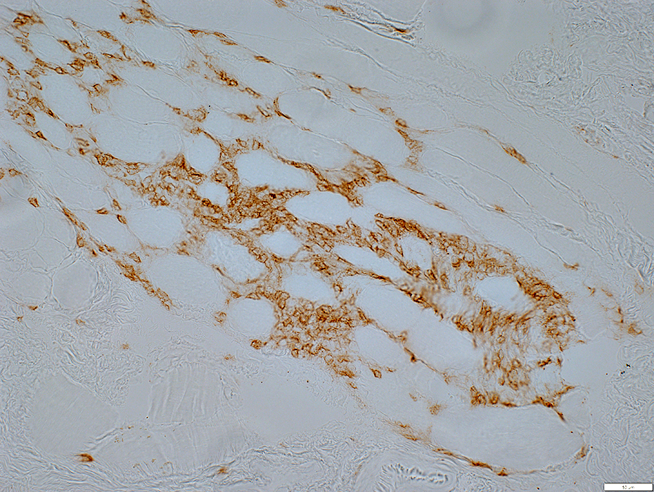
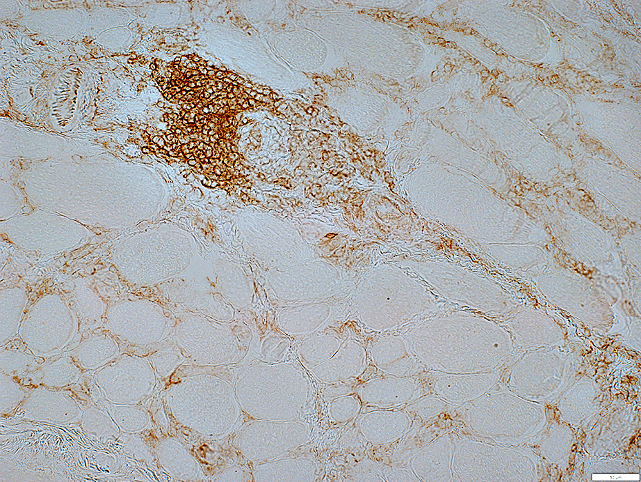
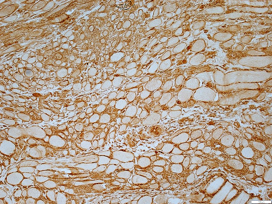
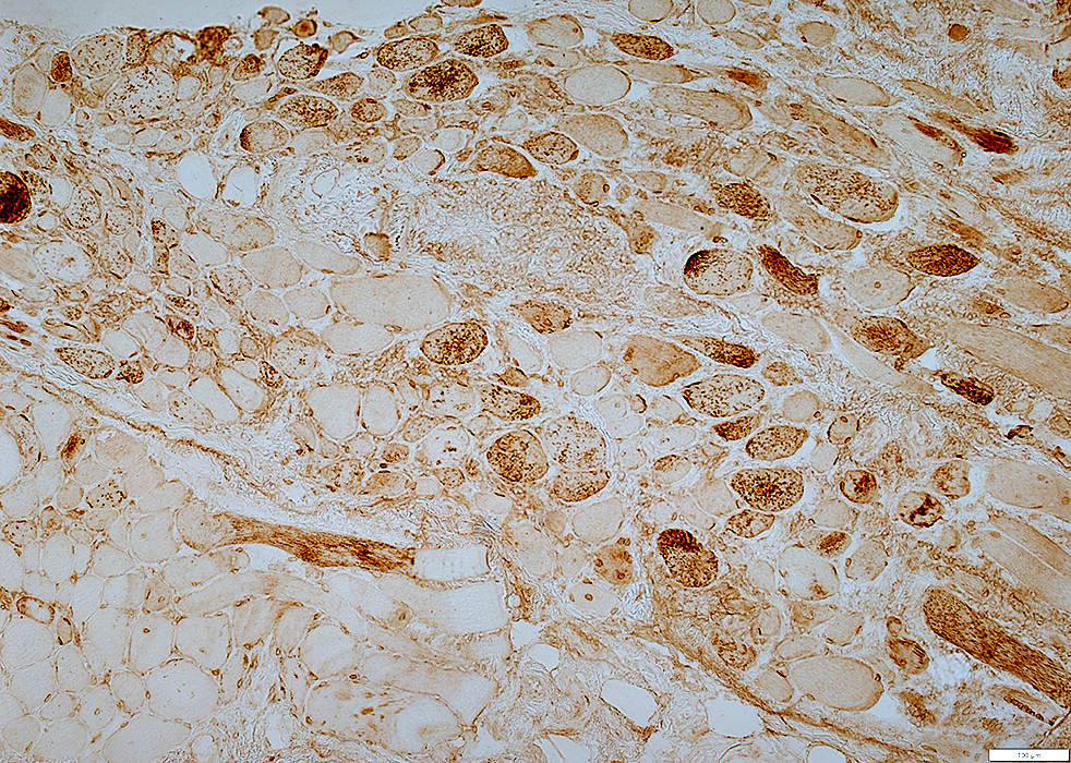
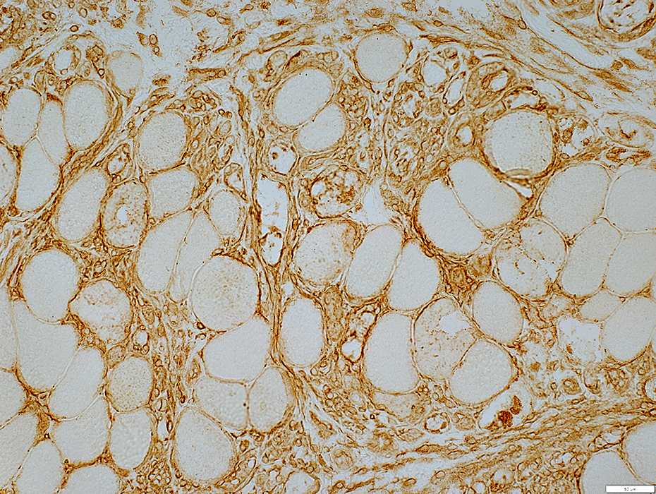
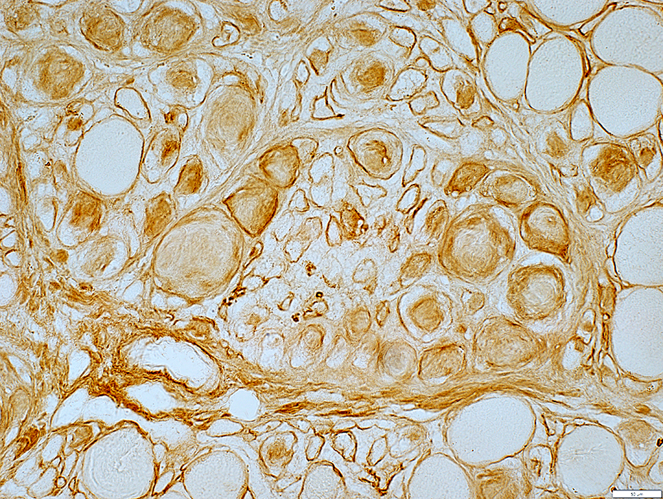
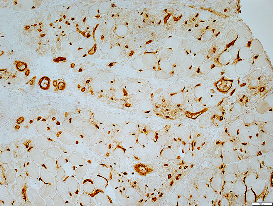
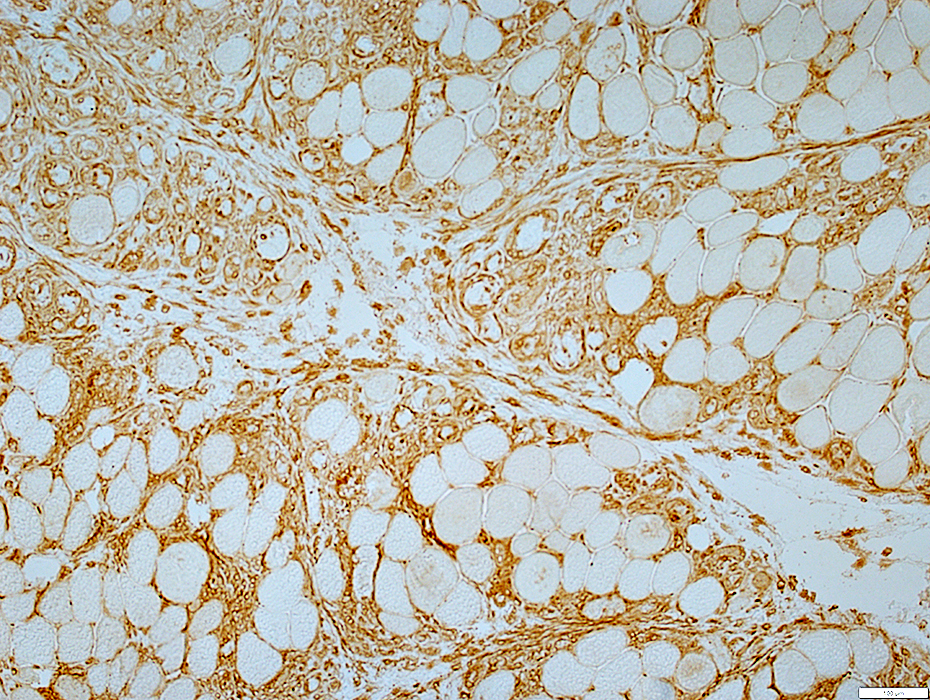 MHC-1 stain |
Muscle fibers: Mild upregulation on muscle fiber surfaces
Endomysial & Perimysial cells: Strong staining
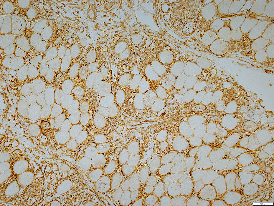 MHC-1 stain |
 MHC-1 stain |
Muscle fibers: Mild upregulation on muscle fiber surfaces
Endomysial & Perimysial cells: Strong staining
 MHC-1 stain |
 MHC-1 stain |
 LC3 stain |
 LC3 stain |
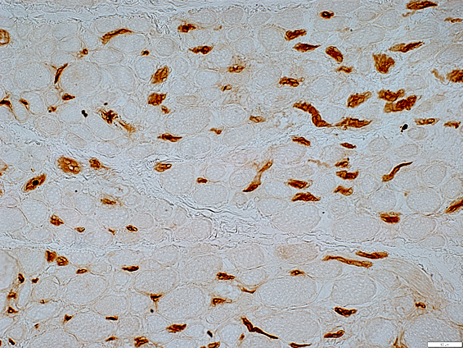
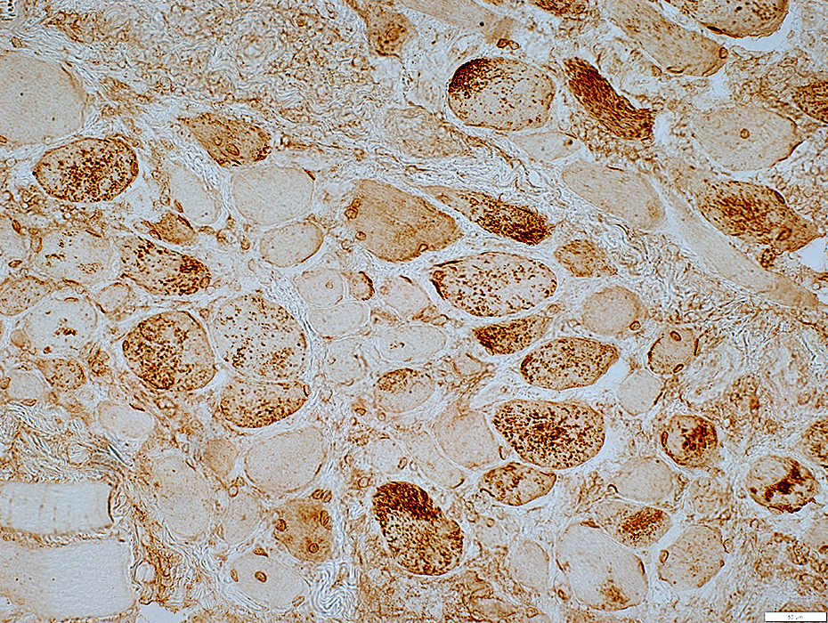
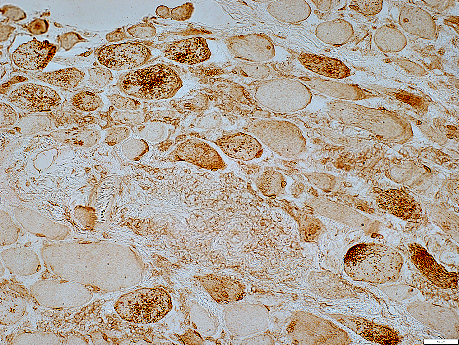
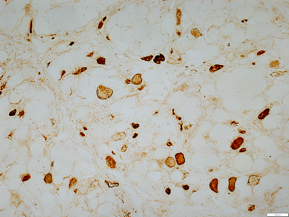
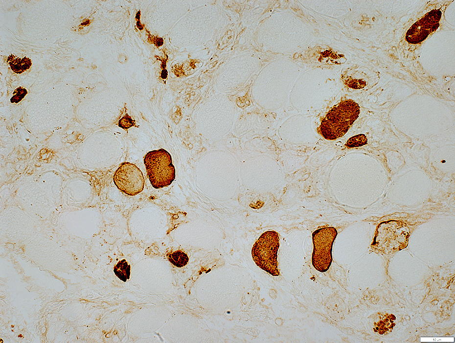
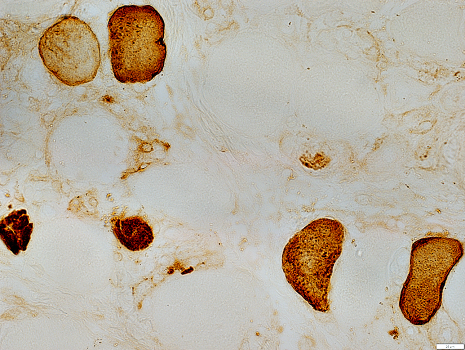
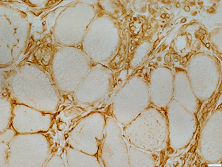
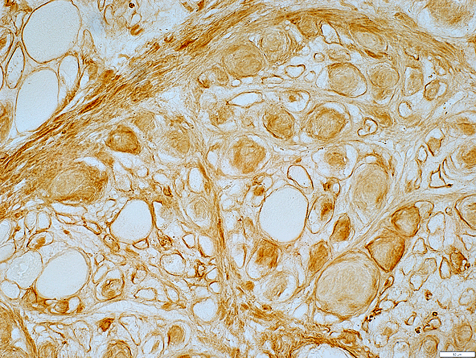
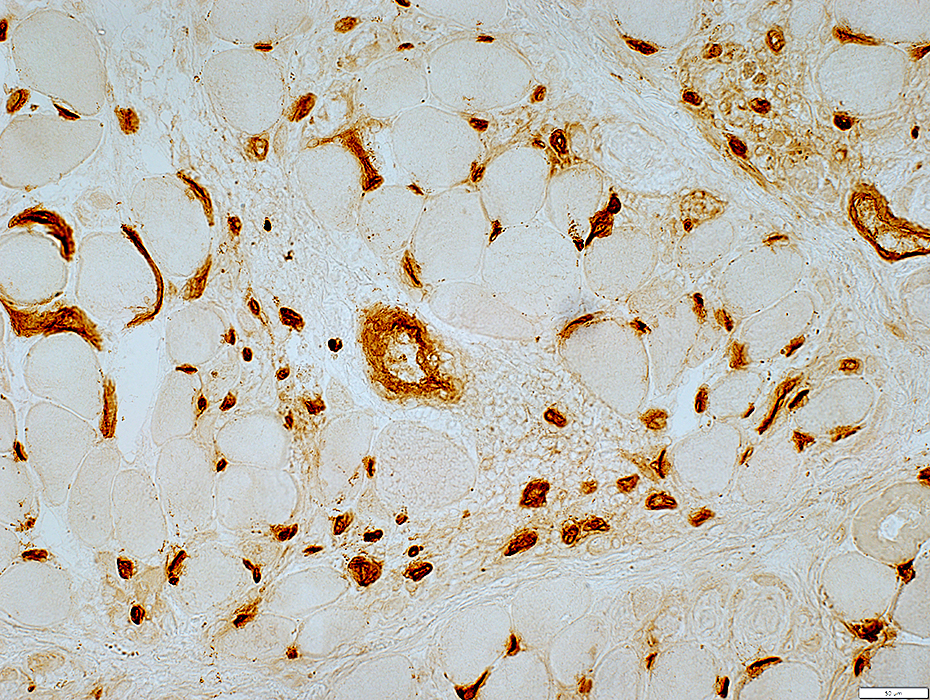
 MxA stain |
Muscle fiber cytoplasm: Mild staining
Endomysial cells: Strong stain
 MxA stain |
Mi-2 myopathy: Immune features
 H&E stain |
Muscle fibers
Varied sizes
Necrosis & Regeneration: Scattered
Multifocal
Inflammation: Focal clusters of cells in endomysium
 Congo red stain |
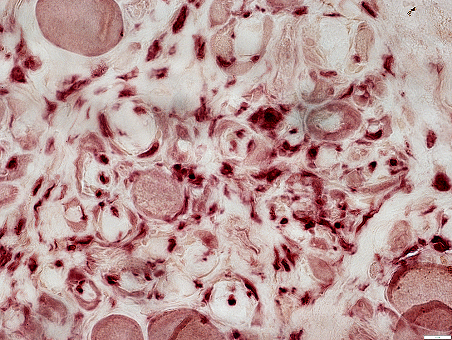
Mi-2 myopathy: Histiocytes
 Acid phosphatase stain |
 Esterase stain |
 Acid phosphatase stain |
Histiocytes
Surround focal, round regions of connective tissue
Scattered in endomysium
 Acid phosphatase stain |
 Esterase stain |
Cells in Endomysium & Perimysium
 H&E stain |
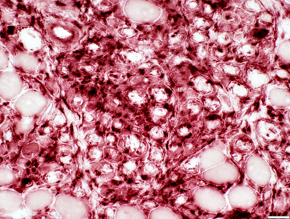
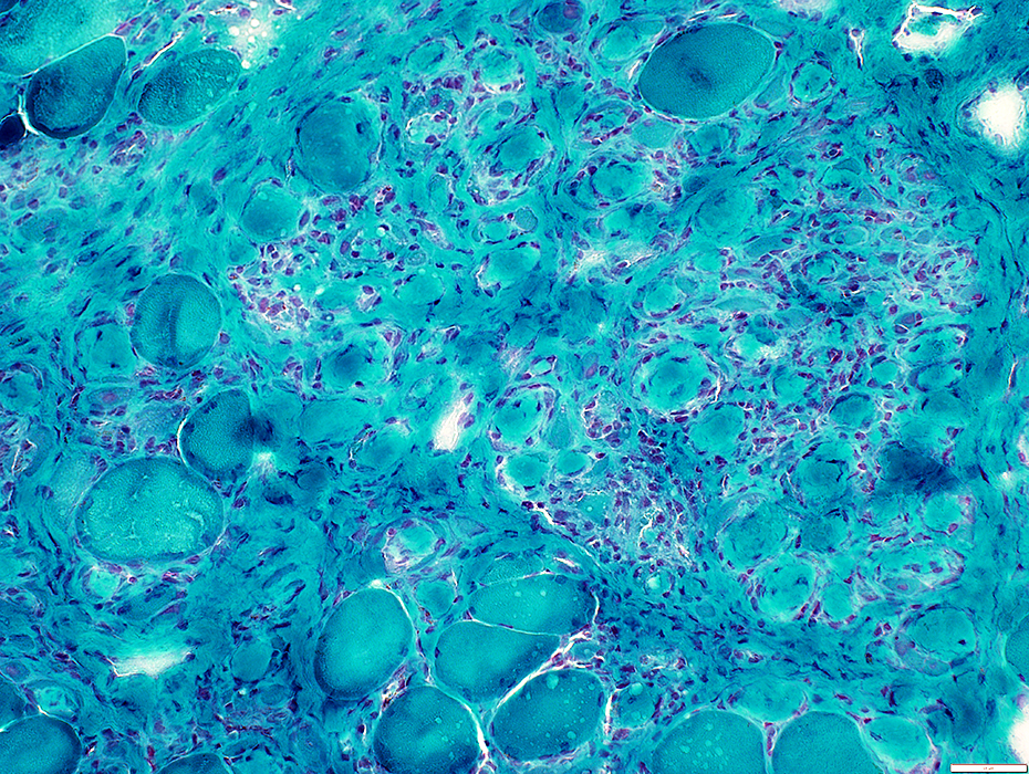
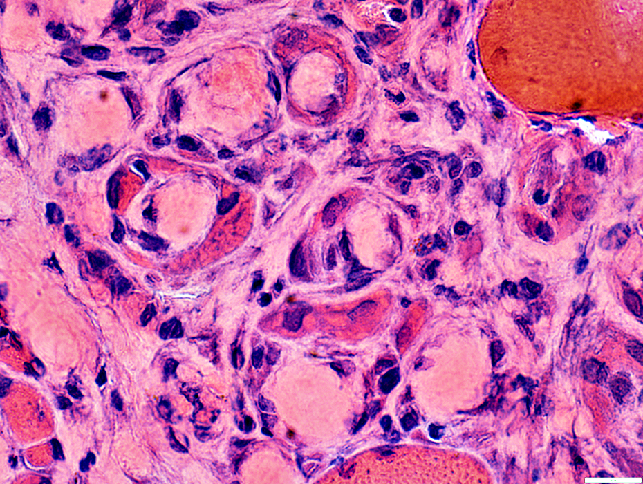
Mi-2 myopathy: Connective tissue pathology
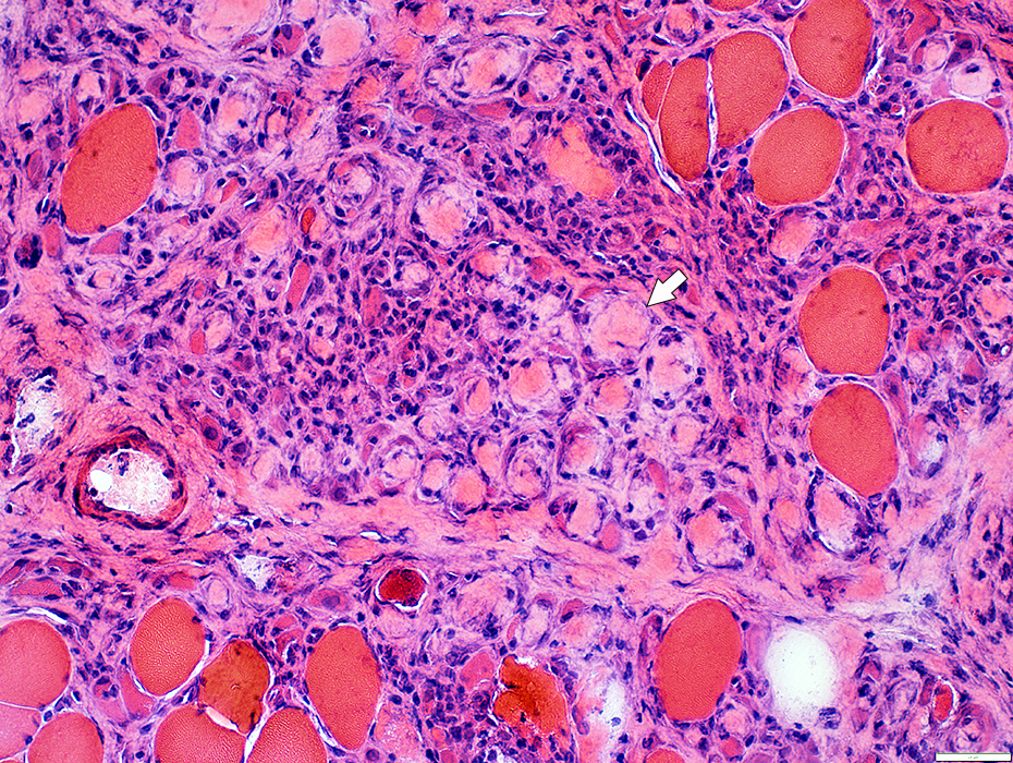 H&E stain |
Increased connective tissue between muscle fibers
Focal, round regions of connective tissue surrounded by thin layer of cells (Arrows)
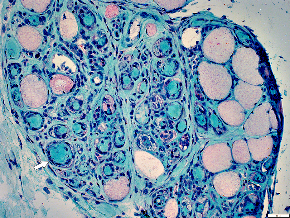
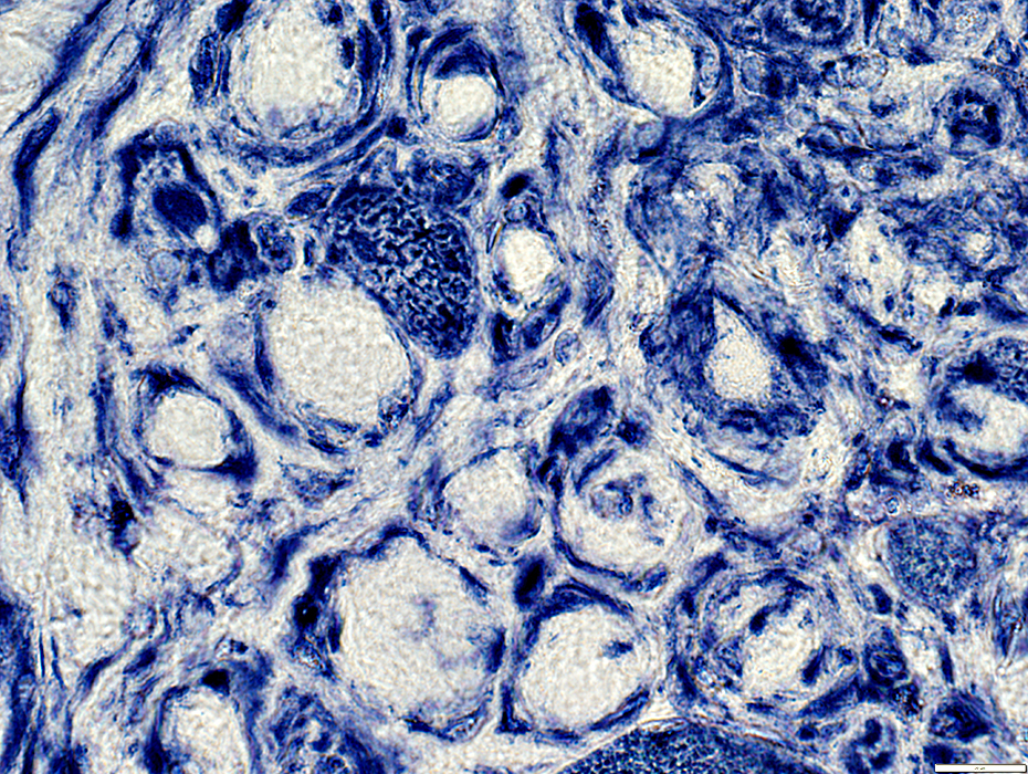
 Alcian blue stain |
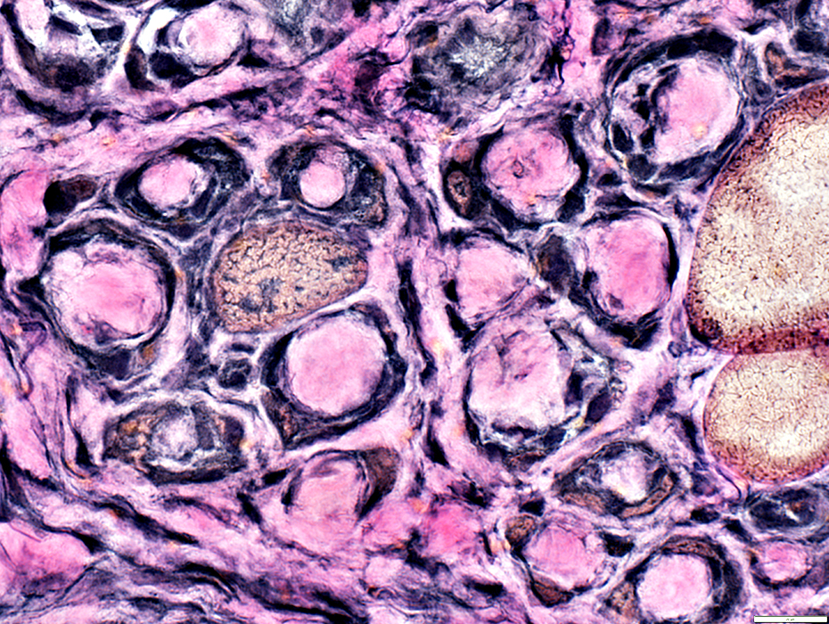
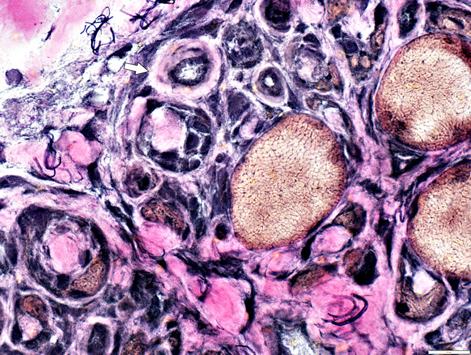
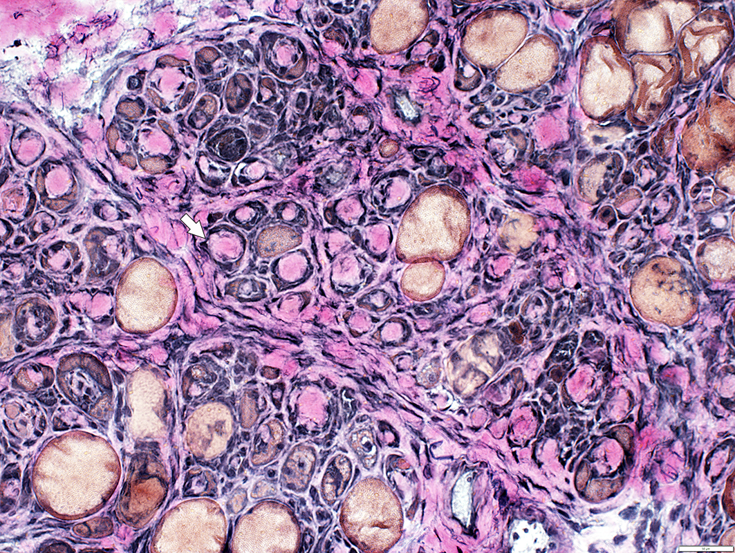 VvG stain |
Increased connective tissue between muscle fibers
Focal, round regions of connective tissue surrounded by thin layer of cells (Arrows)
 Alcian blue stain |
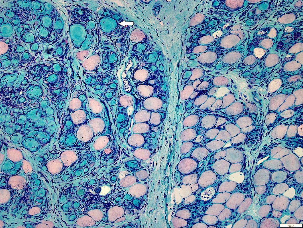
 Gomori trichrome stain |
 H&E stain |
Increased connective tissue between muscle fibers
Focal, round regions of connective tissue surrounded by thin layer of cells (Arrows)
 VvG stain |
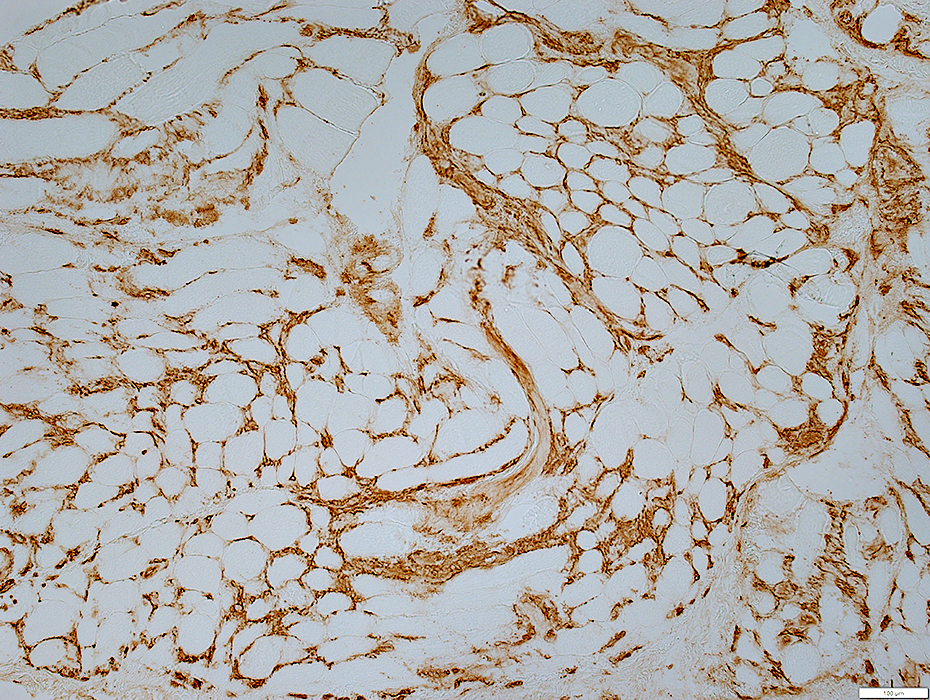
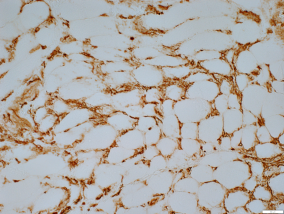
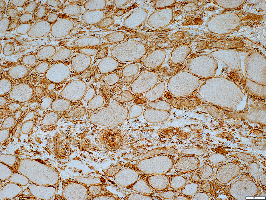
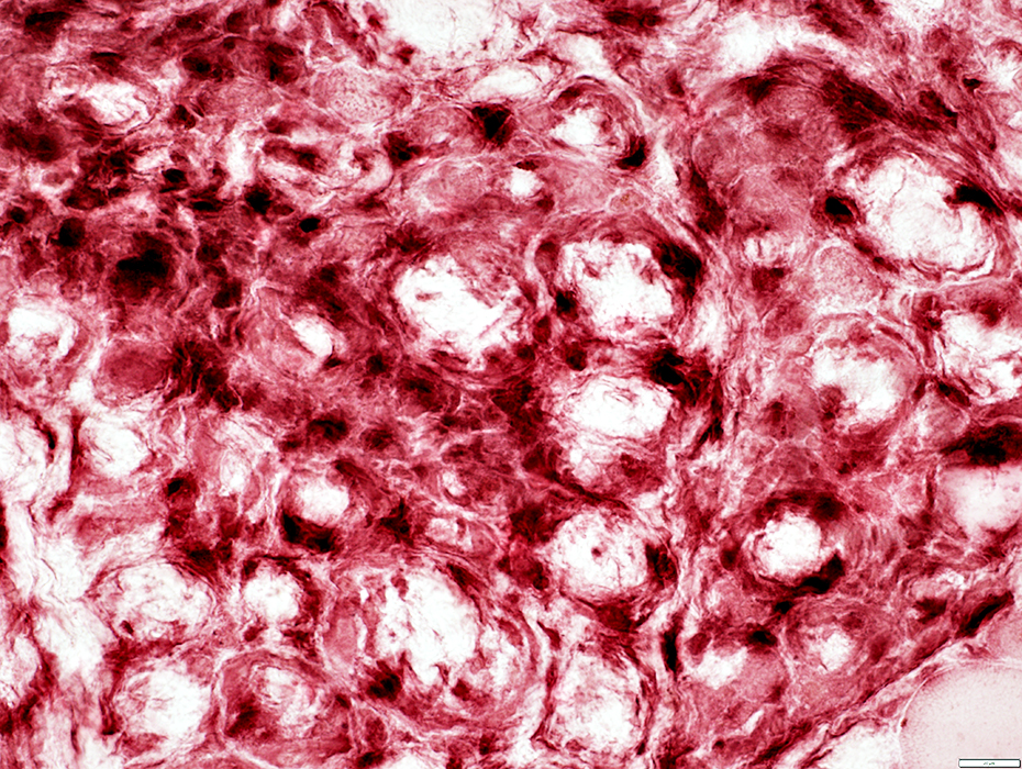
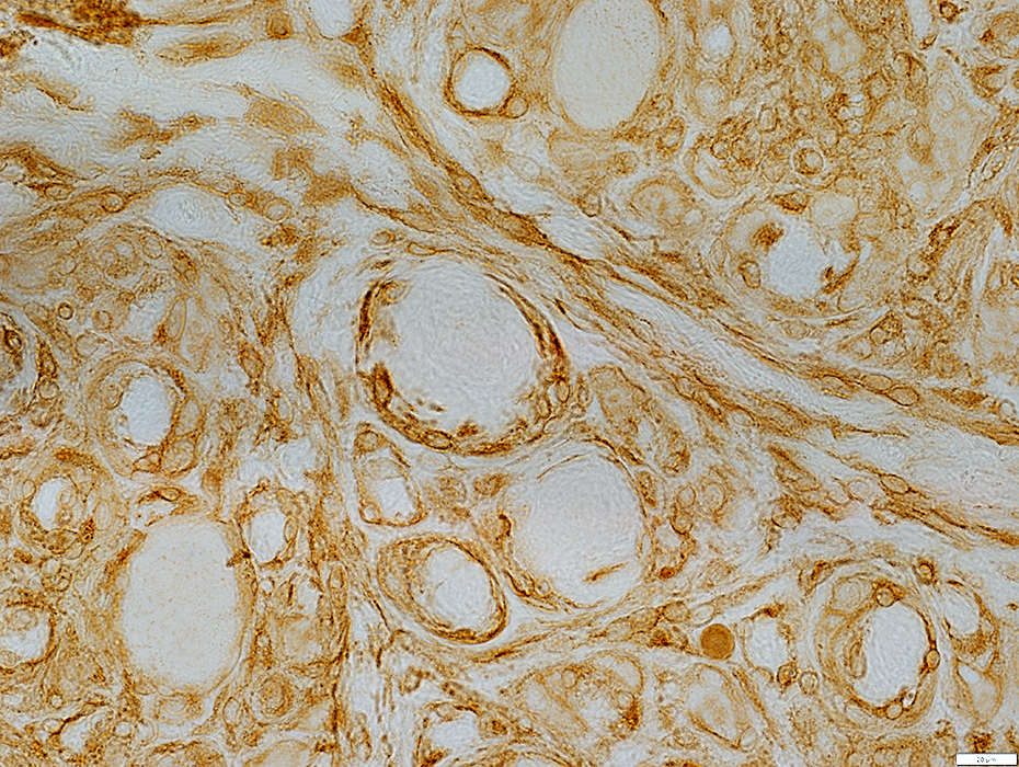
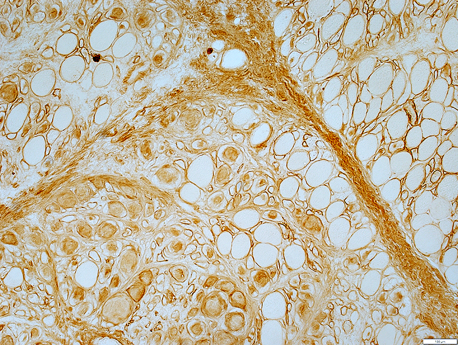 Decorin stain |
Round structures contain chondroitin-SO4
 Decorin stain |
 Decorin stain |
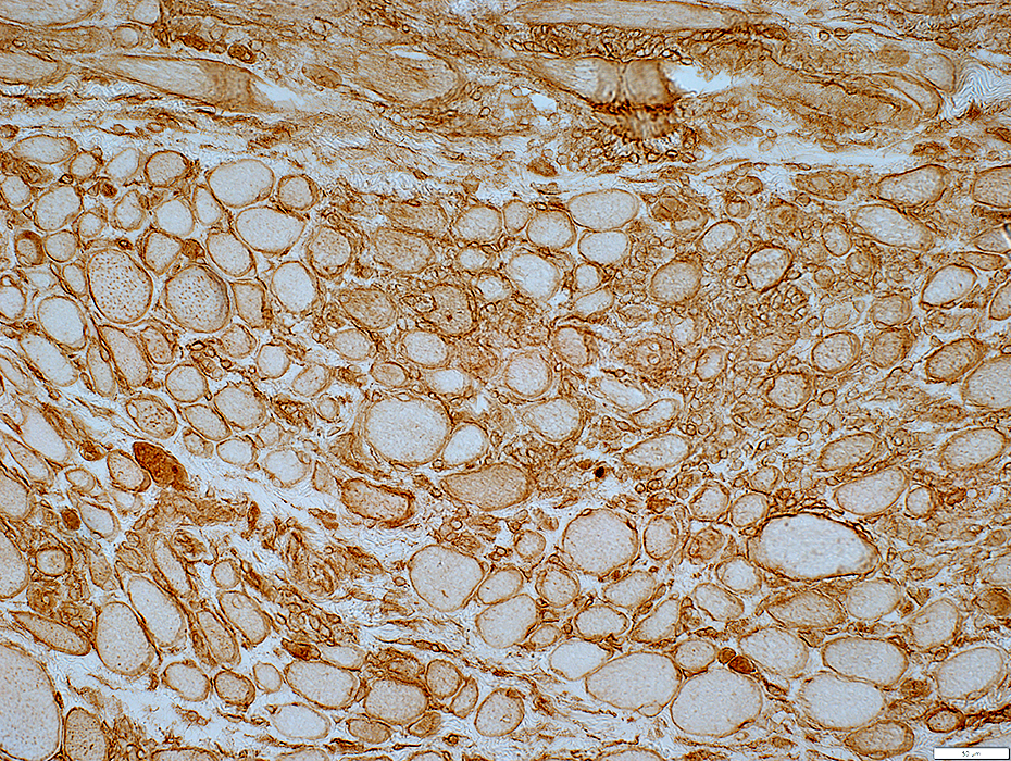
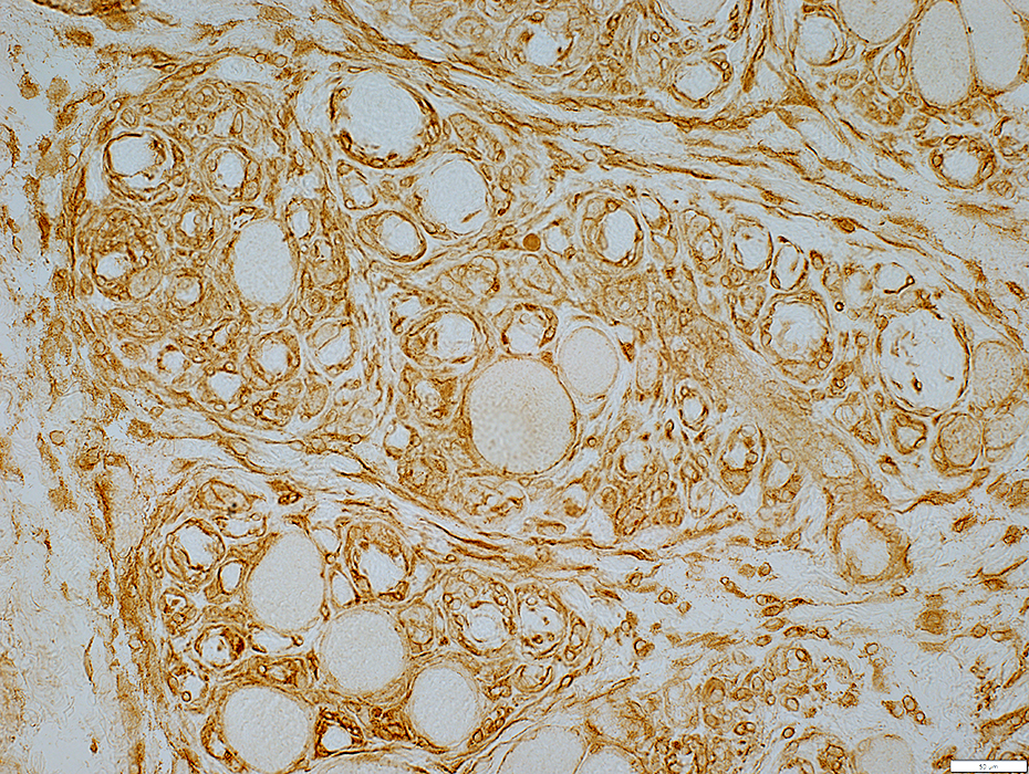
 MHC-I stain |
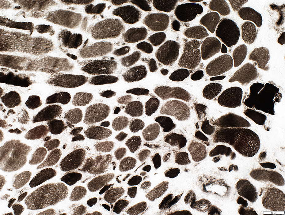
 NADH stain |
Perimysial Connective Tissue Damage
 H&E stain |
Fragmented structure
Contains histiocytes (Scattered large cells)
Perimysial Connective Tissue Damage
 Alkaline phosphatase stain |
 Alkaline phosphatase stain |
Capillary Pathology
 VvG stain |
Large size
thick walls
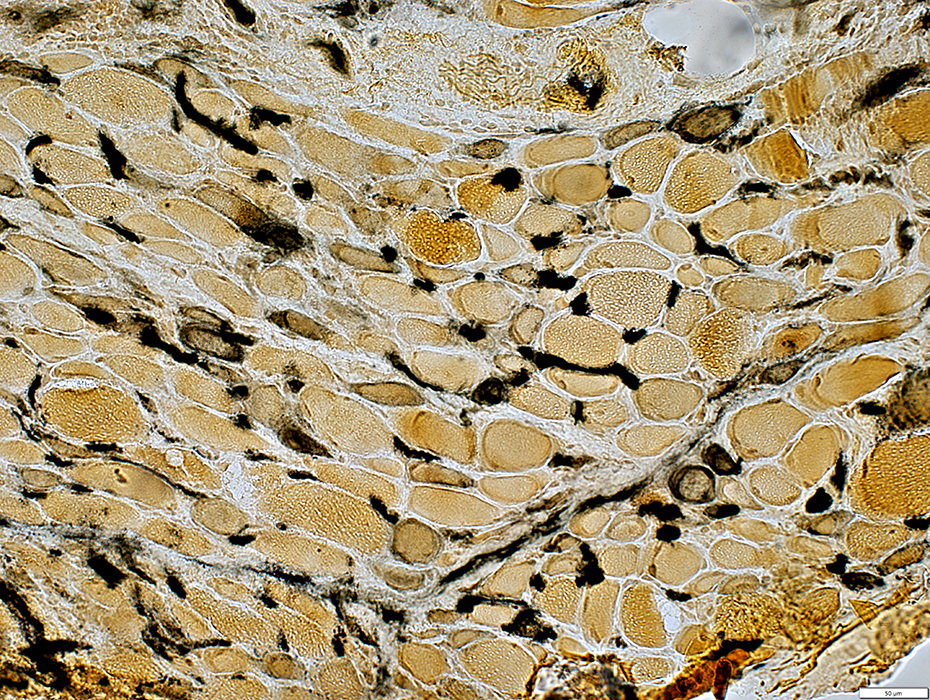
 ATPase pH 4.3 stain |
Endothelium abnormally stains for ATPase
 ATPase pH 4.3 stain |
UEA I stain |
Size: Large
 UEA I stain |
Size: Large
 UEA I stain |
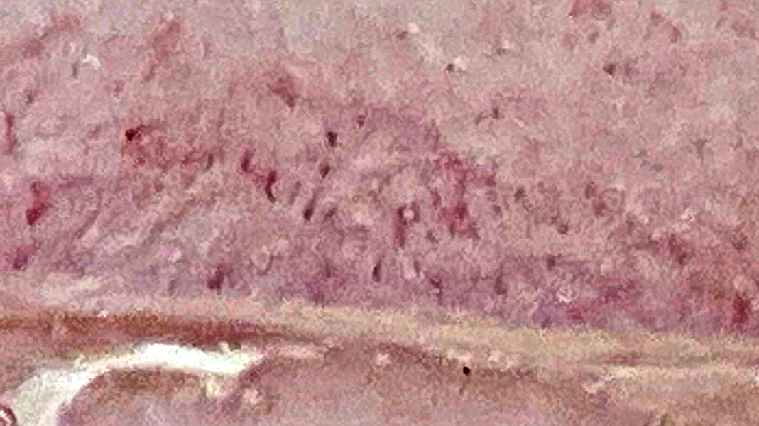
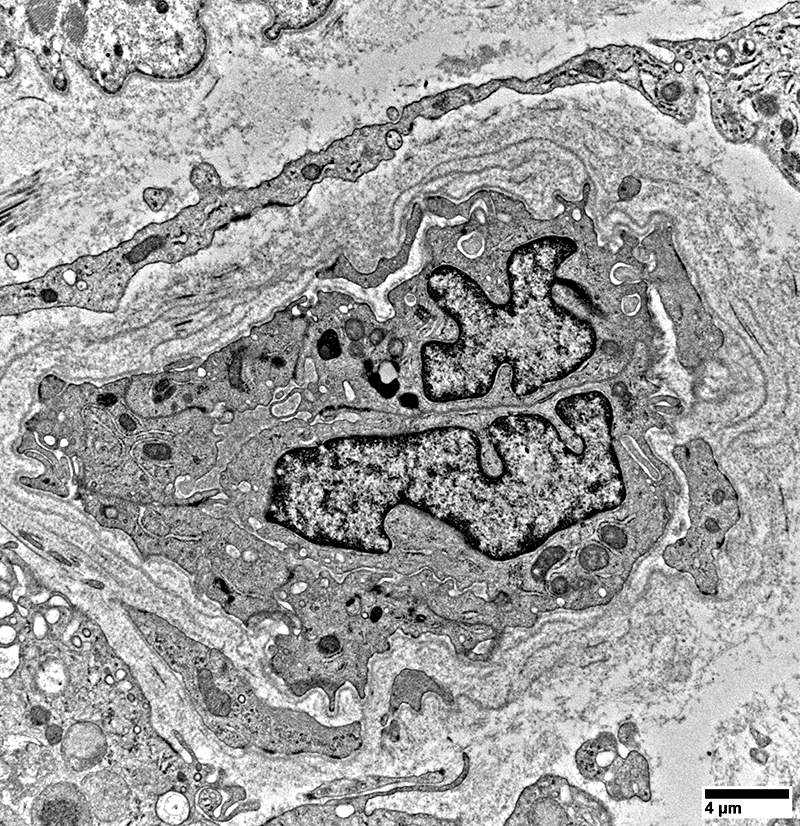
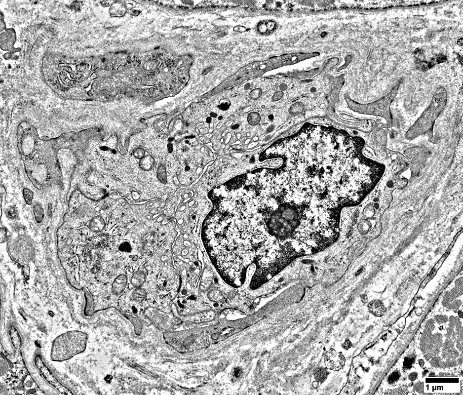 From R Schmidt |
Size: Large
Endothelial cells
Size: Large
May contain tubuloreticular profiles (Below; Arrow)
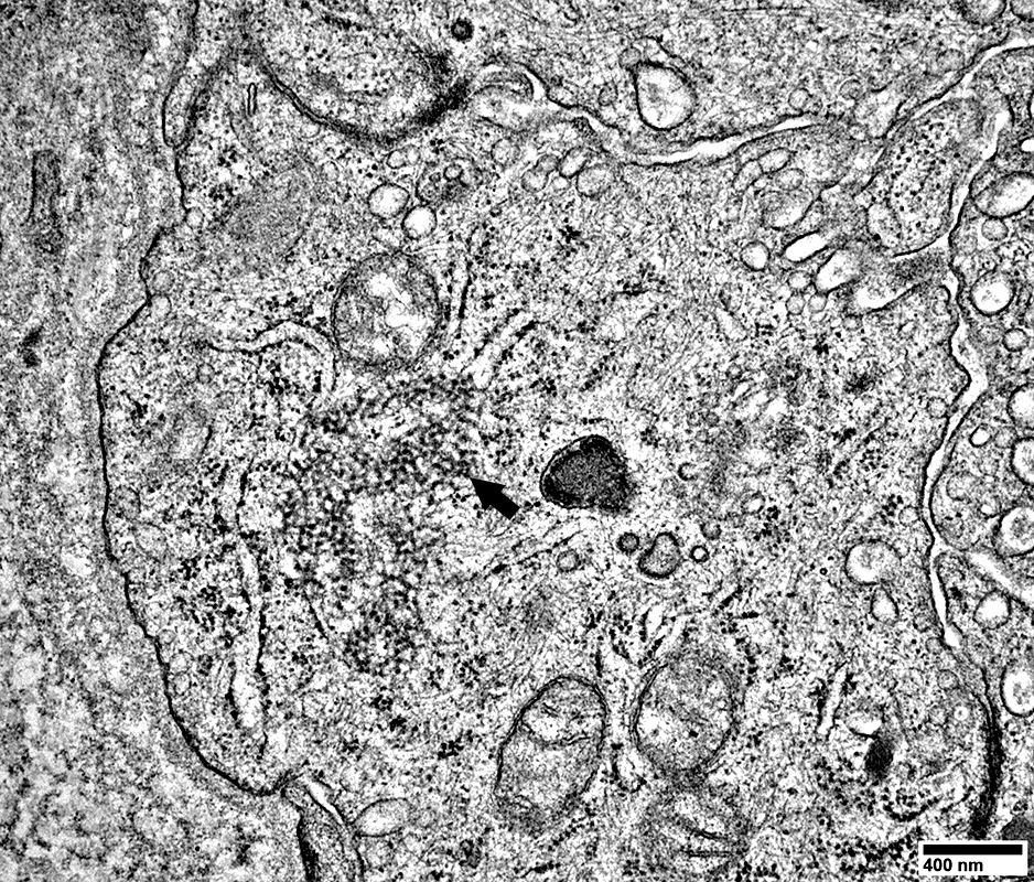 From R Schmidt |
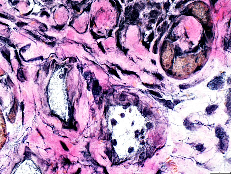 VvG stain |
Walls: Reduced numbers of elastin fibrils
 H&E stain |
Walls
Elastin fibrils: Reduced numbers
Connective tissue: Thick
Associated Lymphocytic cells
In Vessel wall
Surrounding vewssel
Eosinophils, Few
 H&E stain |
Perimysial Vessel
Connective Tissue Wall: Thick
Endothelial cells: Large
Surrounded by: Cell processes
 From R Schmidt |
Return to: Mi-2 antibodies
Return to: Neuromuscular Home Page
3/23/2026