LONP1: Ataxia+
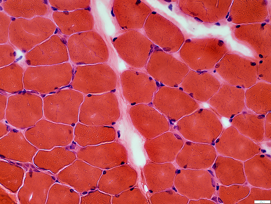 H&E stain |
Morphology: Normal
Mild freeze artefact
 VvG stain |
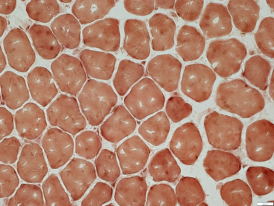
Muscle Fibers
Internal Architecture: Normal
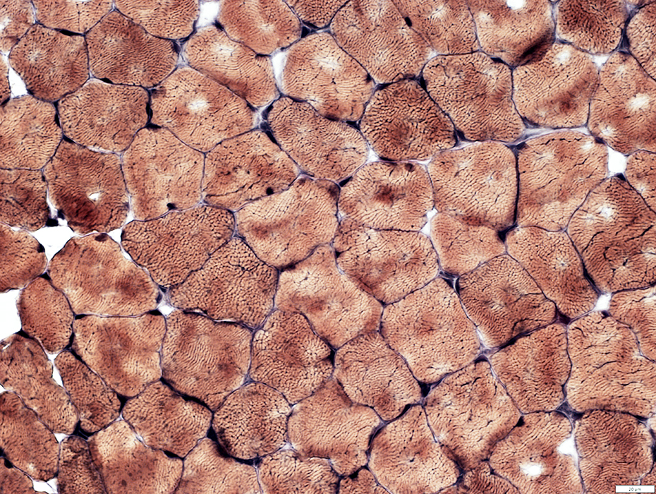
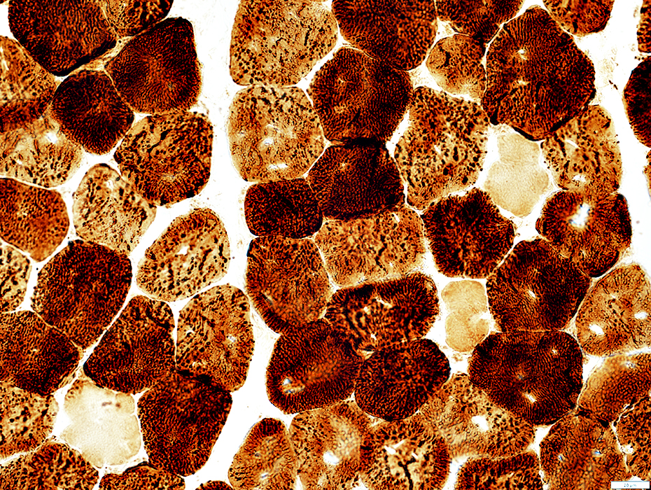
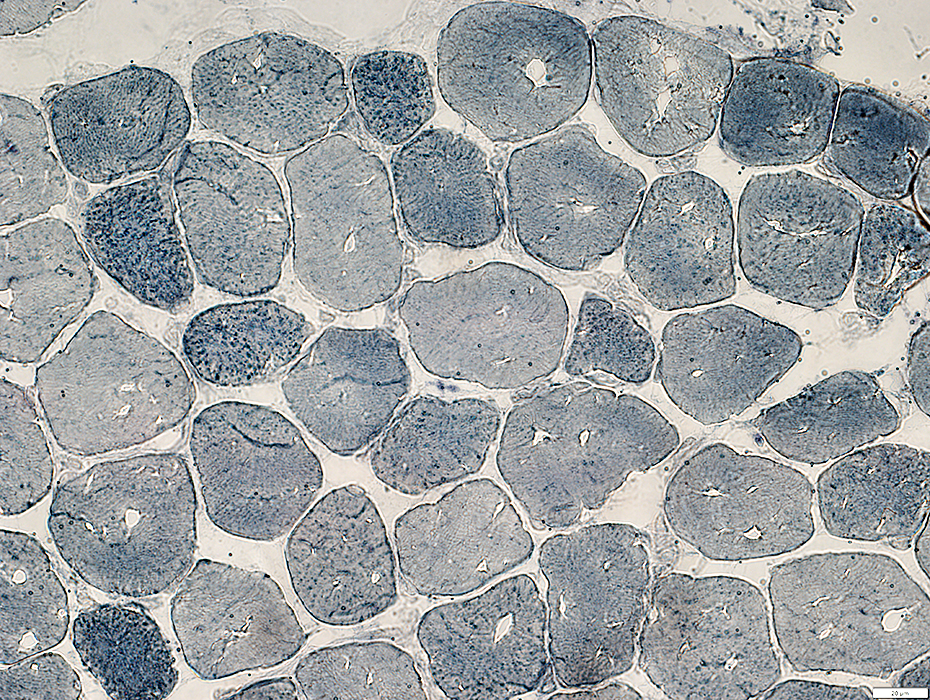
 NADH stain |
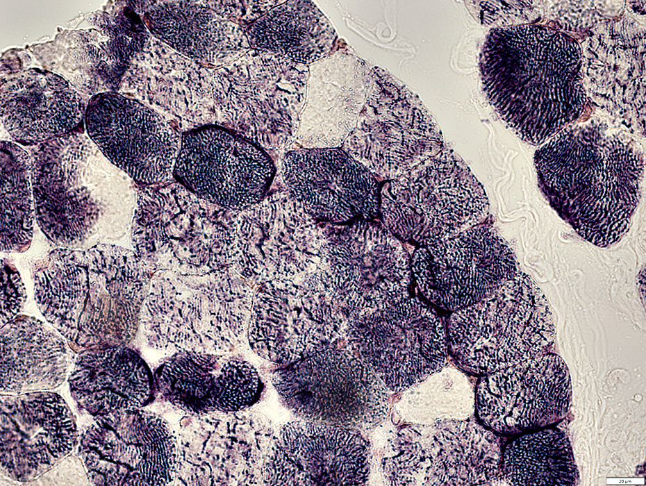
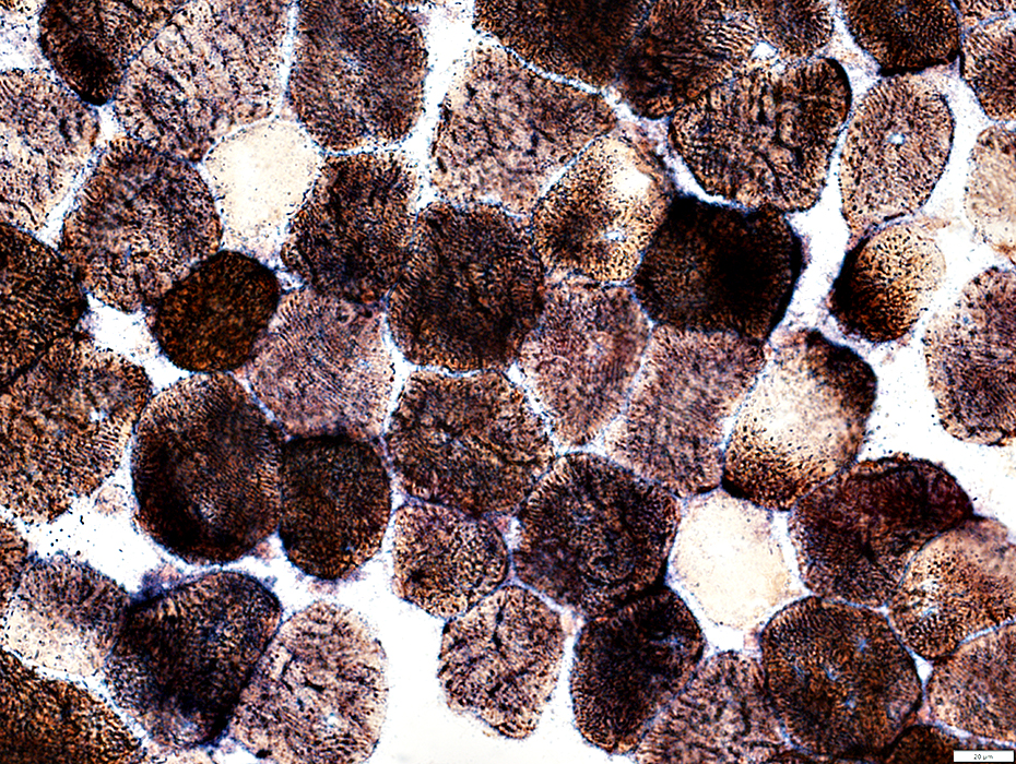
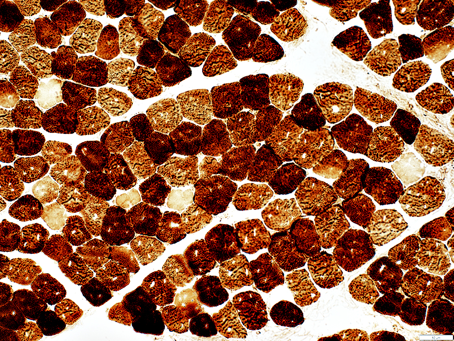 Cytochrome oxidase (COX) stain |
COX negative muscle fibers: Scattered
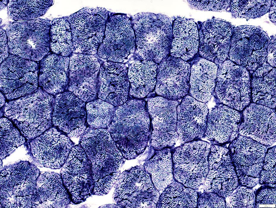
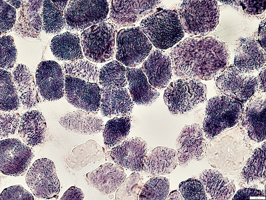
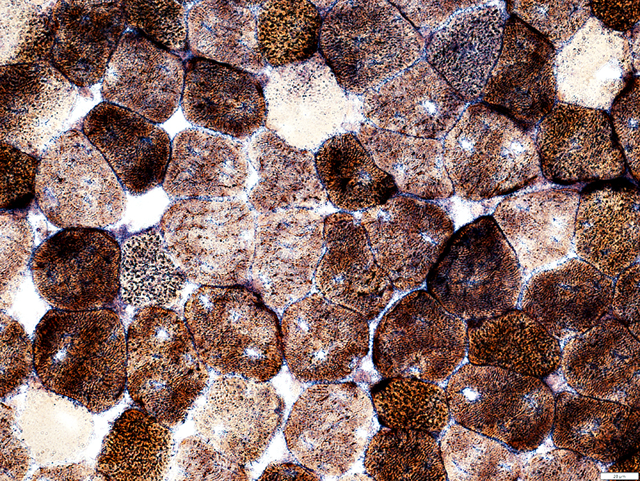
 Succinate Dehydrogenase (SDH) stain |
SDH negative muscle fibers: Scattered
 Succinate Dehydrogenase (SDH) stain |
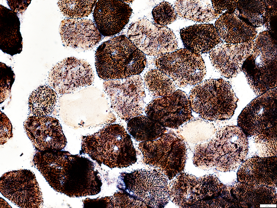
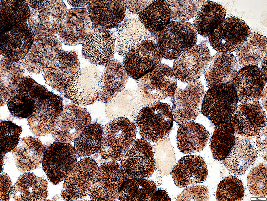
 SDH + COX stain |
SDH & COX: Negative in same muscle fibers
 SDH + COX stain |
 SDH + COX stain |
SDH & COX: Negative in same muscle fibers
 SDH + COX stain |
 SDH + COX stain |
 Sudan stain |
Small lipid droplets in some muscle fibers
 Sudan stain |
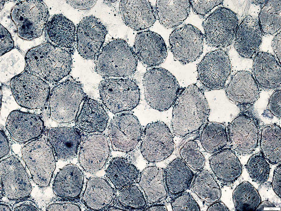
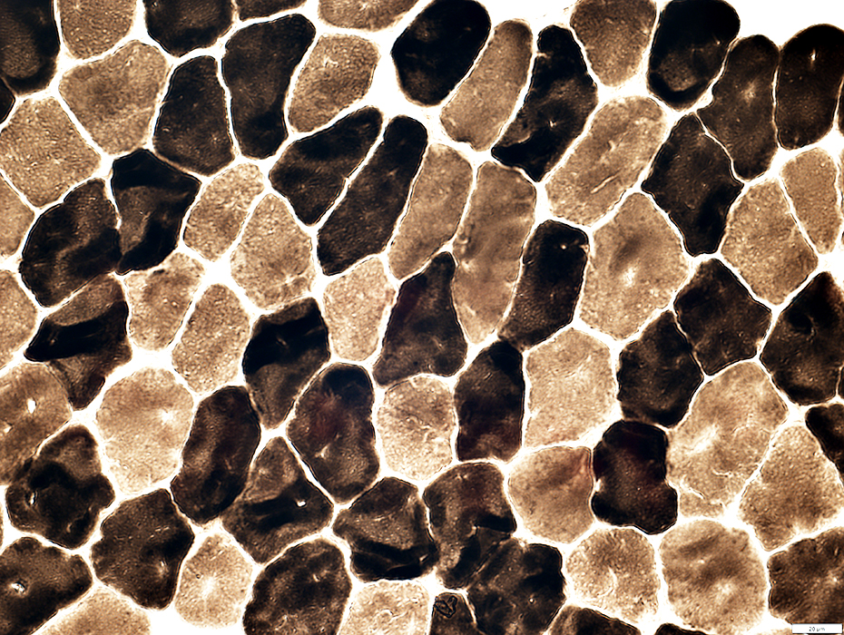 ATPase pH 9.4 stain |
No fiber type grouping
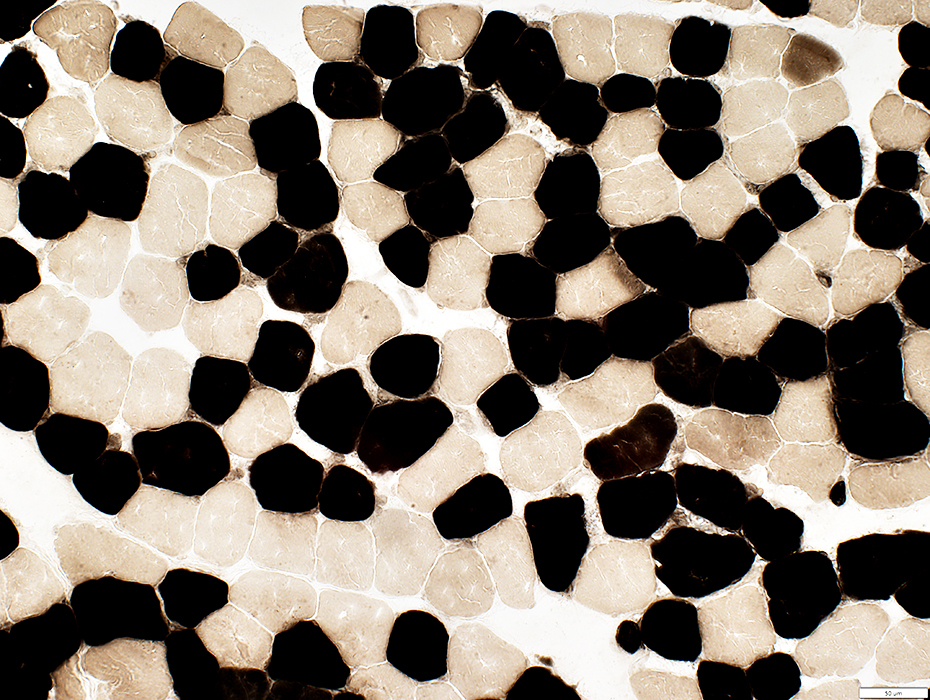 ATPase pH 4.3 stain |
Few intermediate-stained type 2C fibers
Endomysial capillaries: Mild staining
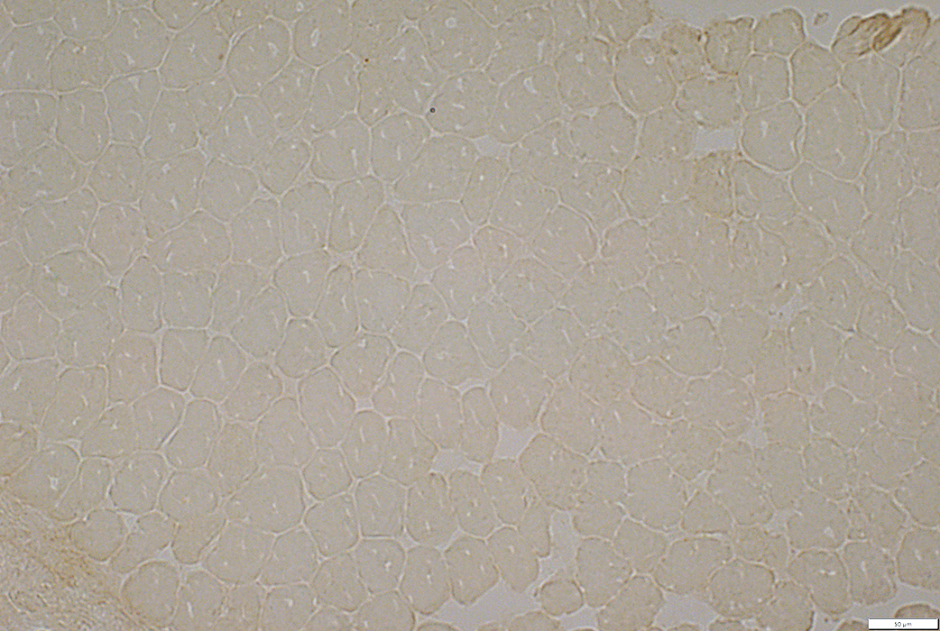 C5b-9 stain |
No necrosis or Inflammation
 Acid Phosphatase stain |
Return to Neuromuscular Home Page
Return to LONP1
7/1/2024