Brancher Deficiency (GSD 3)
Images from: Tahseen Mozaffar; Mari Perez-Rosendahl
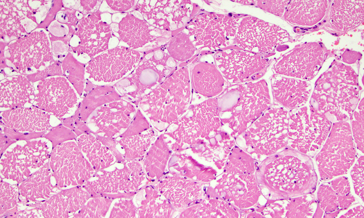 H&E stain |
Size: Varied
Contain: Vacuoles, Multiple, Variably-sized
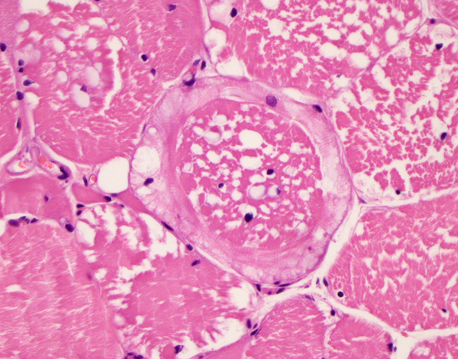 H&E stain |
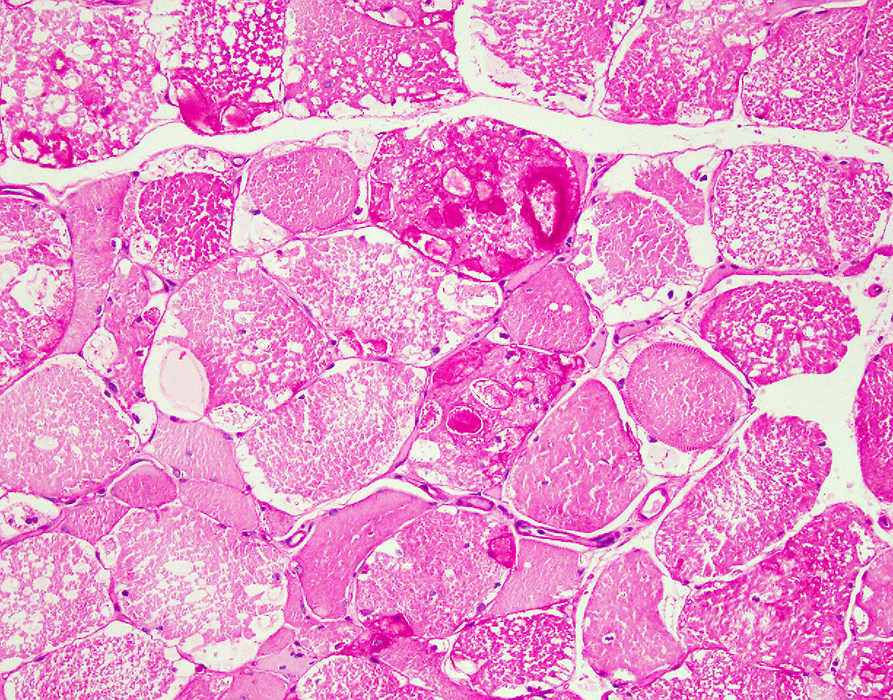
 H&E stain |
 PAS stain |
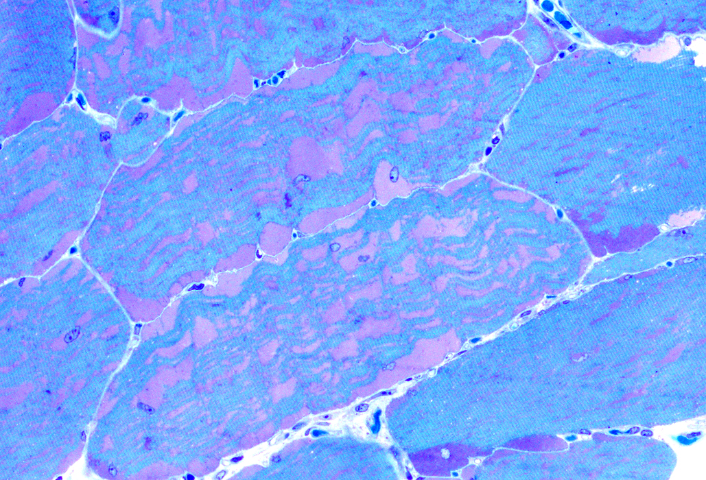 Toluidine blue-stained Plastic section |
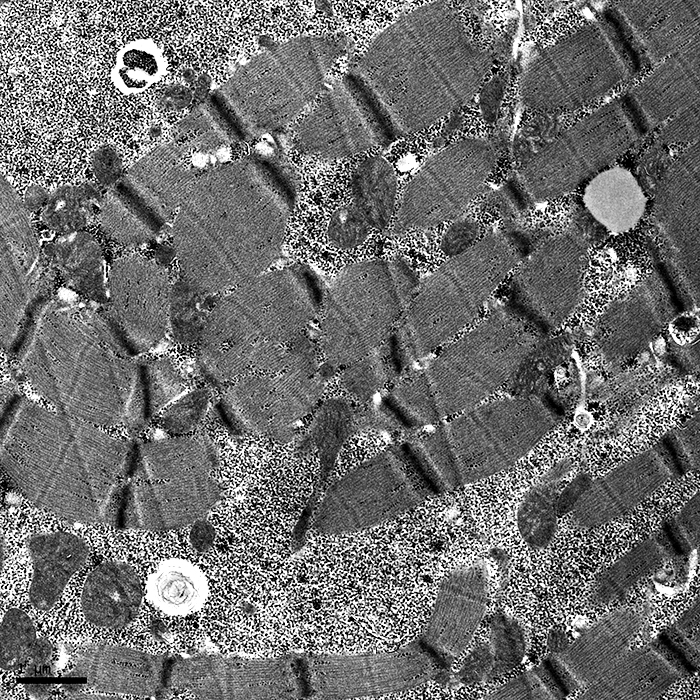
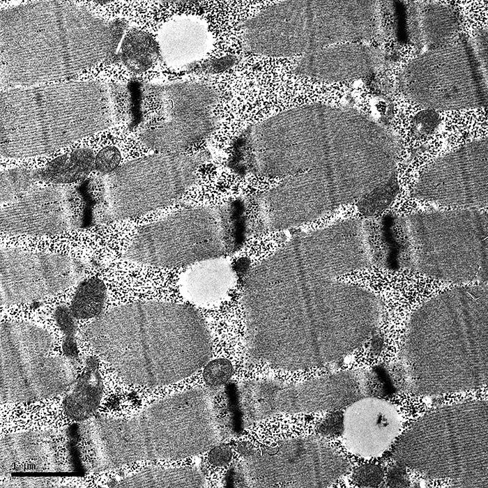
GSD3: Muscle Ultrastructure
|
Deposits: Large, Nonmembrane bound, Sarcoplasmic
Contents: Glycogen granules, Normal
|
Return to GSD3
8/10/2020