Focal Myositis
|
Morphology, General Perimysium Muscle fibers Calcification Spindles |
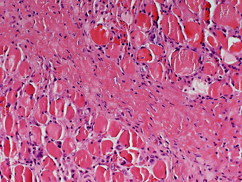
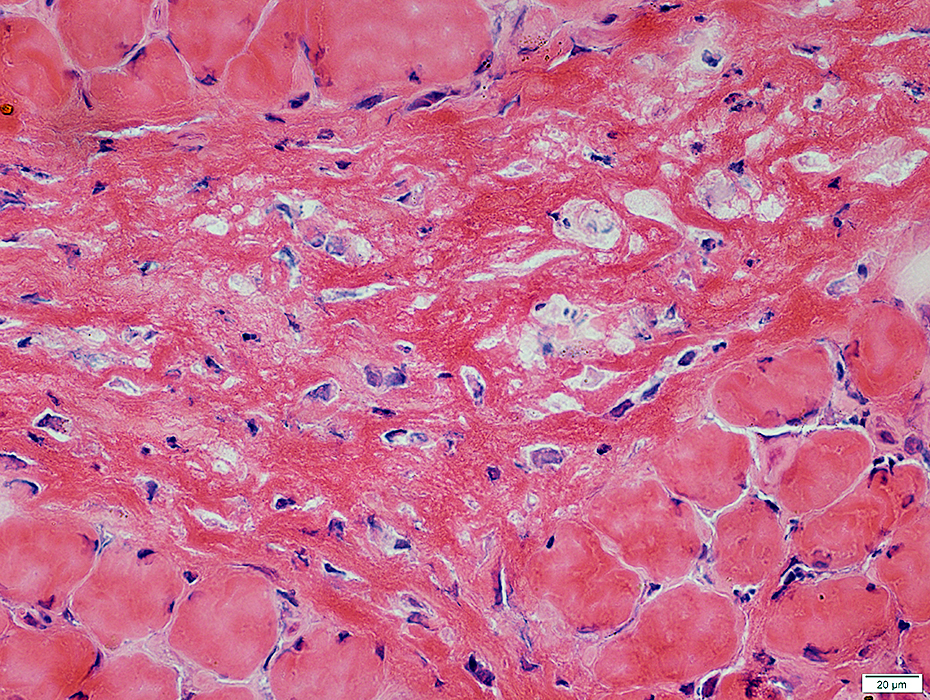
Focal Myositis: Muscle morphology & Hemorrhage
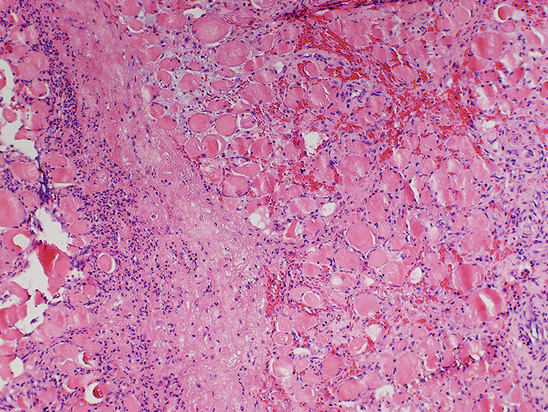 H&E stain |
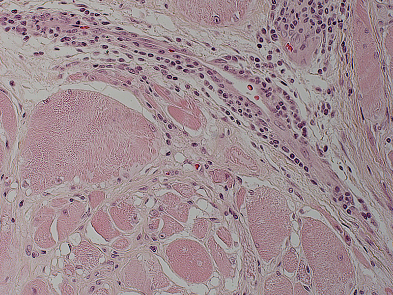
Perimysium: Wide; Cellular in some areas
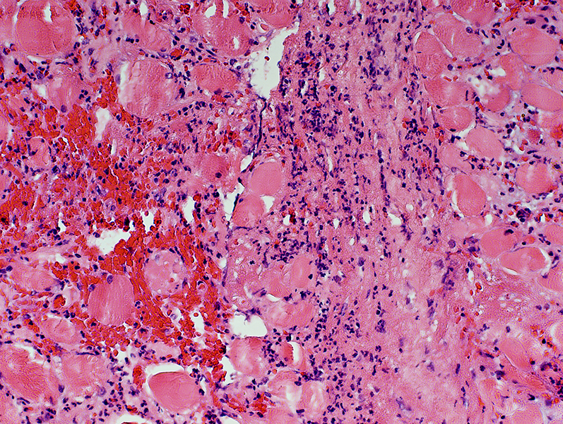
Endomysium: Hemorrhage
Muscle fibers: Varied size; No necrosis
 H&E stain |
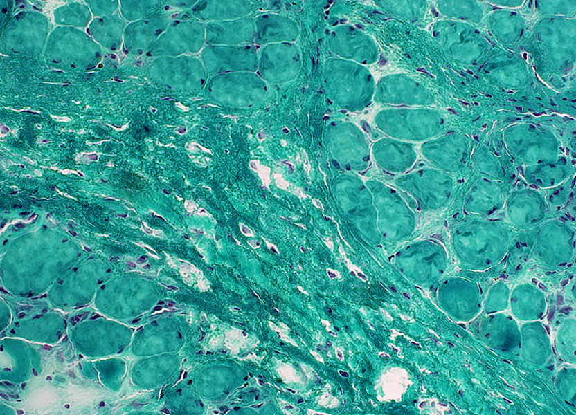
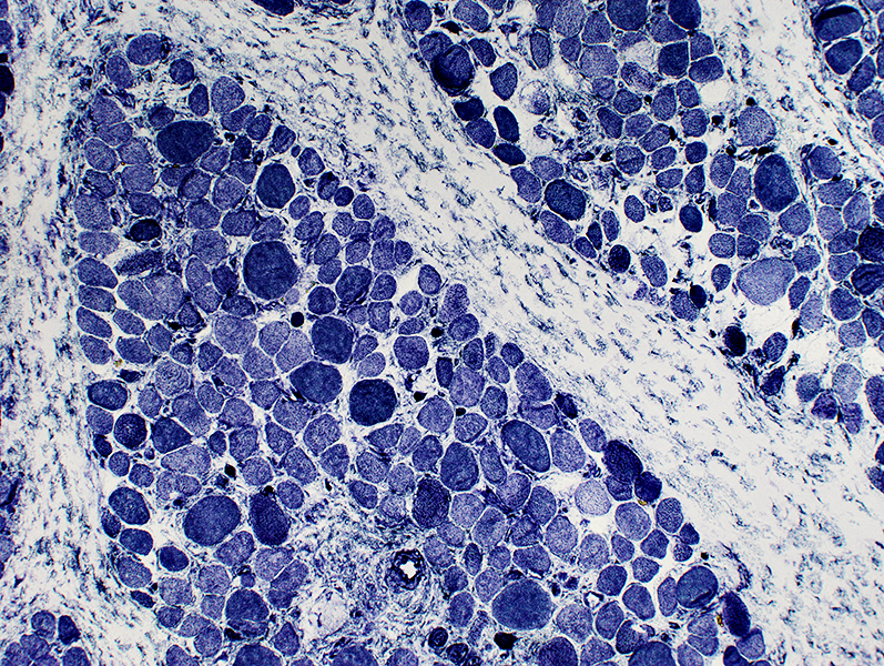
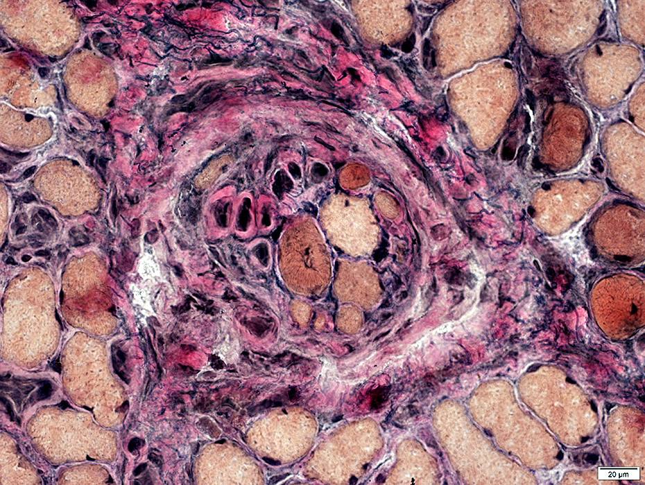
Focal myositis: Endomysium
Hemosiderin depositis
Mildly wide between muscle fibers
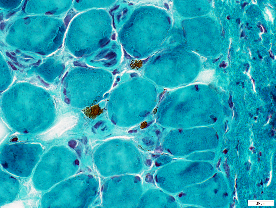 Gomori trichrome stain |
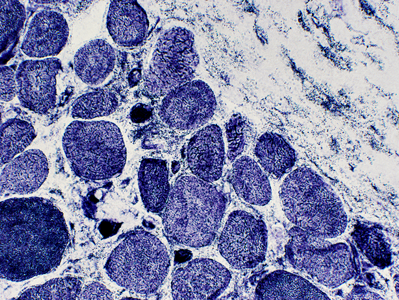
Focal Myositis: Perimysium
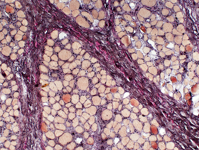 VvG stain |
| Perimysium: Wide; Abundant connective tissue |
 Gomori trichrome stain |
 H&E stain |
 H&E stain Some areas of connective tissue are sworled or disrupted. |
 H&E stain |
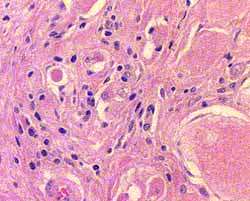
| Perimysium Some areas contain scattered large cells with large nuclei Diffuse staining of perimysium & endomysium with alkaline phosphatase |
 Alkaline phosphatase stain |
Histiocytic cells: Scattered in perimysium & endomysium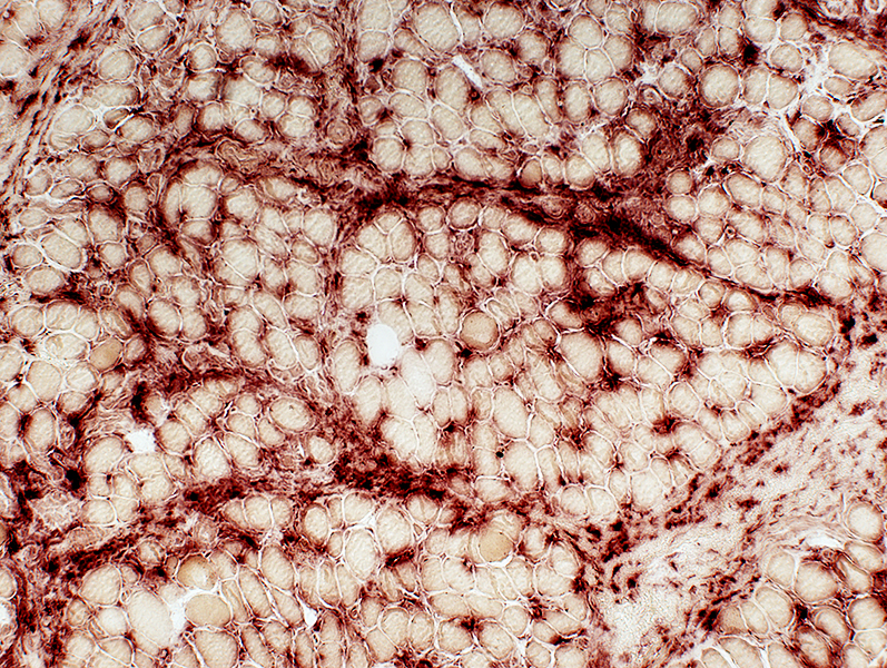 Acid phosphatase stain |
 H&E stain |
 H&E stain |
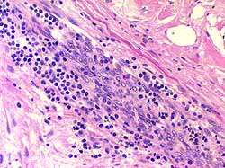
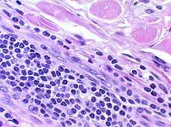
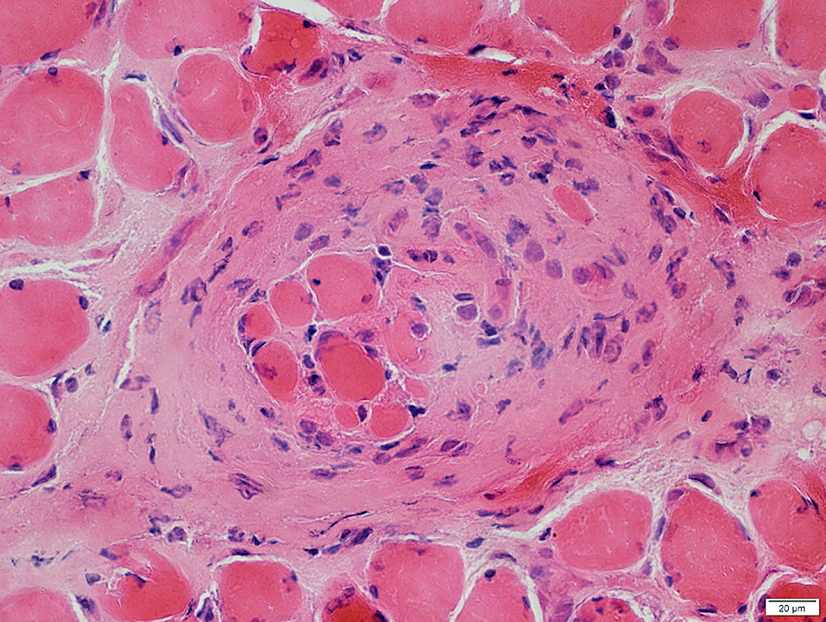
| Lymphocytes: Occasional focal accumulations, perimysial or pervascular |
|
Focal myositis: Muscle fibers
 H&E stain |
|
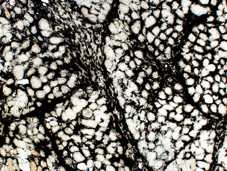
Muscle fibers Varied size Many 2C fibers No necrosis |
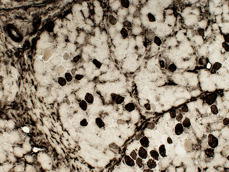 ATPase pH 4.3 stain |
 NADH stain |
|
Muscle fibers May be smaller at edges of fascicles Coarse internal architecture |
 NADH stain |
|
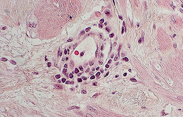
Endomysial capillaries: May be large 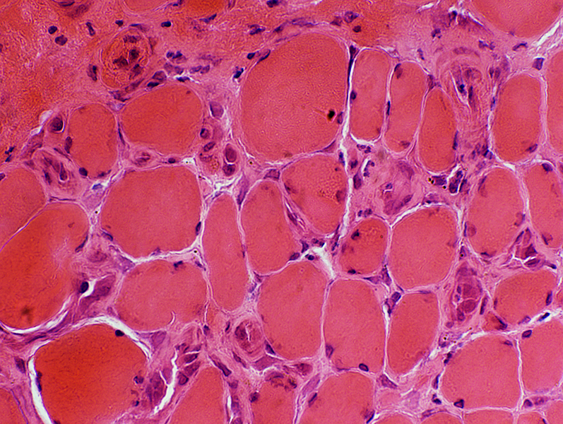 H&E stain |
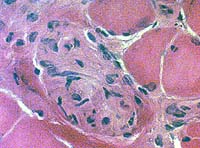
Focal Myositis: Spindles
 H&E stain |
 Gomori trichrome stain |
Focal myositis: Calcification
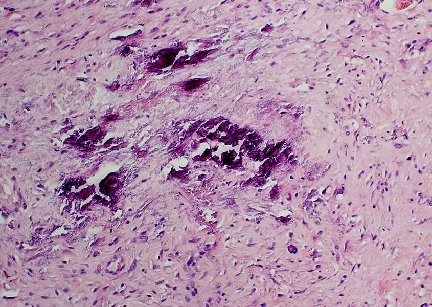 H&E stain |
Return to Neuromuscular Home Page
Return to Inflammatory myopathies
5/22/2017