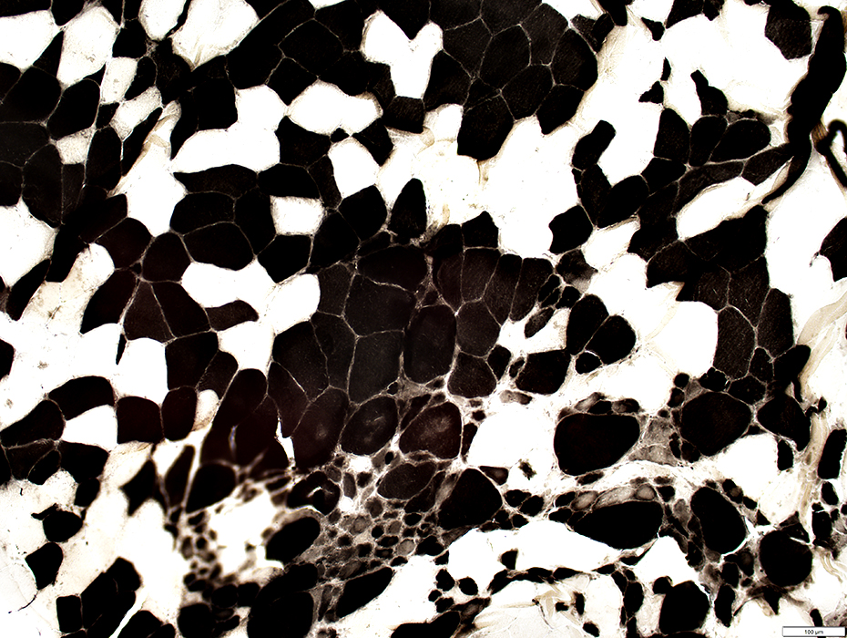
SLONM with Sarcoplasmic pads & Grouped muscle fiber atrophy
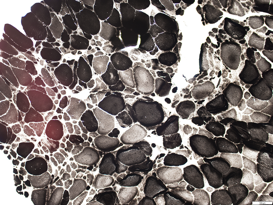
| Ultrastructure |
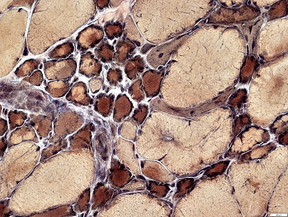
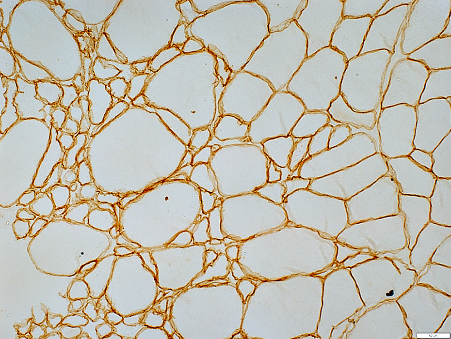
 VvG stain |
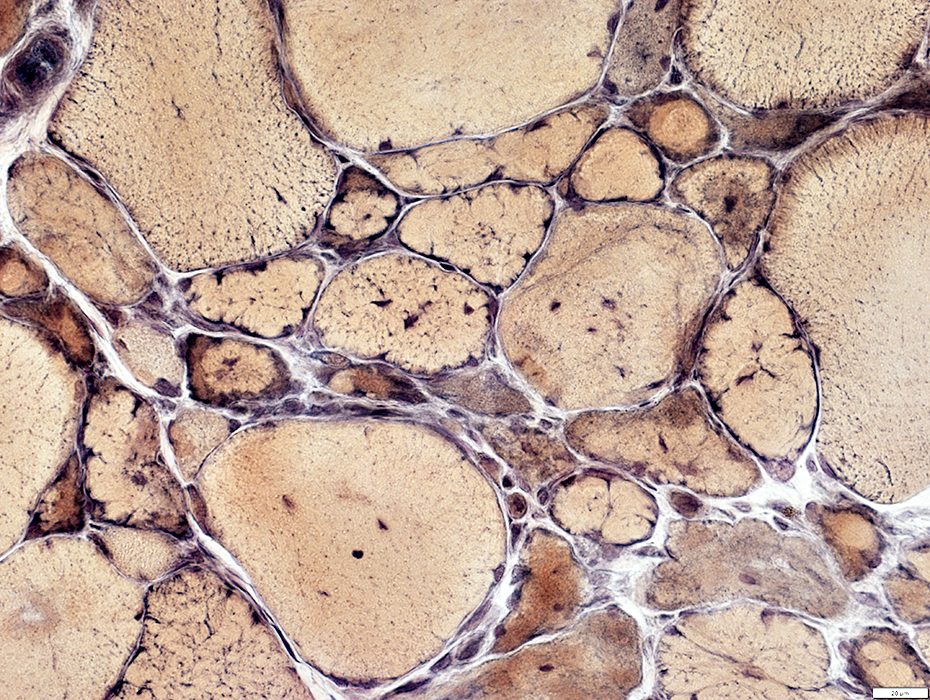
Fiber sizes: Varied
Small muscle fibers: Present in clusters
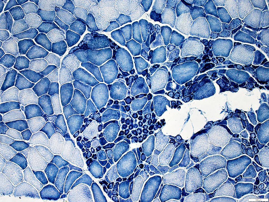 NADH stain |
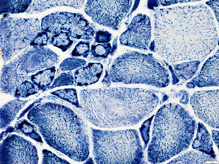
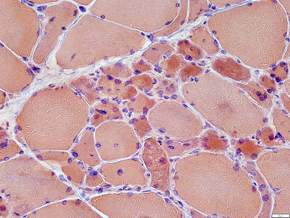
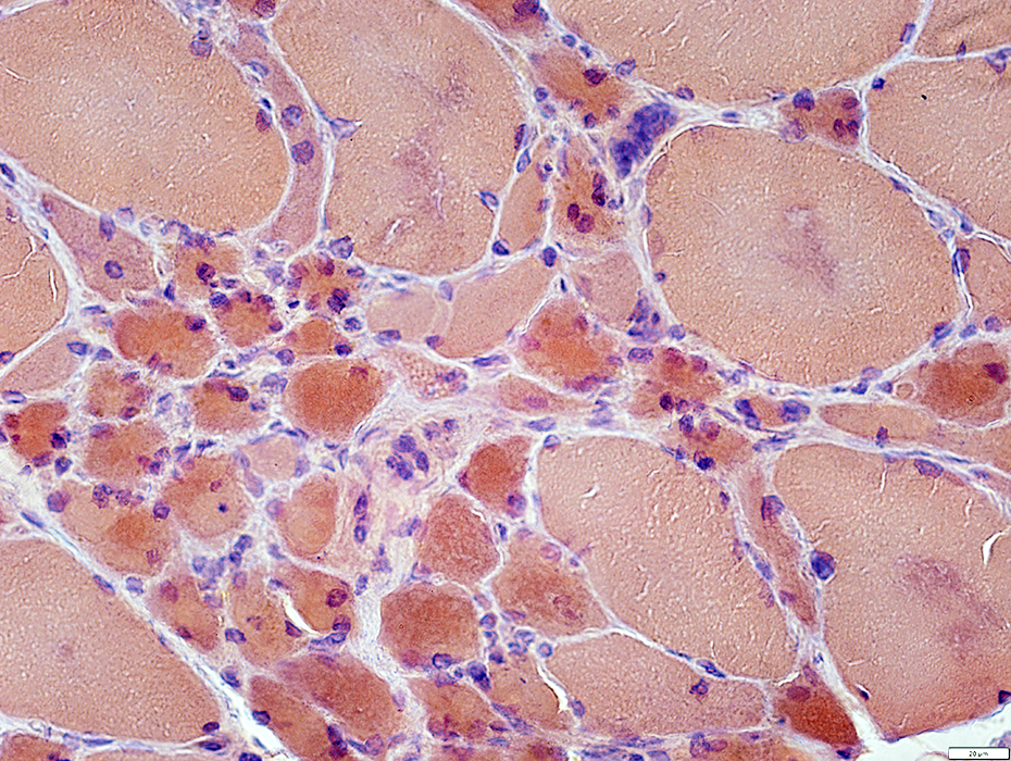
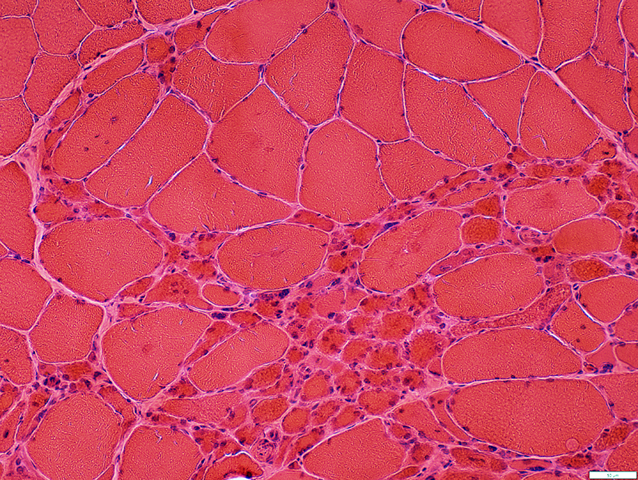 H&E stain |
Varied pathology in different regions
Some areas have many small fibers
Small fibers have dark staining and large nuclei
Other regions only have a few intermediate sized fibers
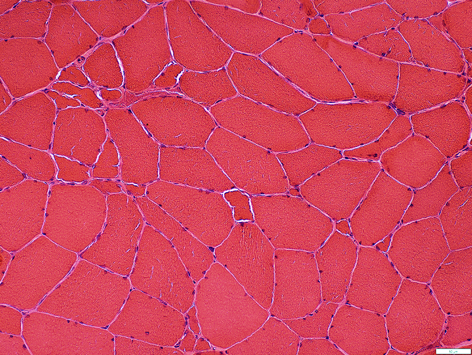 H&E stain |
 VvG stain |
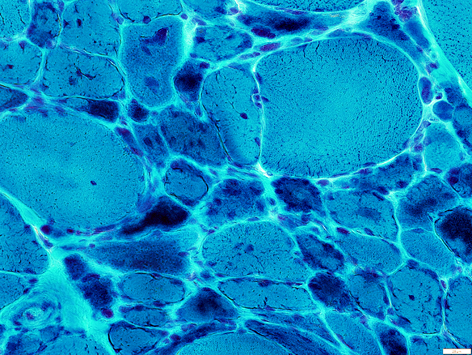
Sarcoplasmic pads around small fibers
Other small & Large fibers have irregular internal archjitecture
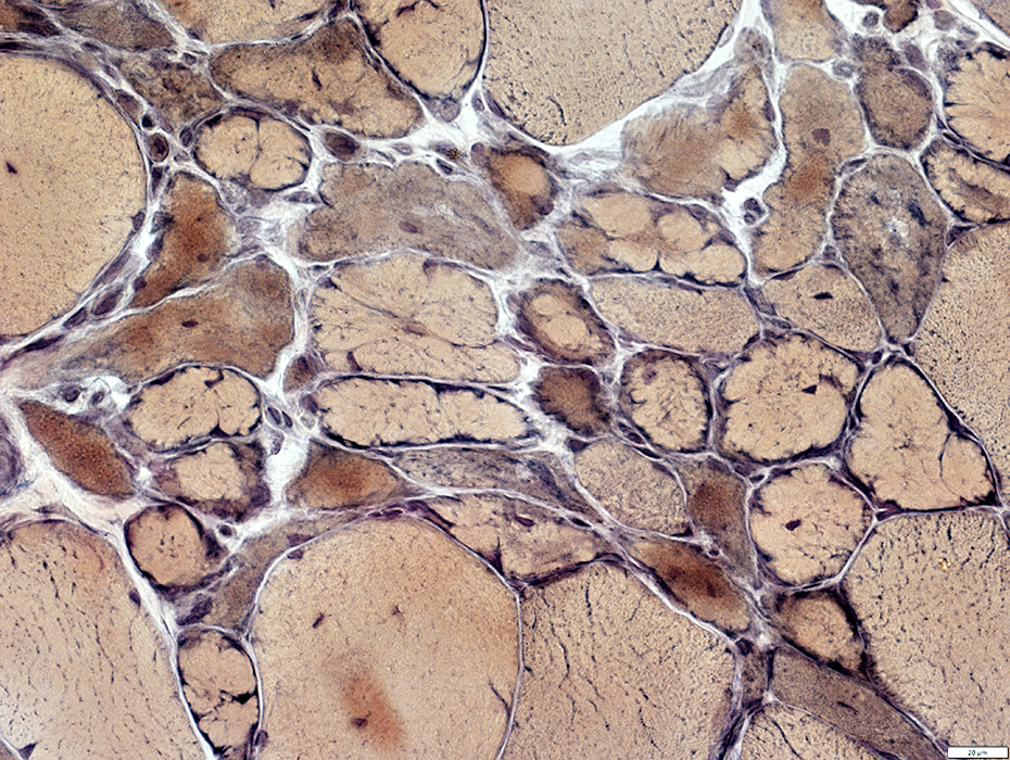
 VvG stain |
Muscle fiber internal architecture
Sarcoplasmic pads around small fibers
Other small & Large fibers have irregular internal archjitecture
Internal nuclei: Present in some muscle fibers
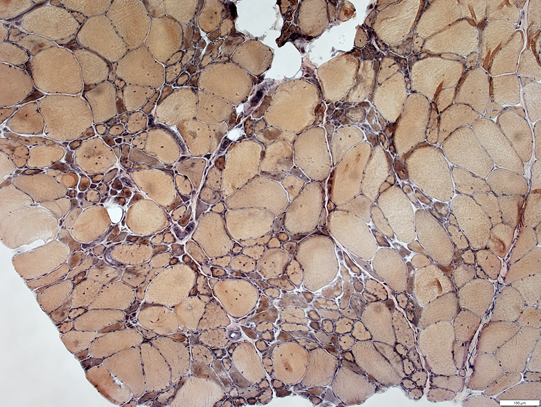
 VvG stain |
 Gomori trichrome stain |
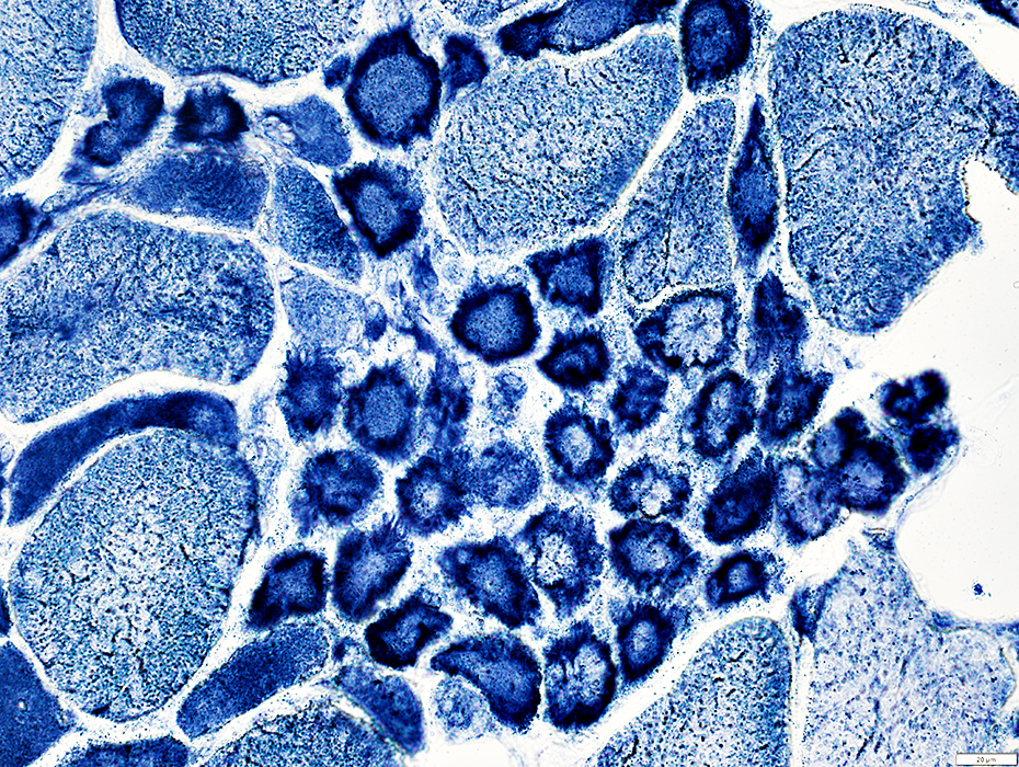
Fiber sizes: Varied
Small fibers: Dark stained
Endomysial connective tissue: Increased between muscle fibers
 Gomori trichrome stain |
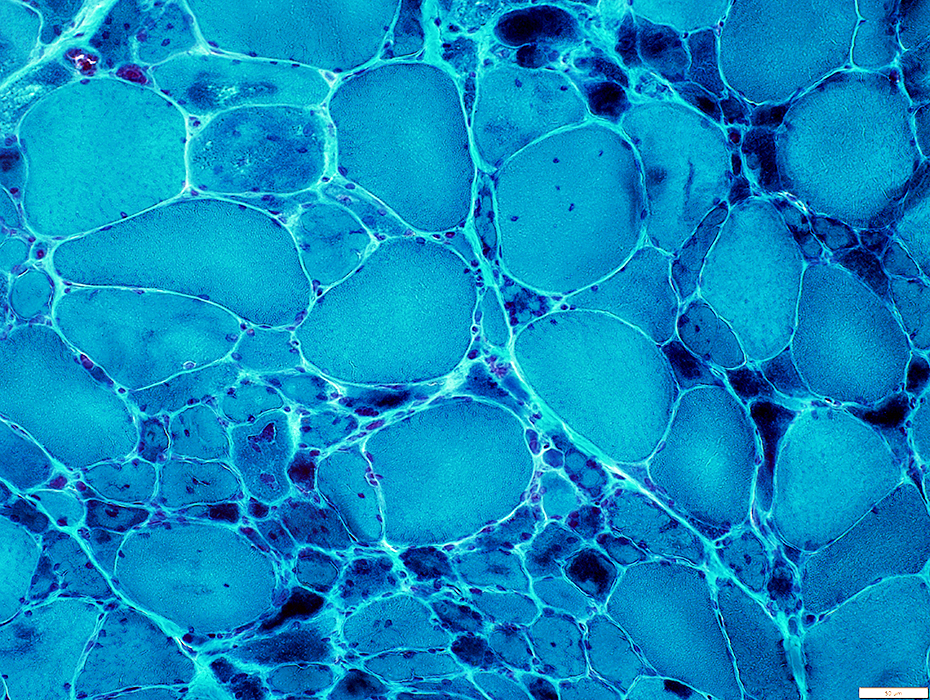
 NADH stain |
Sarcoplasmic pads around small fibers
Other small muscle fibers have irregular internal archjitecture
 NADH stain |
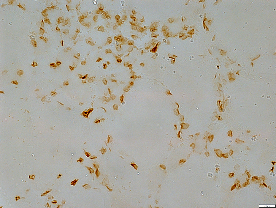
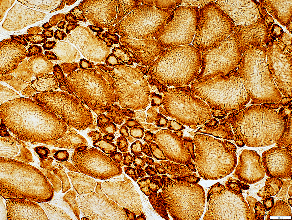 COX stain |
Abundant in sarcoplasmic pads
 COX stain |
Mitochondria
Abundant in sarcoplasmic pads
 SDH stain |
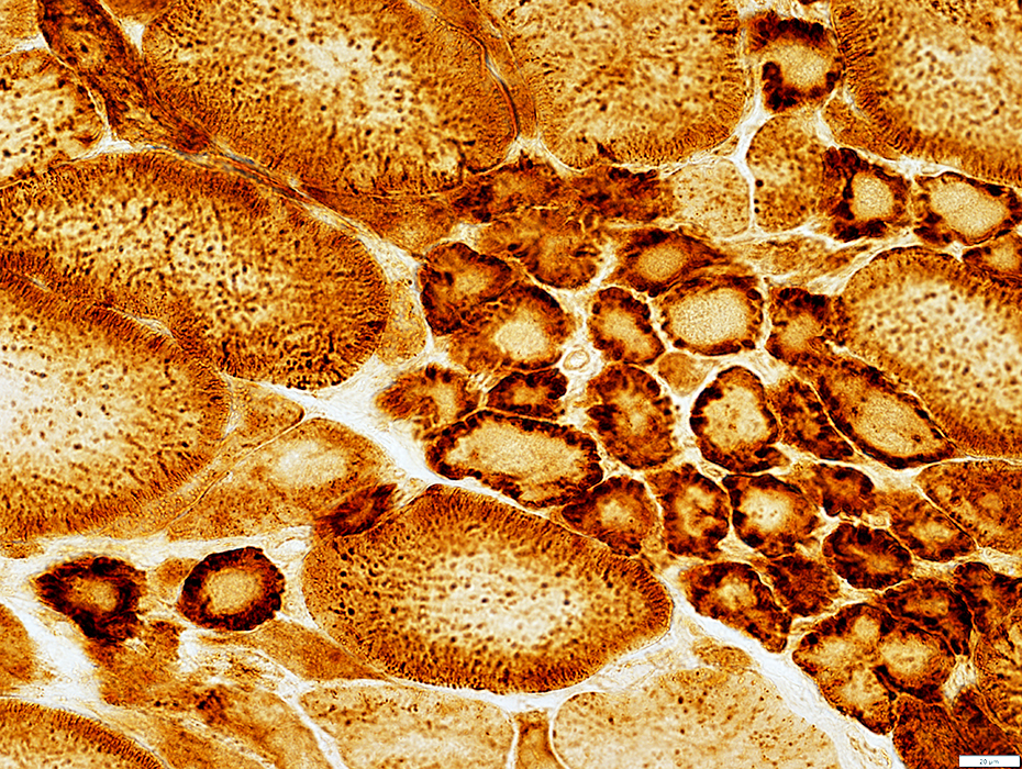
 Desmin stain |
Increased in cytoplasm of small muscle fibers
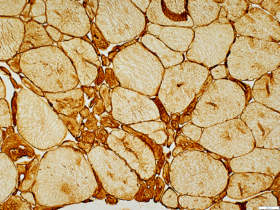
 Dys1 stain |
Small fibers have moderately thick sarcolemma
 Congo red stain |
Large & irregular shapes in small fibers
 Congo red stain |
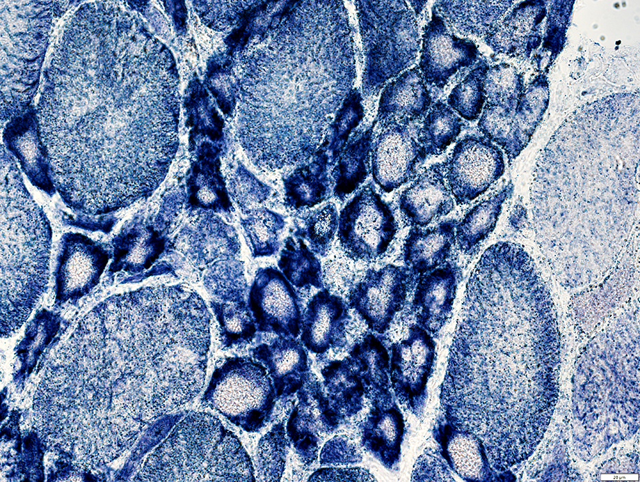
 Emerin stain |
Large & irregular shapes in small fibers
 ATPase pH 9.4 stain |
Small fibers: Mixed types; Many 2C
No fiber type grouping
 ATPase pH 4.3 stain |
Sarcoplasmic Pads: After muscle trauma
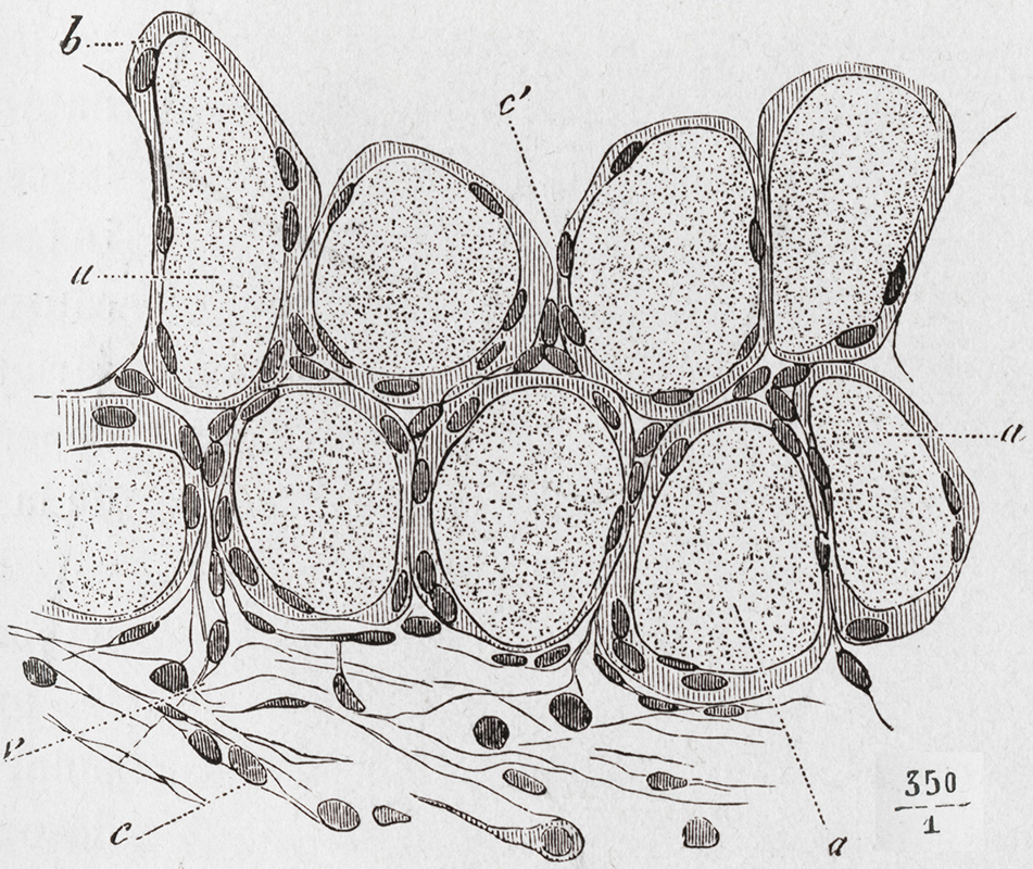 D>urant 1902
|
Return to: Filamin C
Return to: Neuromuscular Home Page
12/17/2025