Danon Disease: Pathology
Images from J Danon
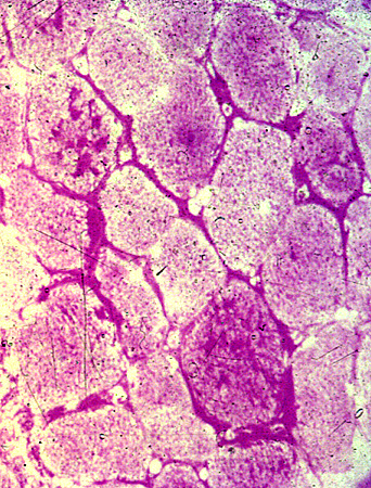
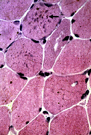 H & E stain Basophilic granules (Arrow) Cytoplasmic, Small |
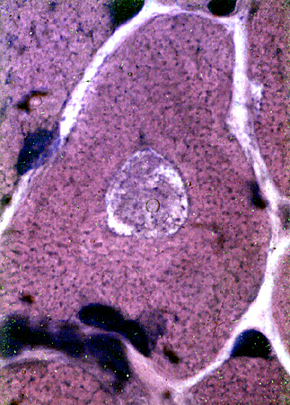 H & E stain Vacuole: Large |
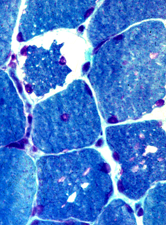 Gomori trichrome stain |
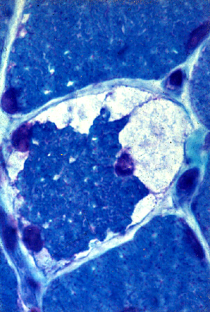 Gomori trichrome stain |
| Vacuoles in muscle fibers | |
 PAS stain |
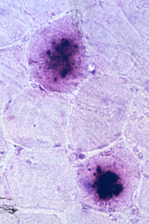 Acid phosphatase stain |
| Glycogen staining in muscle fibers | Acid phosphatase stains vacuoles |
|
|
|
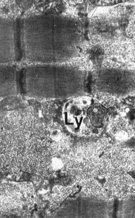
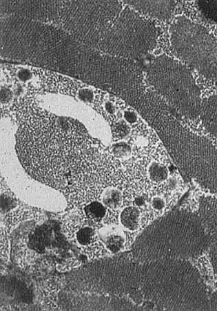
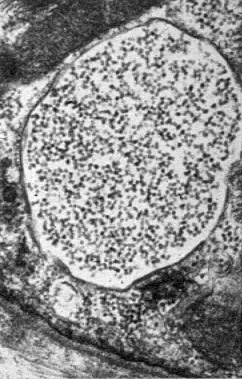
| Glycogen & Lysosome | Autophagic vacuoles | Membrane-bound glycogen |
Return to Danon disease
Return to Neuromuscular Home Page
10/25/2019