Myopathy with deficient Chondroitin Sulfate C in Connective tissue
|
Myopathy Myonuclei Δ Atrophy Inflammation Perimysium Histiocytes Alkaline phosphatase Chrondroitin SO4 C |
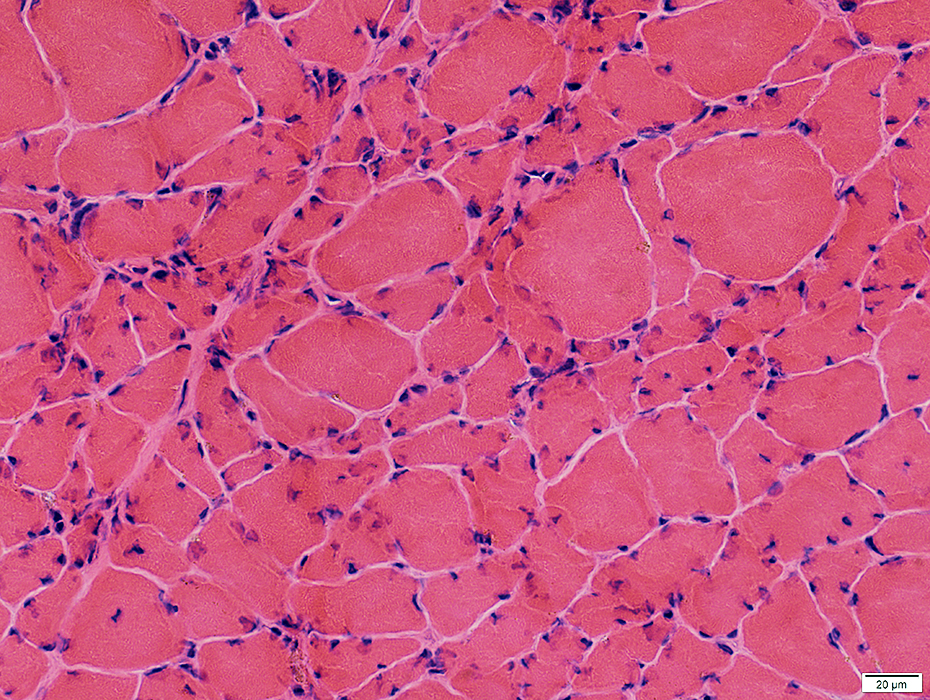 H&E stain |
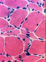 H&E stain |
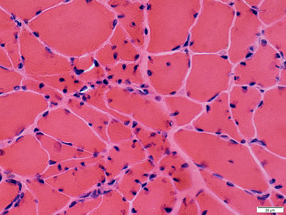
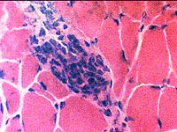
- Fiber size: Varied
- No active myopathic changes
- Myonuclei: Irregular shapes
 H&E stain |
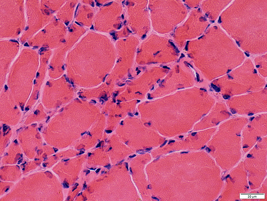 H&E stain |
Myonuclei with irregular shapes
Varied fiber size
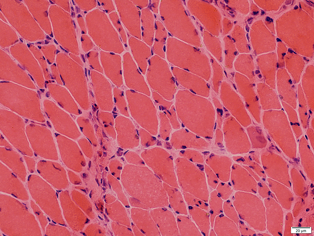
 H&E stain |
 Congo red stain |
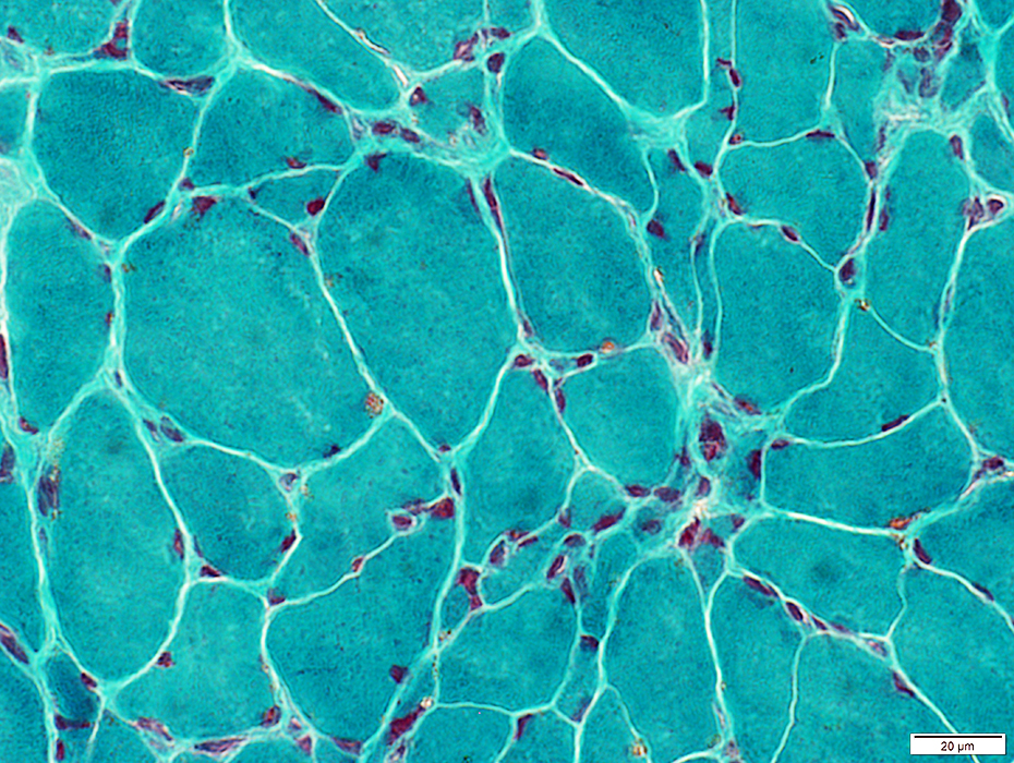 Gomori trichrome stain |
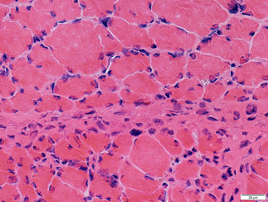 H&E stain |
Type 2 muscle fibers: Very small
Type 1 muscle fibers: Mild atrophy; Varied size
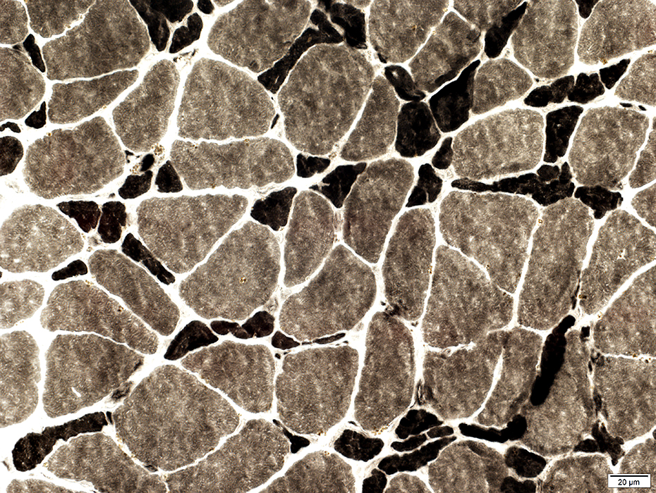 ATPase pH 9.4 stain |
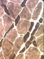 ATPase pH 9.4 stain |
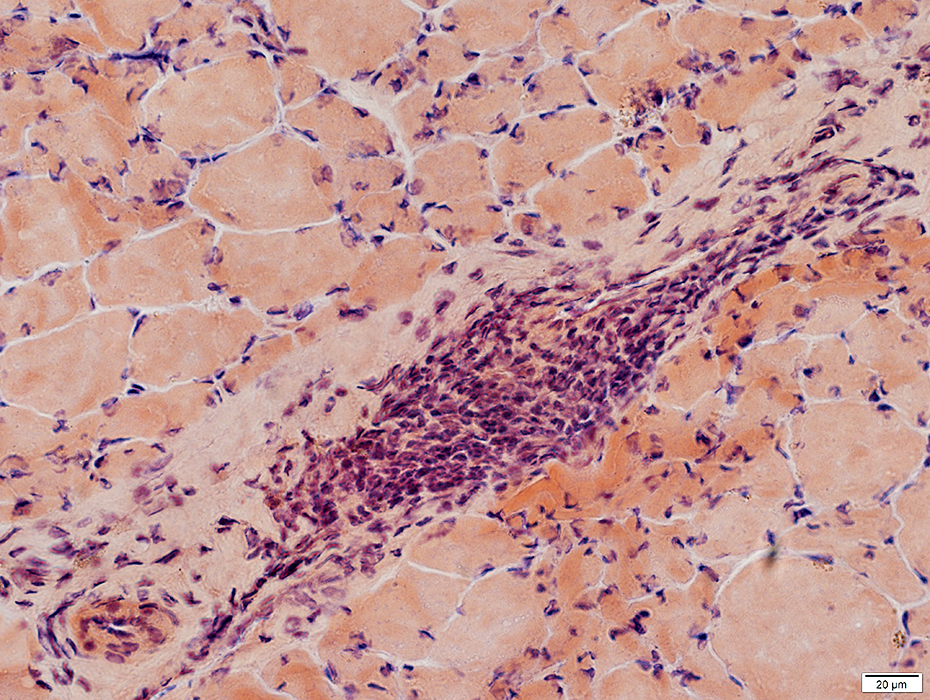 Congo red stain |
Cells near & around Perimysial vessel
 Congo red stain |
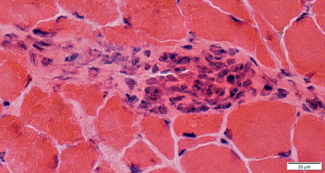 H&E stain |
 H&E stain |
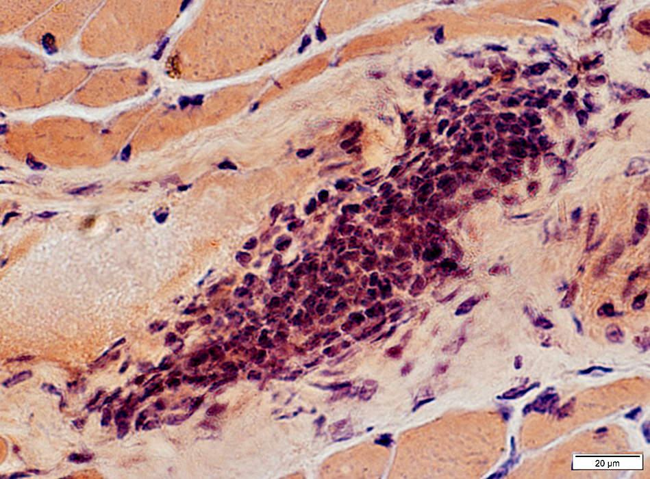
 Acid phosphatase stain |
Acid phosphatase positive histiocytic cells
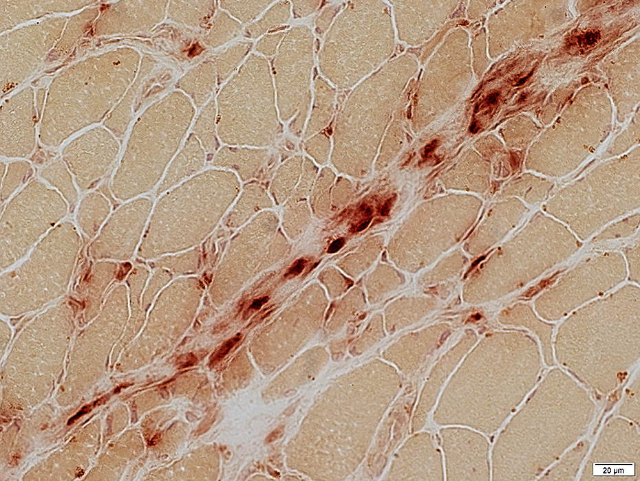 Acid phosphatase stain |
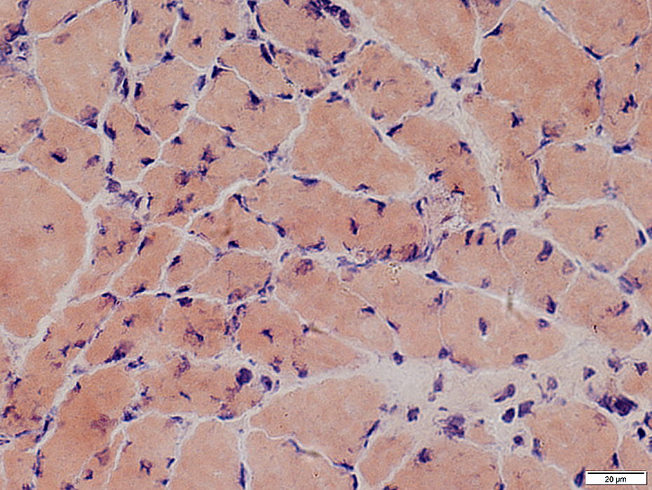
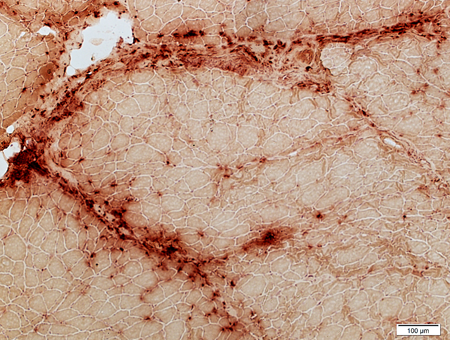
 Alkaline phosphatase stain |
 Alkaline phosphatase stain |
Increased staining by alkaline phosphatase
 Alkaline phosphatase stain |
- Perimysial cells staining with acid phosphatase
- Perimysium staining with alkaline phosphatase
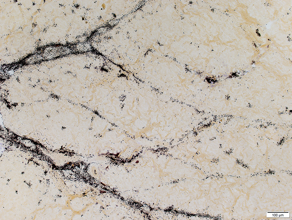
Chondroitin sulfate C: Patterns of Staining
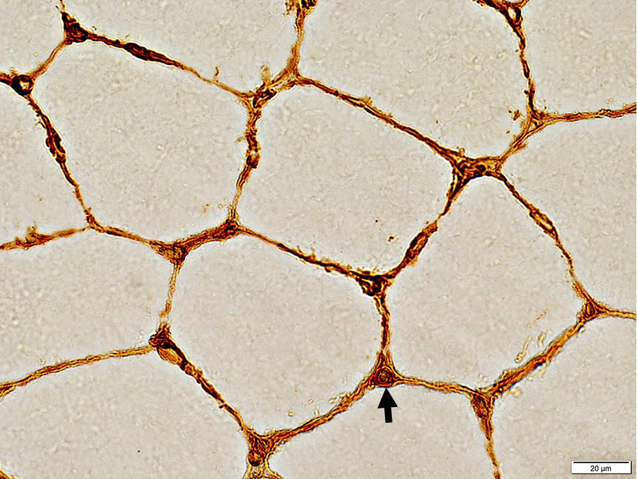 Chondroitin sulfate C stain |
Dark staining on endomysum & capillaries (Arrow)
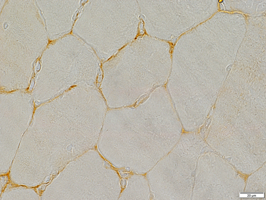 Chondroitin sulfate C stain |
Reduced staining on endomysum & capillaries
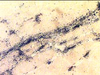
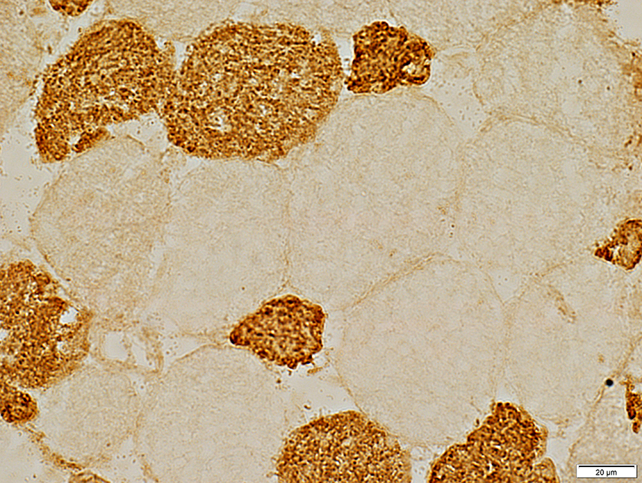 Chondroitin sulfate C stain |
Reduced staining on endomysum & capillaries; Increased staining in muscle fiber cytoplasm
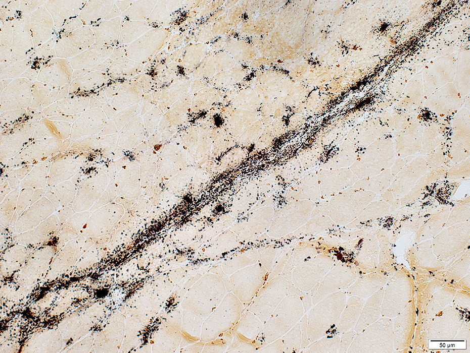
 Chondroitin sulfate C stain Patient muscle: Pre-Rx |
 Chondroitin sulfate C stain Patient: Post steroid Rx |
- Normal: Abundant on skeletal muscle endomysium & endomysial capillaries
- Myopathy: Absent in patient muscle before treatment
- After corticosteroid treatment: Returns to near normal
Return to Neuromuscular Home Page
Return to Inflammatory myopathies
1/28/2019