Congenital Hypomyelinating Neuropathy
4 y.o. Female: PMP-22 point mutation
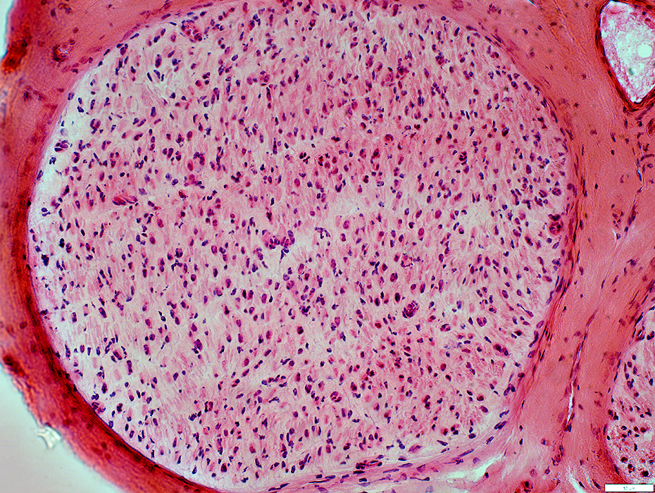 H&E stain |
No large myelinated axons
Cellular
Some increased connective tissue space
Perineurium & Endoneurium: Normal
 H&E stain |
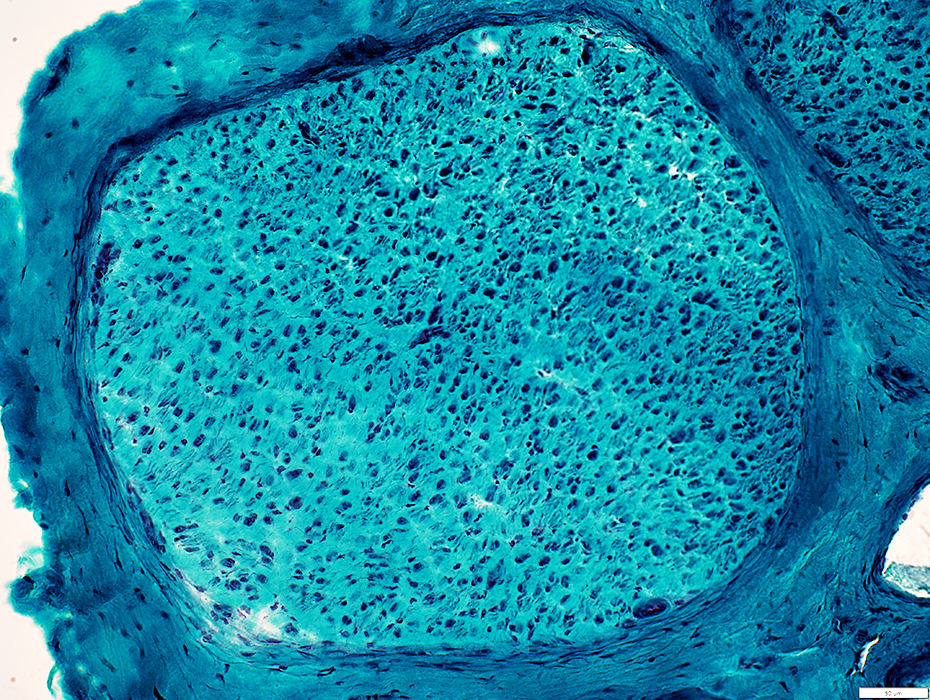 Gomori trichrome stain |
No large myelinated axons
Perineurium & Endoneurium: Normal
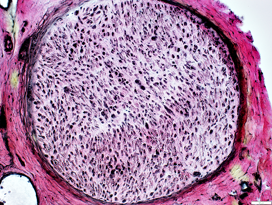 VvG stain |
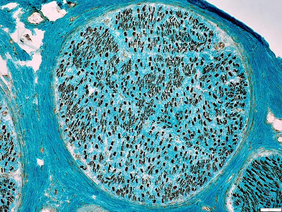 Neurofilament stain |
Large axons: Many
Small axons: Normal to Reduced Numbers
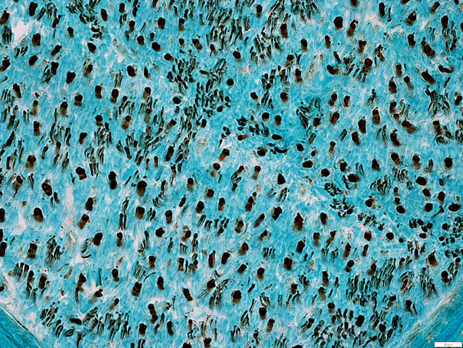 Neurofilament stain |
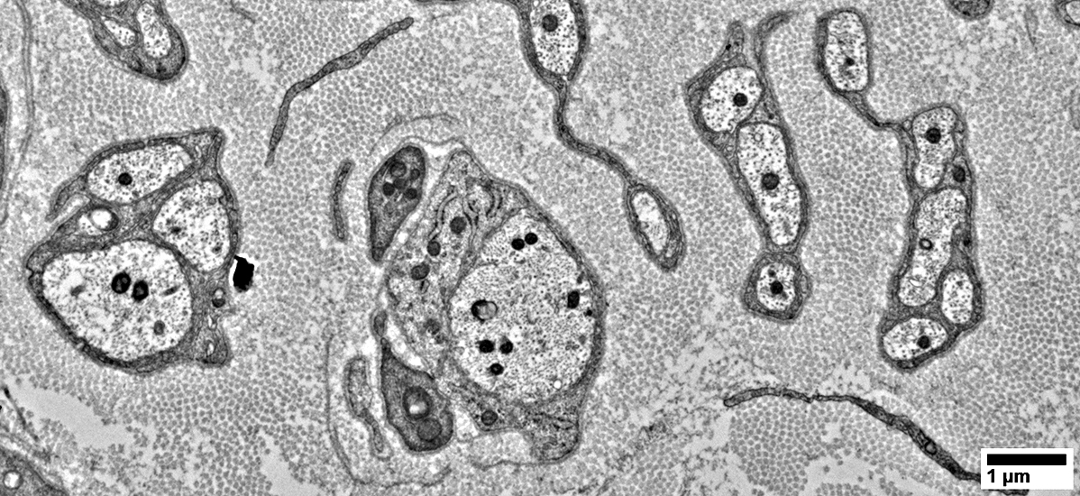
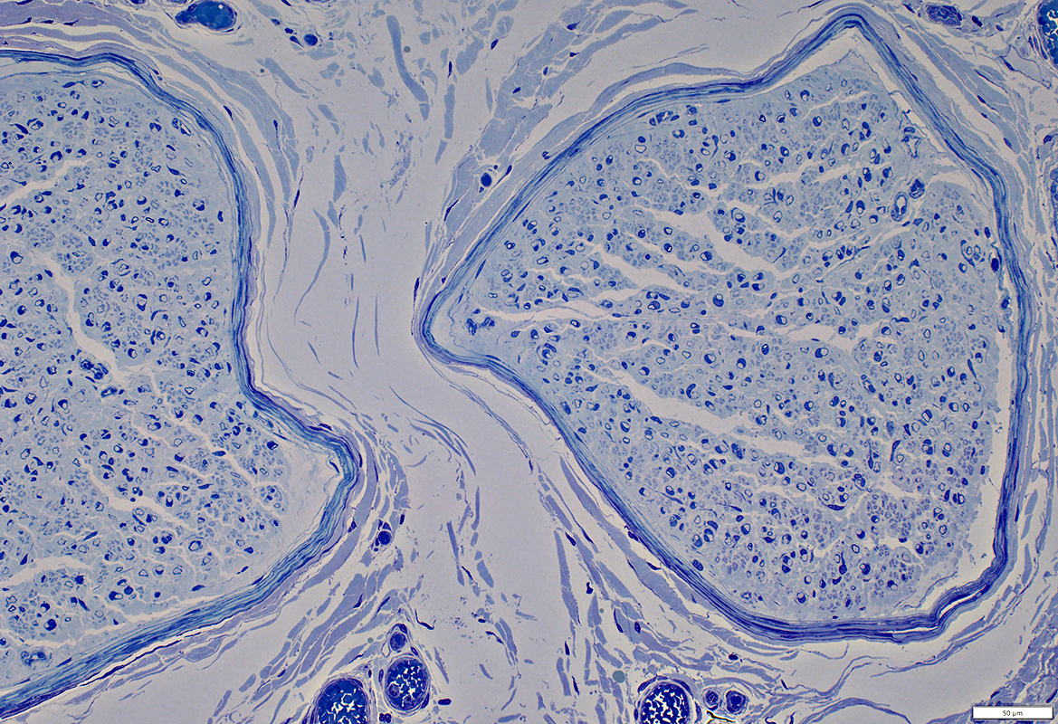 Toluidine blue stain |
Large axons: Thinly myelinated; Reduced numbers
BLOBs: Increased space around axons (Arrow; Below)
Fibroblasts: Scattered; Long processes
 Toluidine blue stain |
 Toluidine blue stain |
Large axons: Thinly myelinated; Reduced numbers
BLOBs: Increased space around axons
Fibroblasts (Arrows)
Scattered
Long processes
Some processes around outside of BLOBs
 Toluidine blue stain |
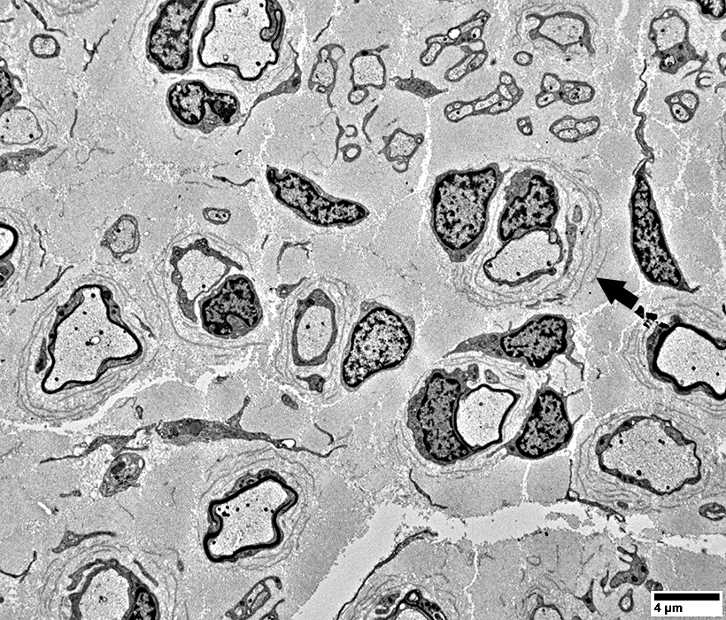
Basal Lamina Onion Bulbs (BLOBs)
BLOB StructureBasal Lamina
Concentric thin fragments (Arrow), Double-layered
Around larger axons
Collagen: Between layers of basal lamina
Schwann cell processes
Few
Within layers of basal lamina
Center of BLOB: May immediately surround axon
Myelin: Present around some axons
Fibroblasts: Process may surround BLOB
 |
BLOB Structure
Basal Lamina
Concentric thin fragments (Arrow), Double-layered
Surround larger axons
Collagen: Between layers of basal lamina
Schwann cell processes
Few
Within layers of basal lamina
Center of BLOB: May immediately surround axon
Myelin: Present around some axons
Fibroblasts: Process may surround BLOB (Arrow)
 |
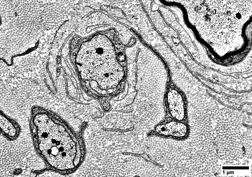
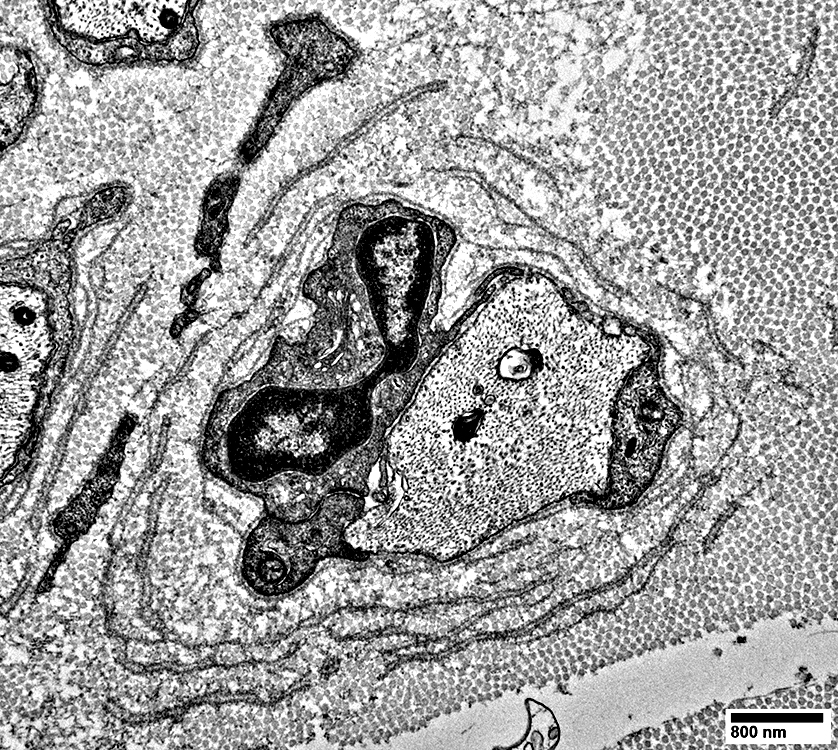
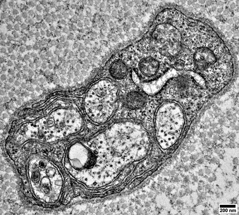
BLOBs: Surround Unmyelinated & Hypomyelinated Axons
6 Axons (Left to Right)1. Far left: Small axon, part, surrounded by Schwann cell process with external basal lamina
2. Left: Small-Intermediate-sized axon surrounded by dark Schwann cell processes
3. Left-Center (BLOB): Intermediate-sized, unmyelinated axon with surrounding layers
Inner: Schwann cell processes (Dark) directly around axon; No associated myelin
Intermediate
Double layers of Schwann cell Basal Lamina; Each extends partly around axon
Two very small Schwann cell processes (Left) are present within individual layers of Schwann cell basal lamina
Outer
Double basal lamina (Above)
Partially contains a Schwann cell process
Schwann cell process extends from a Schwann cell that surrounds 2 unmyelinated axons
Fibroblast process,partial (Left of Basal lamina layers)
4. & 5. Right-Center: 2 Small axons: Surrounded by Schwann cell processes
6. Right Upper: Larger axon surrounded by
Thin myelin sheath layer
BLOB: Double layers of circumferential basal lamina
 |
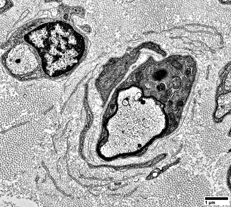
BLOBs: Surround Unmyelinated Axons
 |
Center: Intermediate-sized, unmyelinated axon with surrounding layers
Inner layer
Schwann cell processes (Dark): Directly around axon; No associated myelin
Schwann cell nucleus: Near axon
Intermediate layers
Double layers of Schwann cell Basal Lamina; Each extends partly around axon
Outer layer
Fibroblast process,partial (Left of Basal lamina layers)
Collagen particles
 |
 |
Center: Intermediate-sized, unmyelinated axon with surrounding layers
Inner layer
Schwann cell processes (Dark): Directly around axon; No associated myelin
Schwann cell nucleus: Near axon
Intermediate layers
Double layers of Schwann cell Basal Lamina; Each extends partly around axon
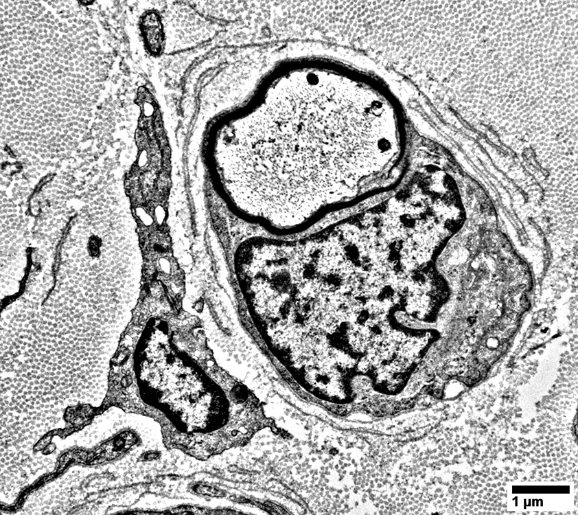
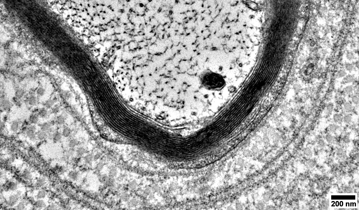
BLOBs: Basal laminal layers around Axon with Incomplete, Thin Myelin Layer
 |
Center: Intermediate-sized axon with surrounding layers
Inner layer
Schwann cell processes (Dark): Directly around axon; Incomplete layer of associated myelin
Intermediate layers
Double layers of Schwann cell Basal Lamina; Each extends partly around axon
Schwann cell processes: Few present (Contain basal lamina)
Outer layer: May contain fibroblast processes (Right)
 |
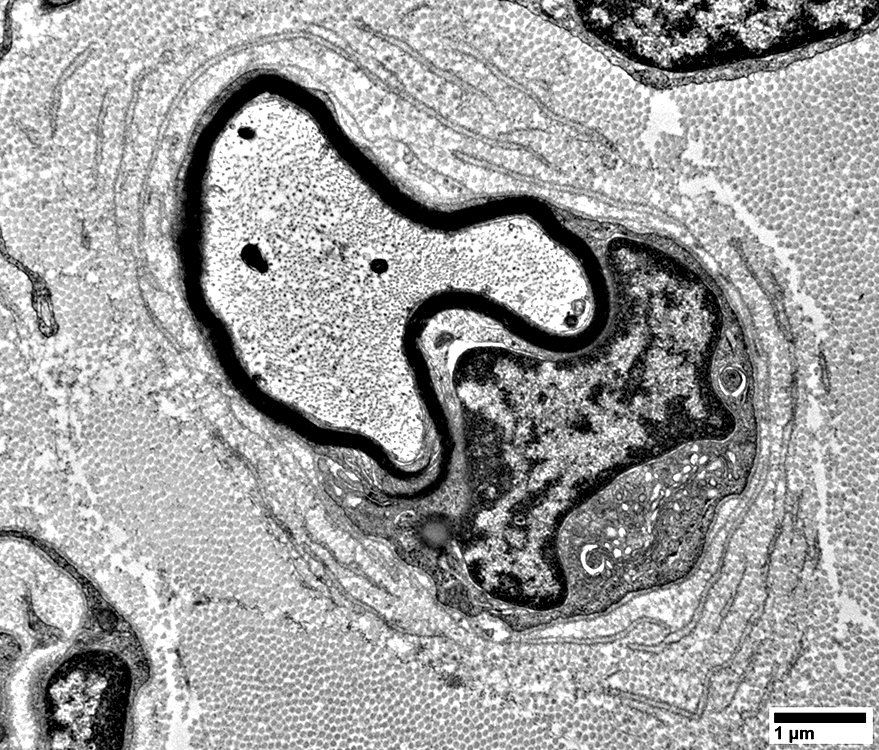
BLOBs: Around Axon with Thin Myelin Layer (Hypomyelination)
 |
Center: Intermediate-sized, Hypomyelinated axon with surrounding layers
Inner layer
Myelin sheath: Thin
Schwann cell cytoplasm: Directly around part of myelin
Schwann cell nucleus: Near axon
Intermediate layers
Double layers of Schwann cell Basal Lamina; Each extends partly around axon
Outer layer
Fibroblast nucleus & processes (Above; Right)
Collagen particles
 |
 |
Center: Intermediate-sized, Hypomyelinated axon with surrounding layers
Inner layer
Myelin sheath: Thin
Schwann cell cytoplasm: Directly around part of myelin
Intermediate layers
Double layers of Schwann cell Basal Lamina; Each extends partly around axon
Schwann cell processes within some basal lamina
Outer layer
Collagen particles
BLOB: Basal Lamina, Double layer, around Thinly Myelinated Axon
 From: R Schmidt |
|
Unmyelinated Axons
 From: R Schmidt |
 From: R Schmidt |
 From: R Schmidt |
Return to Neuromuscular Home Page
4/28/2024