Acute Nutritional Neuropathy
|
General Axons Myelin Loss Schwann cell pathology Myelin pathology Unmyelinated Wallerian Degeneration Cell processes |
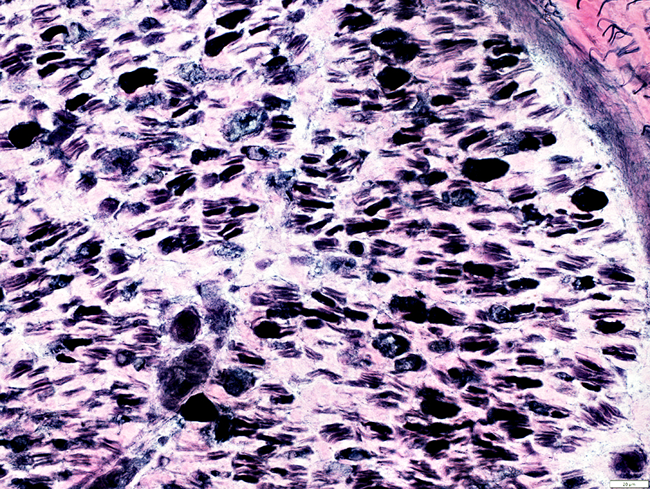
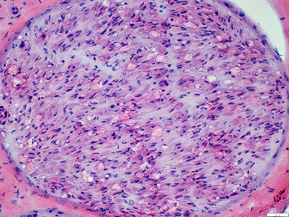 H&E stain |
Reduced number
Damaged myelin (Loss of staining)
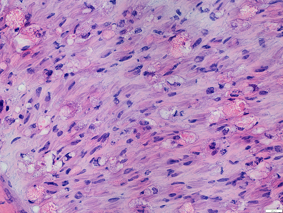 H&E stain |
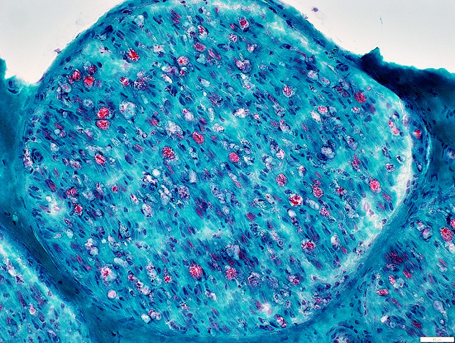
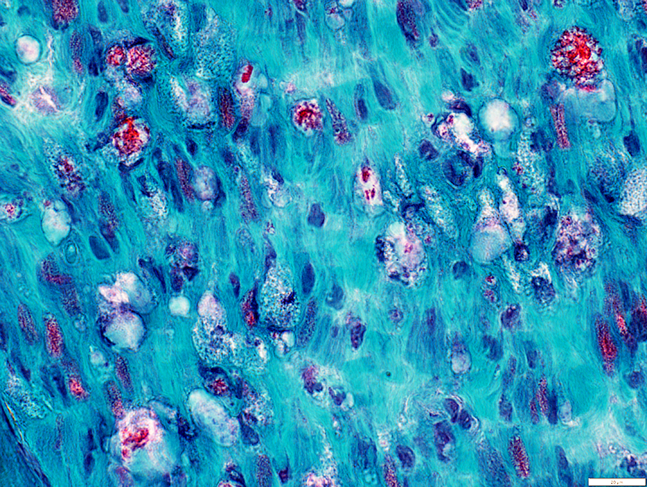
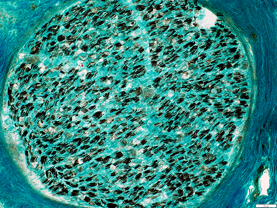
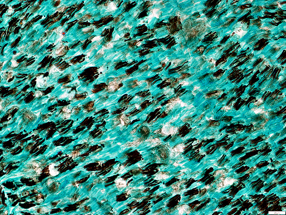
Gomori trichrome stain |
Reduced number
Damaged myelin (Loss of red color)
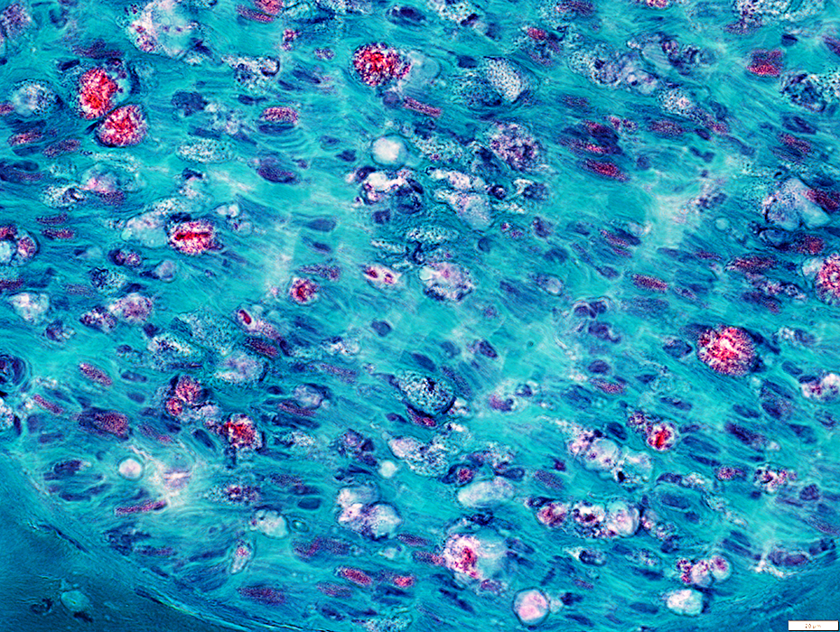
Gomori trichrome stain |
Gomori trichrome stain |
Reduced number
Damaged myelin (Loss of staining)
VvG stain |
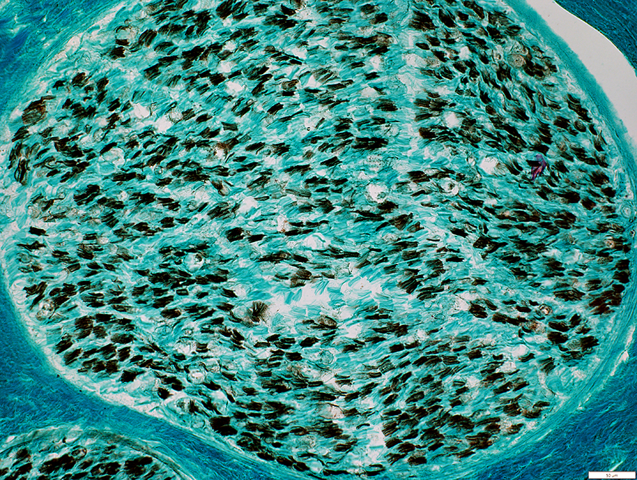
Acute Nutritional Neuropathy: Wallerian Degeneration
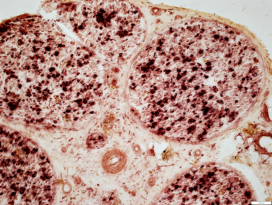
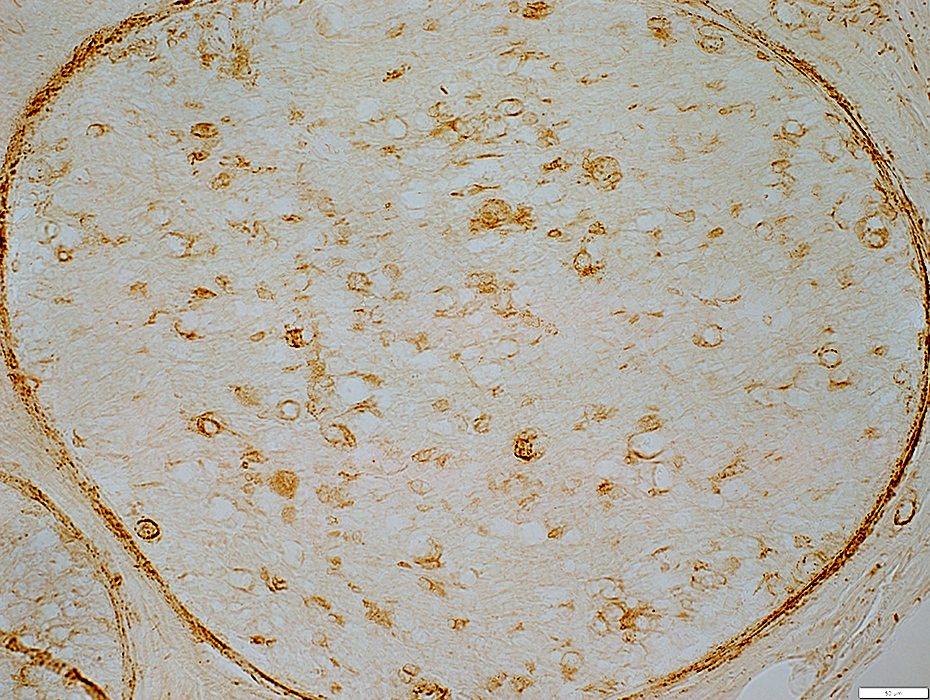
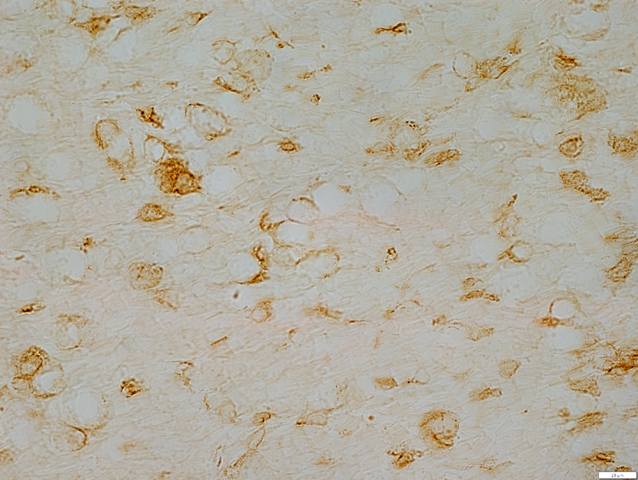
Acid phosphatase stain |
Diffusely stained by acid phosphatase
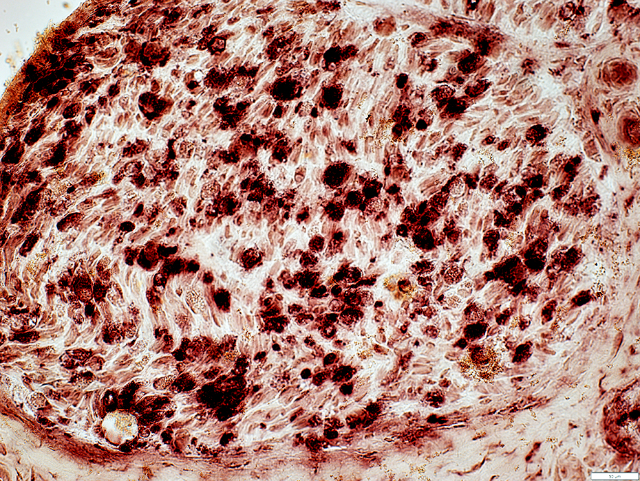 Acid phosphatase stain |
 Acid phosphatase stain |
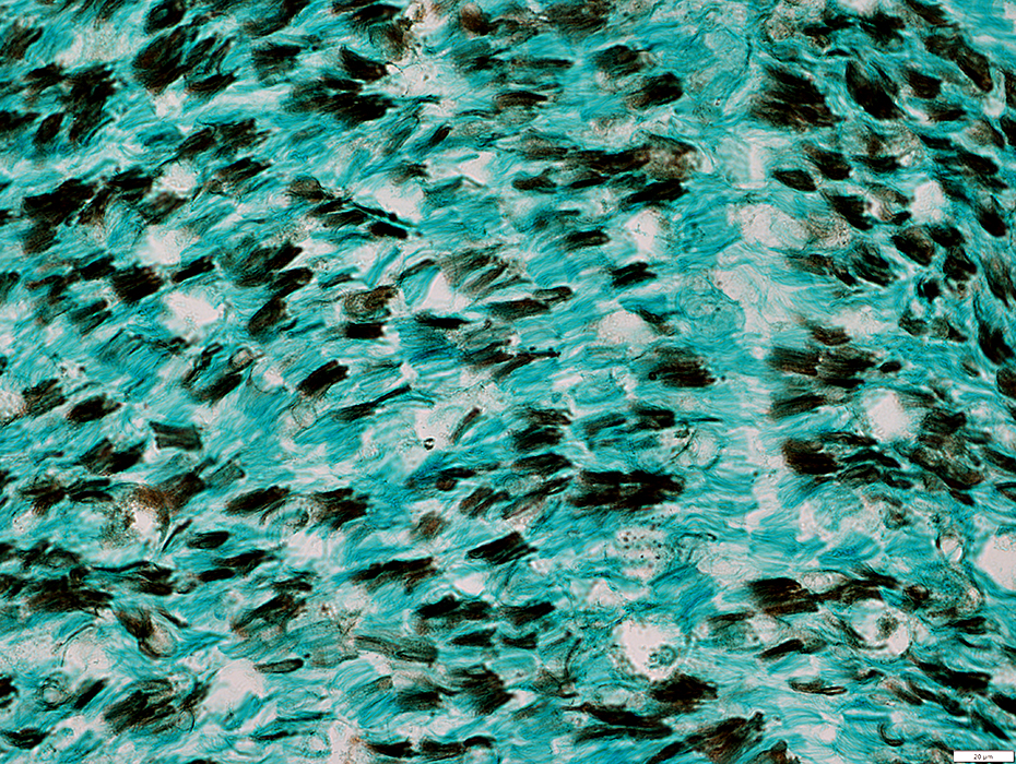
Neurofilament stain |
Loss of neurofilament stain
Neurofilament stain: Diffuse through some myelin sheaths
Unmyelinated Axons
Preserved numbers
Neurofilament stain |
MBP(r)_07sm.jpg)
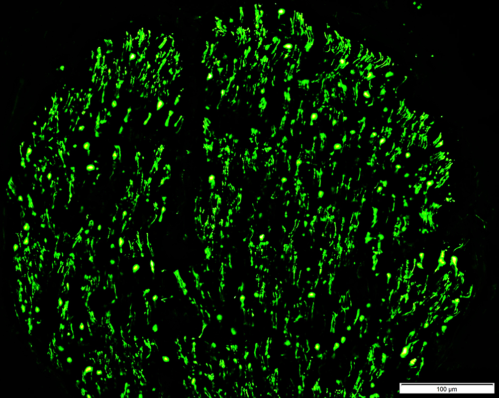
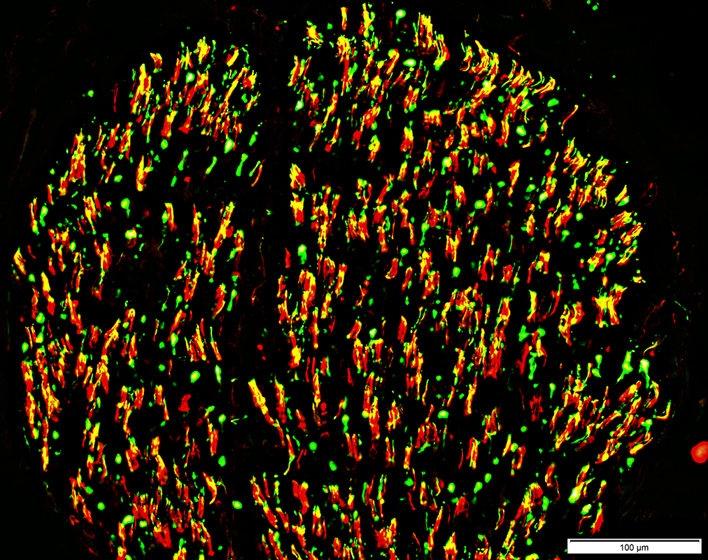
Neurofilament (Green), Myelin basic protein (Red) stain |
Lost within many MBP stained myelin sheaths (Red)
Unmyelinated Axons: preserved numbers (Green)
MBP(r)_01sm.jpg)
Neurofilament (Green), Myelin basic protein (Red) stain |
)MBP(r)_01sm.jpg)
Neurofilament (Green), Myelin basic protein (Red) stain |
Lost within some MBP stained myelin sheaths (Red)
Unmyelinated Axons: preserved numbers (Green)
MBsm.jpg)
Neurofilament (Green), Myelin basic protein (Red) stain |
Acute loss: MBP-stained myelin sheaths without axons
Chronic loss: Reduced numbers of MBP-stained myelin sheaths
)MBP(r)_05sm.jpg)
Neurofilament (Green), Myelin basic protein (Red) stain |
)MBP(r)_07sm.jpg)
Neurofilament (Green), Myelin basic protein (Red) stain |
C5b-9 stain |
C5b-9 stains in & around myelin sheaths
C5b-9 stain |
From: R Schmidt |
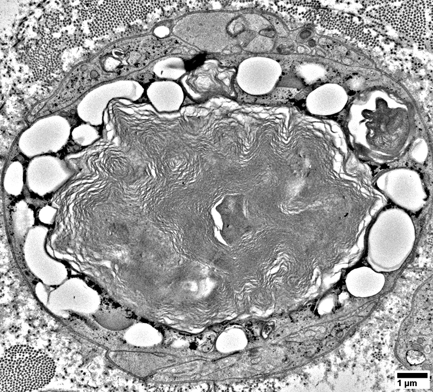
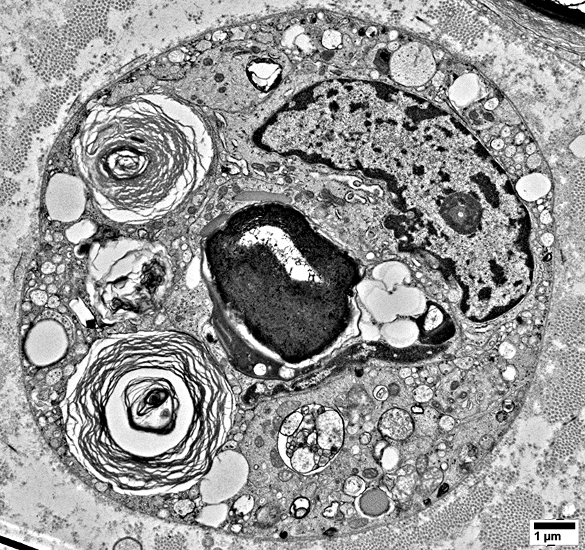
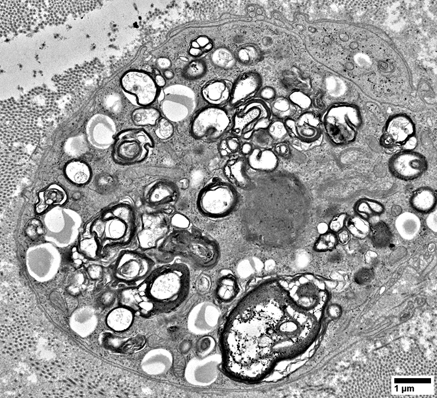
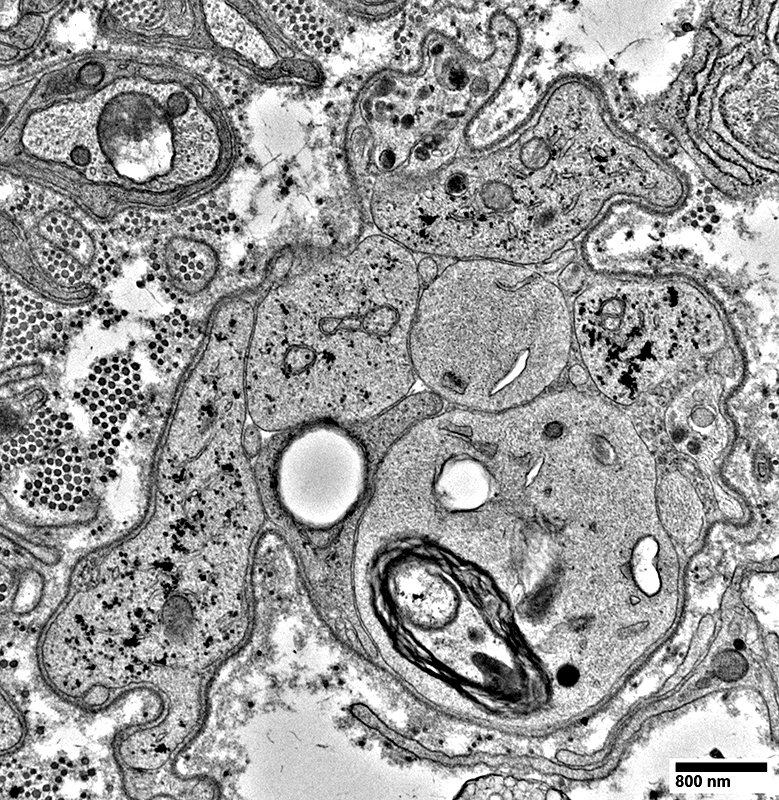
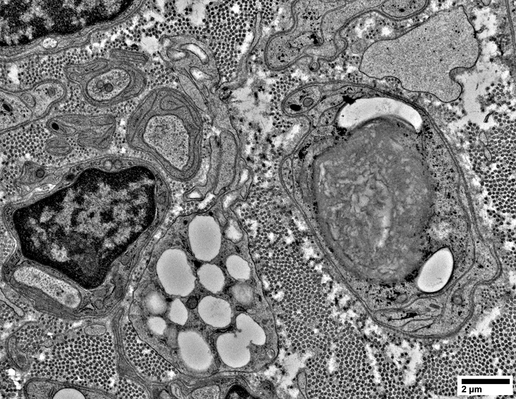
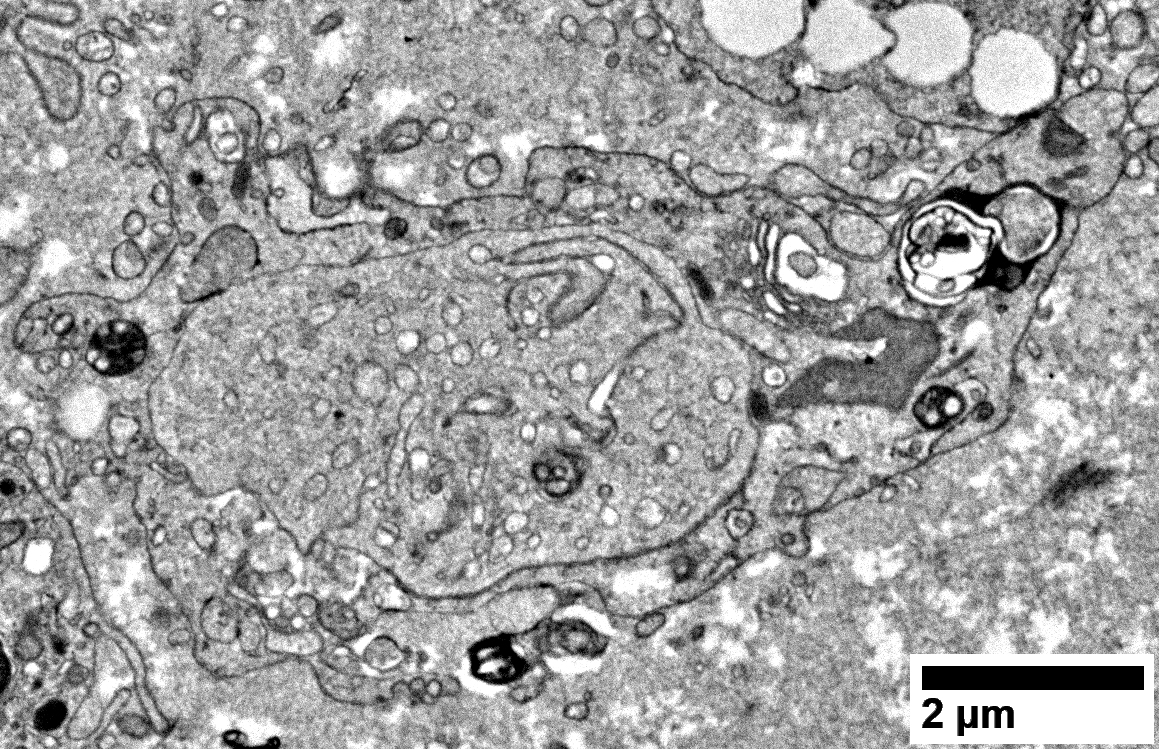
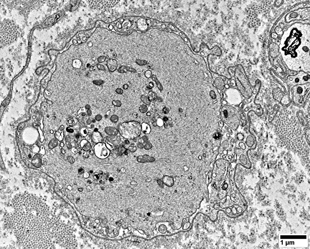
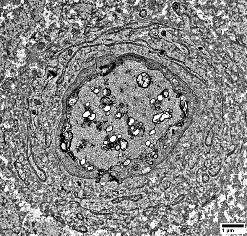
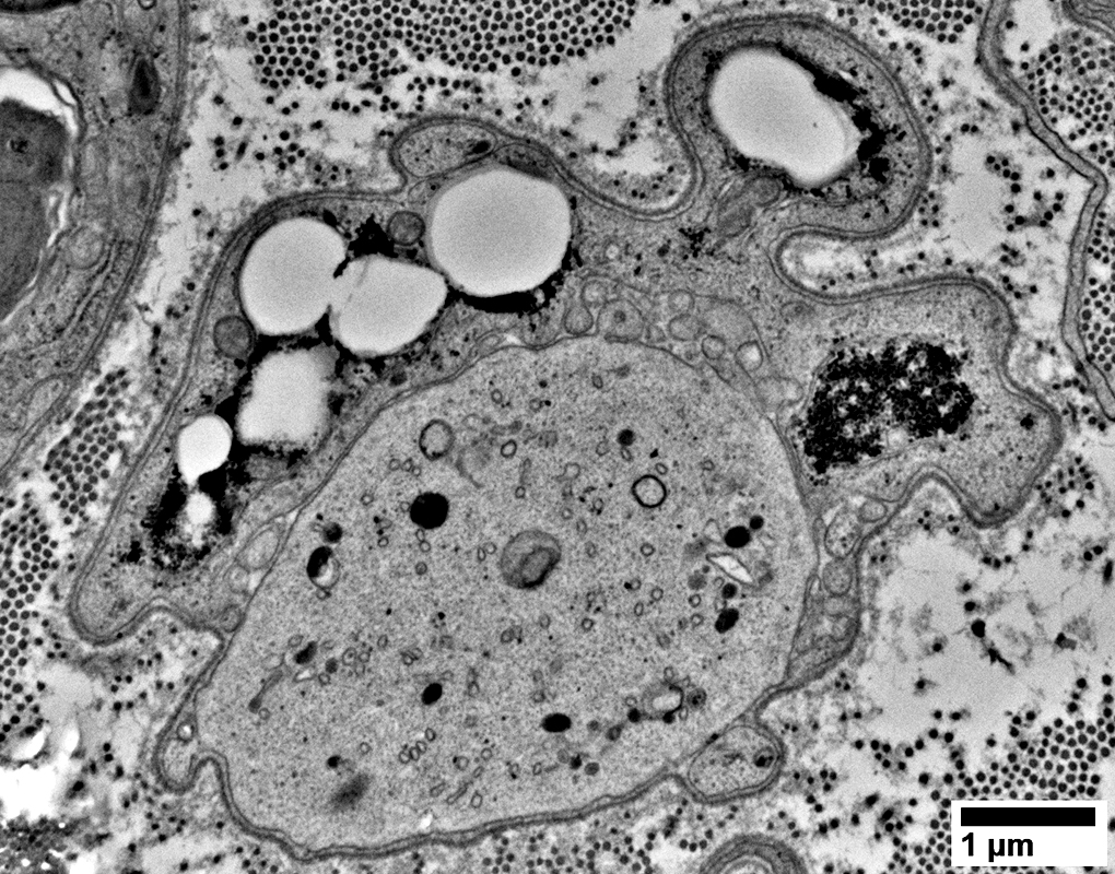
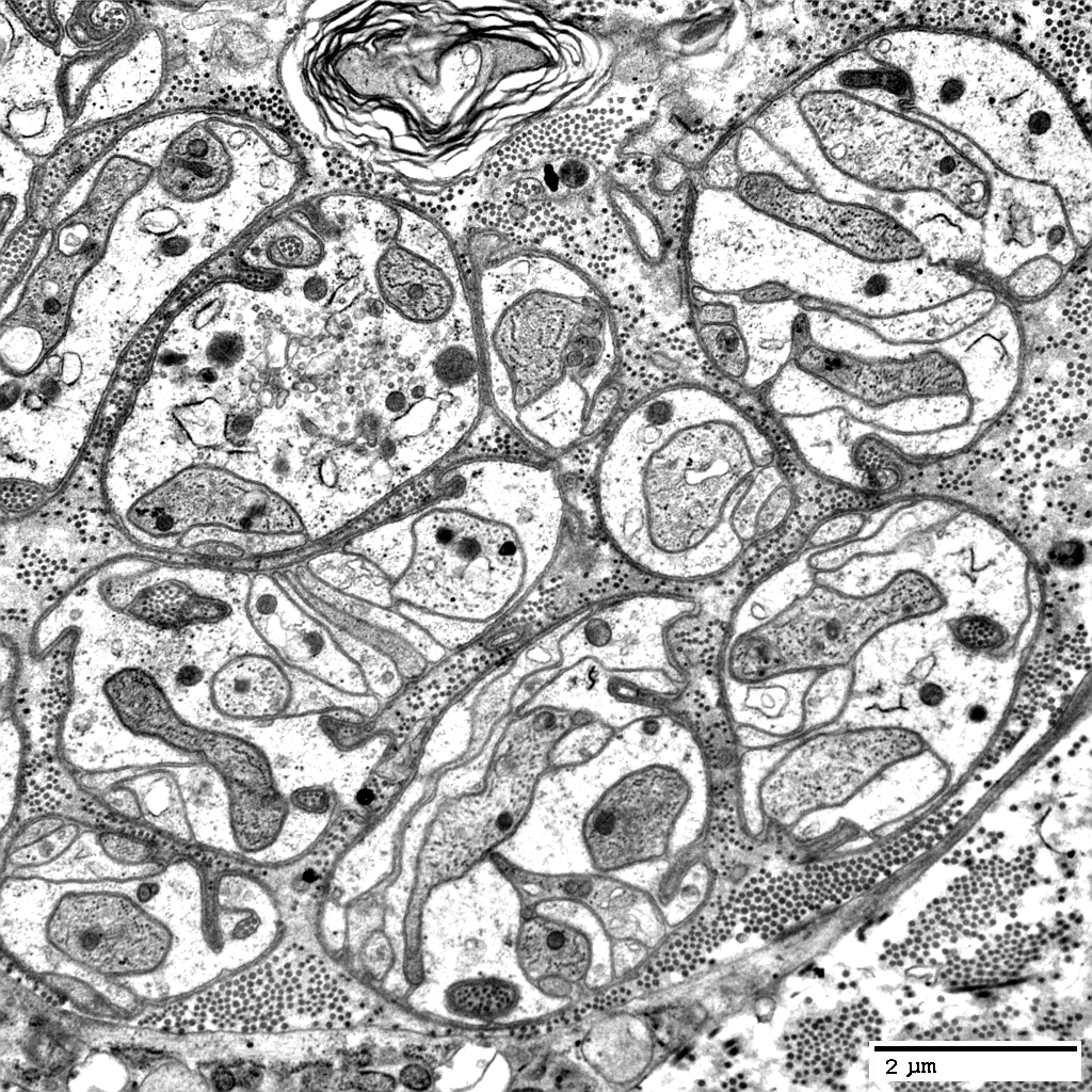
Schwann Cells
Some contain Lipid debris
From: R Schmidt |
From: R Schmidt |
From: R Schmidt |
From: R Schmidt |
From: R Schmidt |

From: R Schmidt |
From: R Schmidt |

From: R Schmidt |
From: R Schmidt |
From: R Schmidt |
From: R Schmidt |
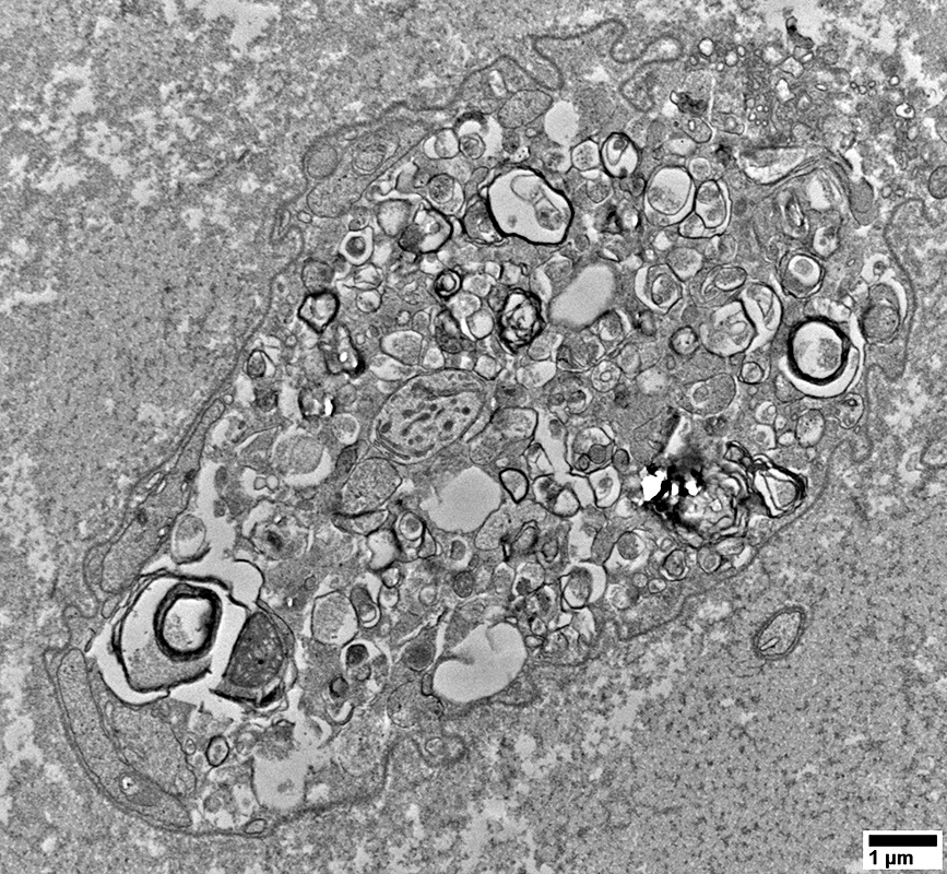
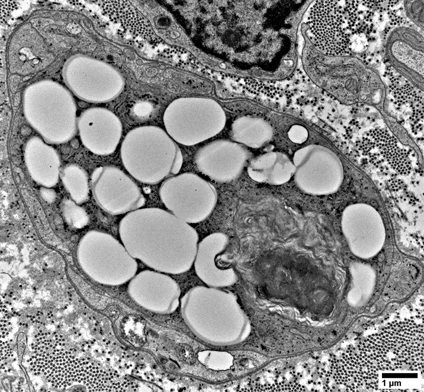
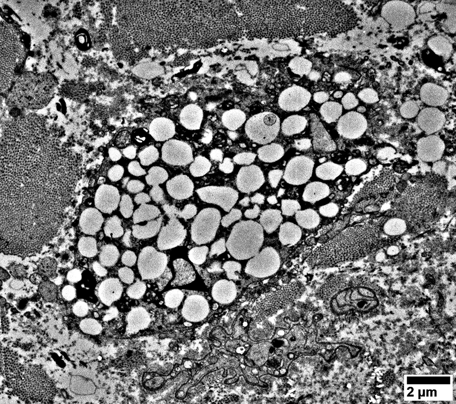
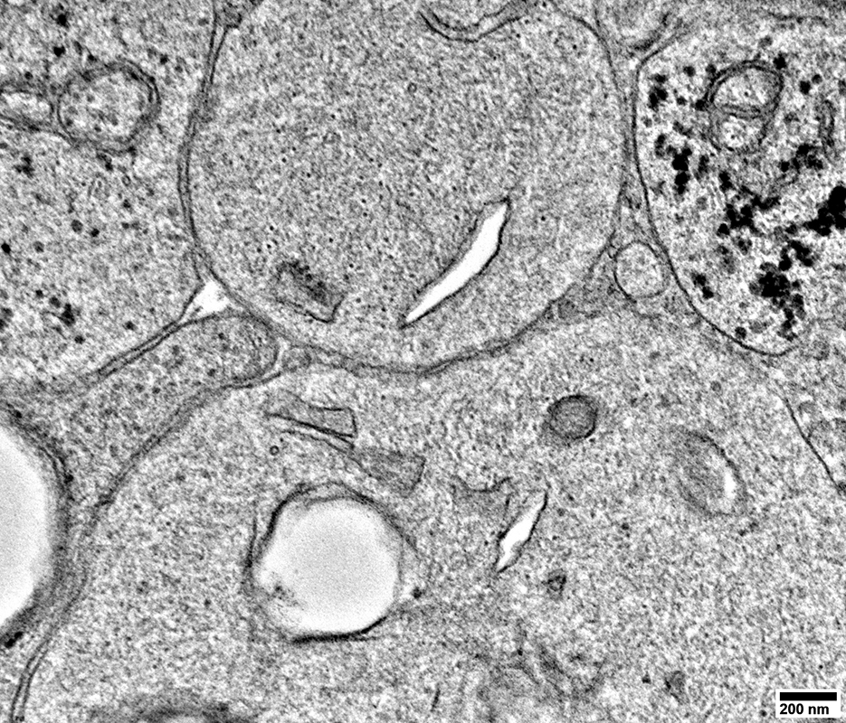
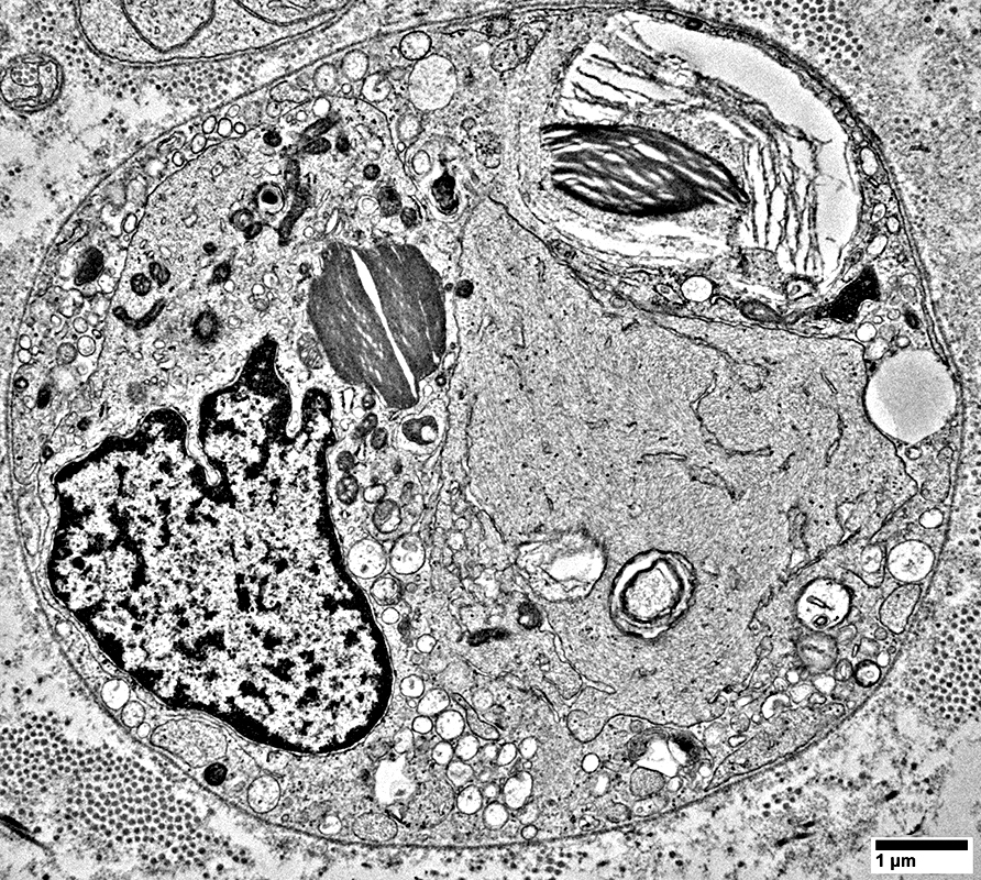
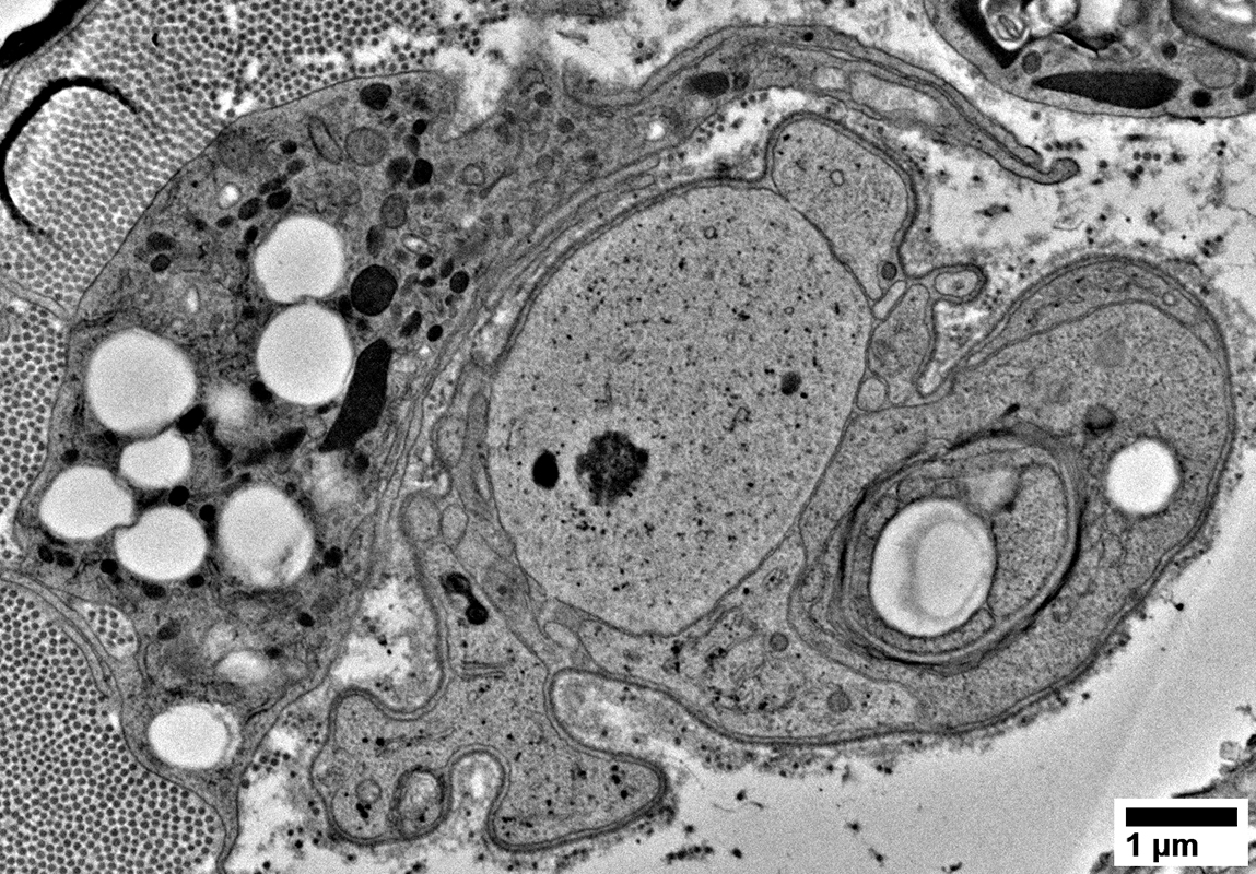
Lipid Droplets in an endoneurial cell
From: R Schmidt |
From: R Schmidt |
From: R Schmidt |
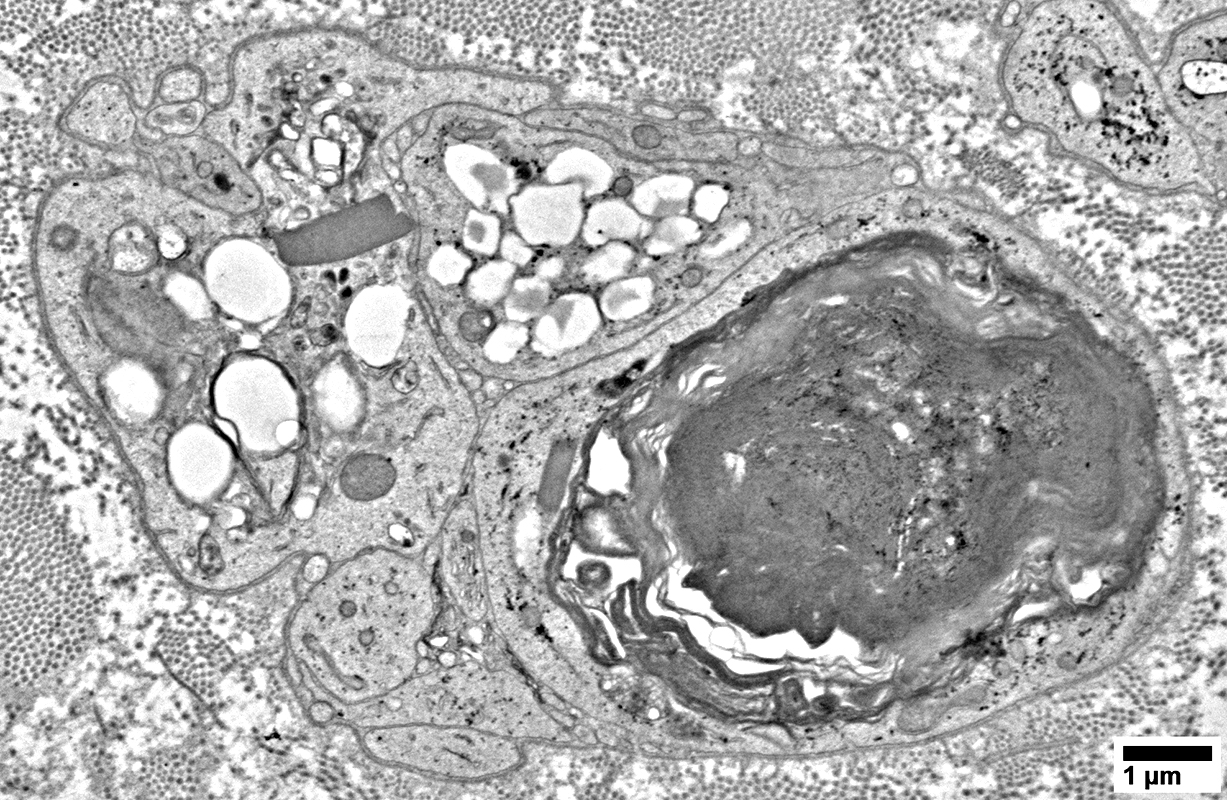
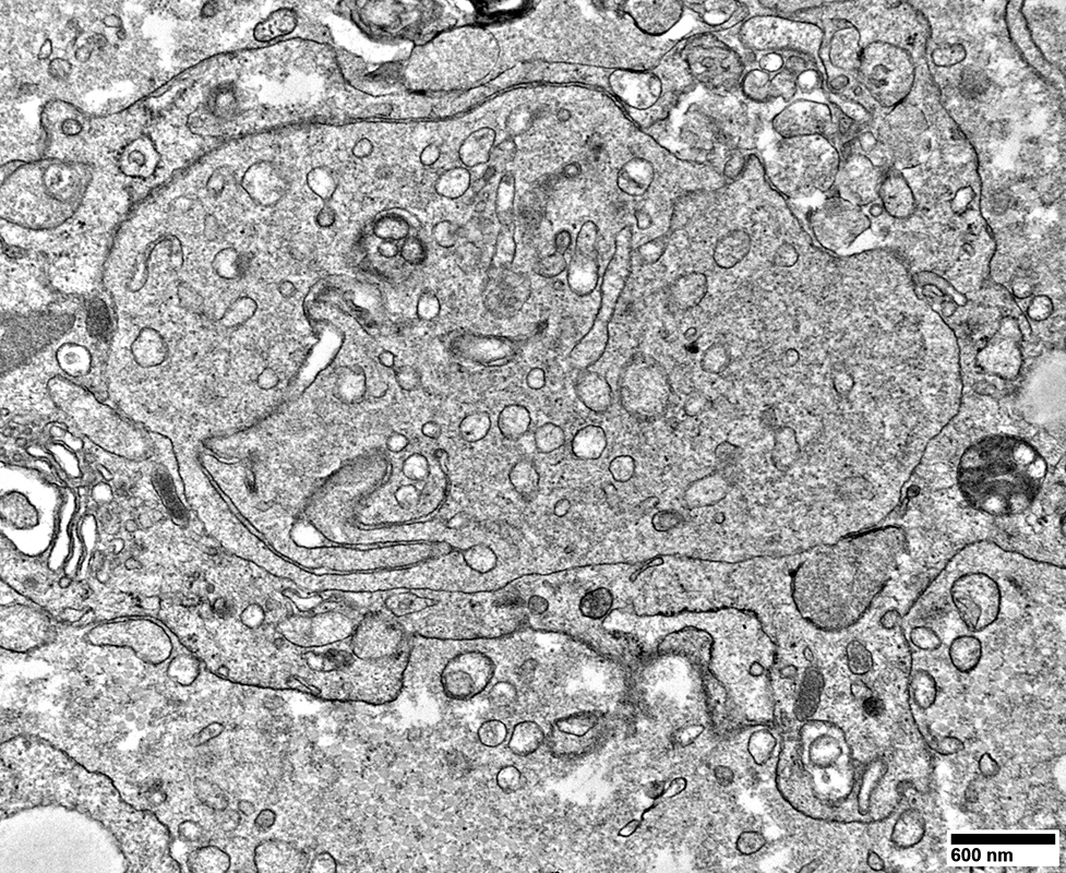
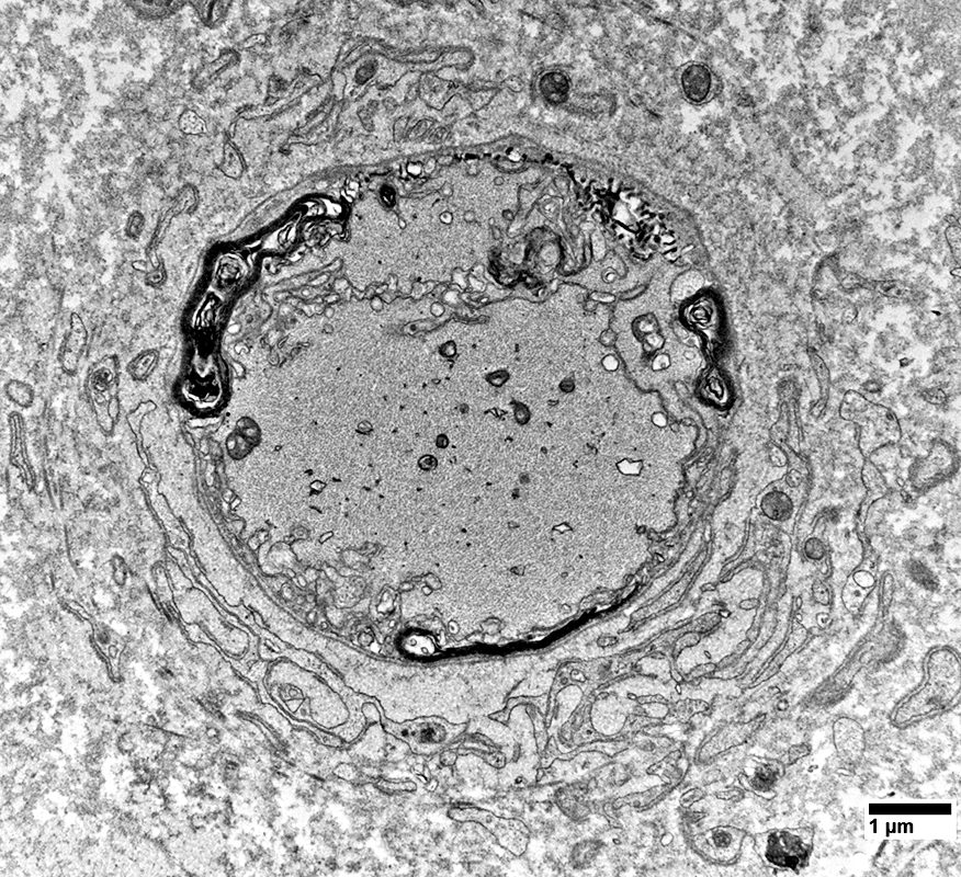
Cell Processes: Other
From: R Schmidt |
From: R Schmidt |
From: R Schmidt |
From: R Schmidt |
From: R Schmidt |
From: R Schmidt |
From: R Schmidt |
From: R Schmidt |
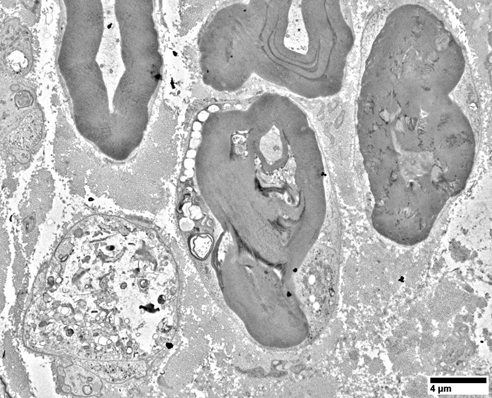
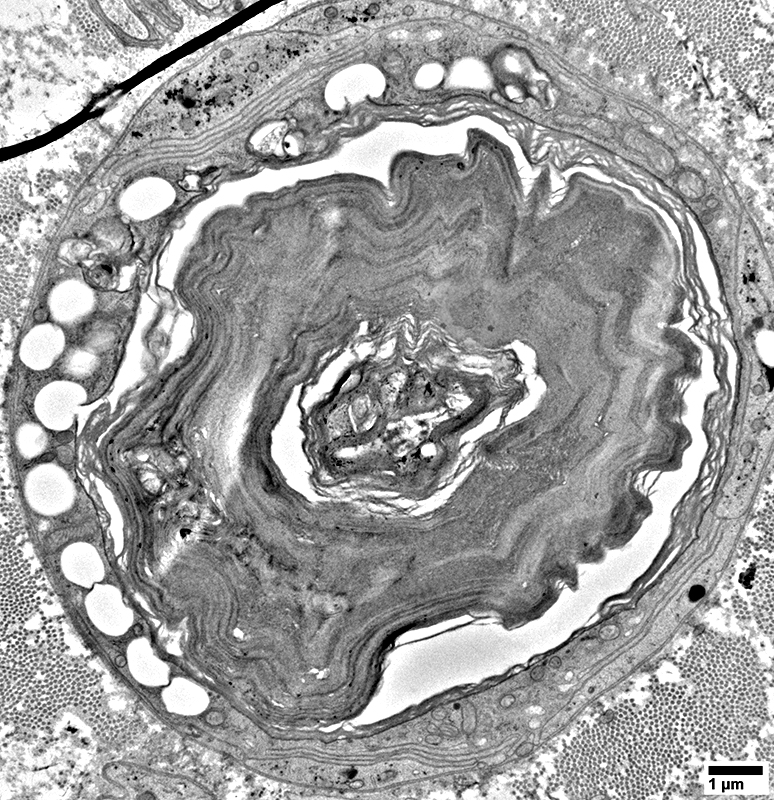
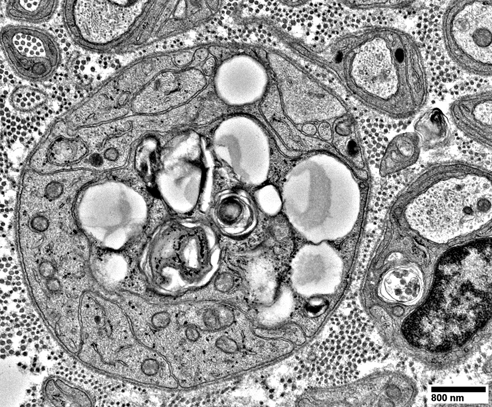
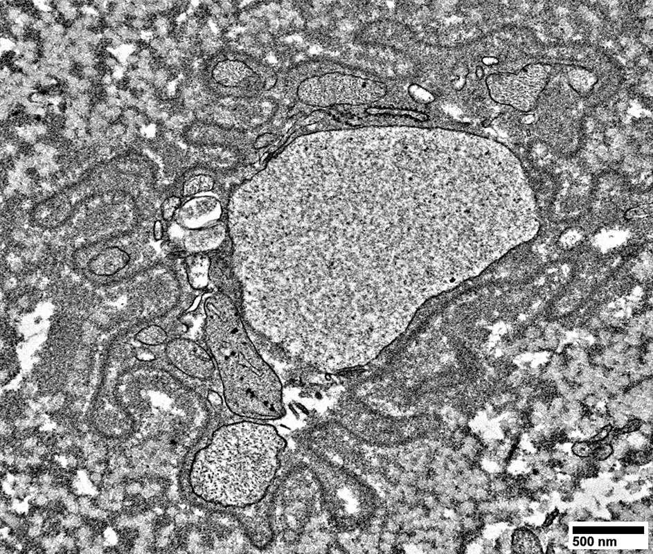
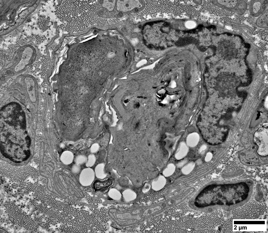
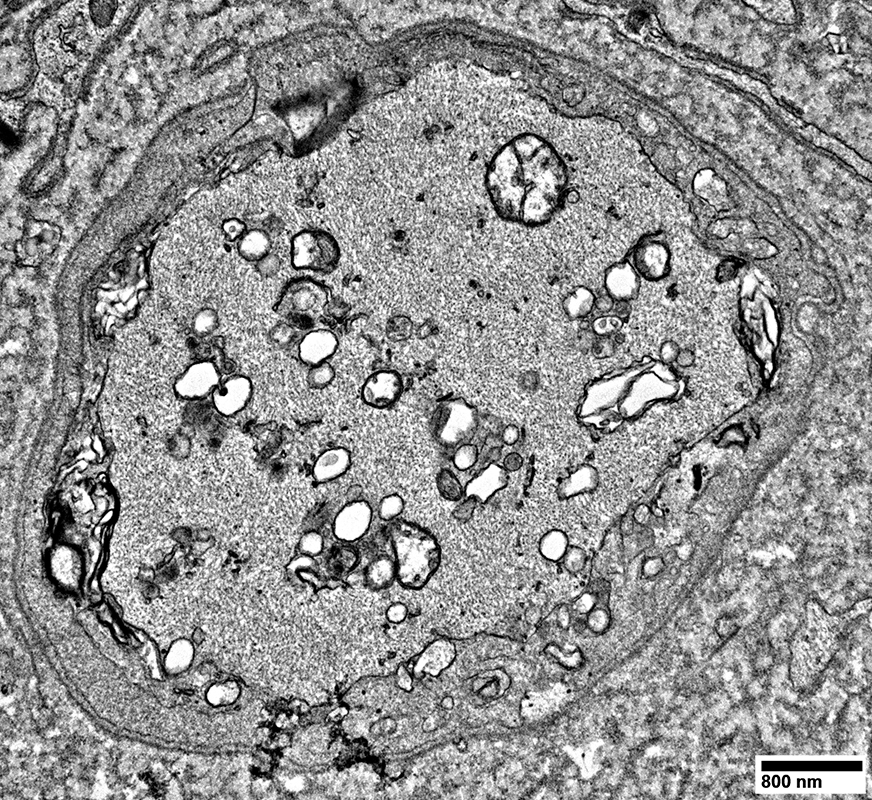
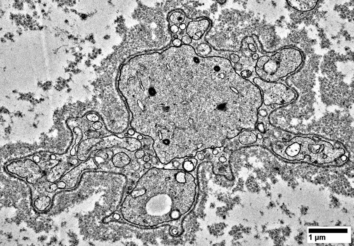
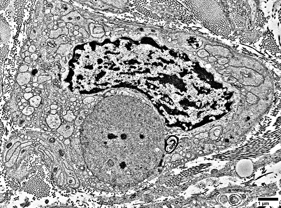
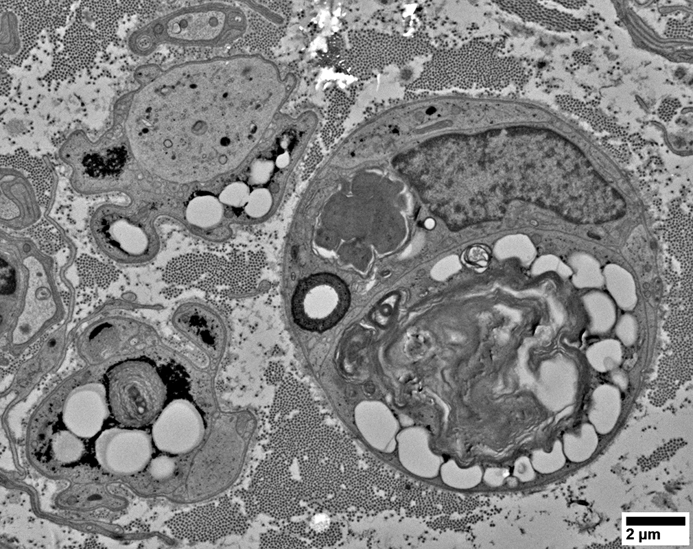
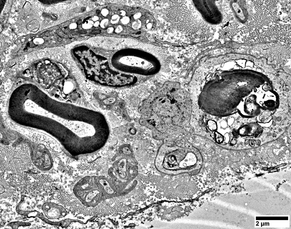
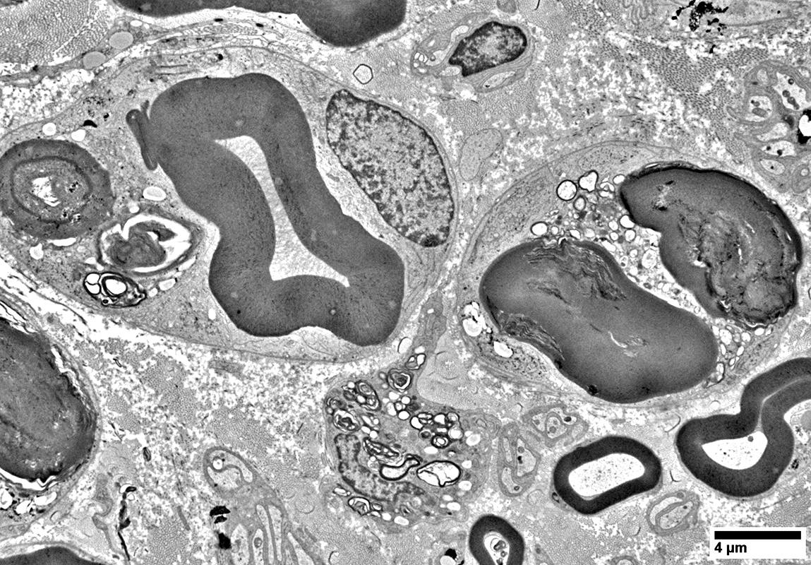
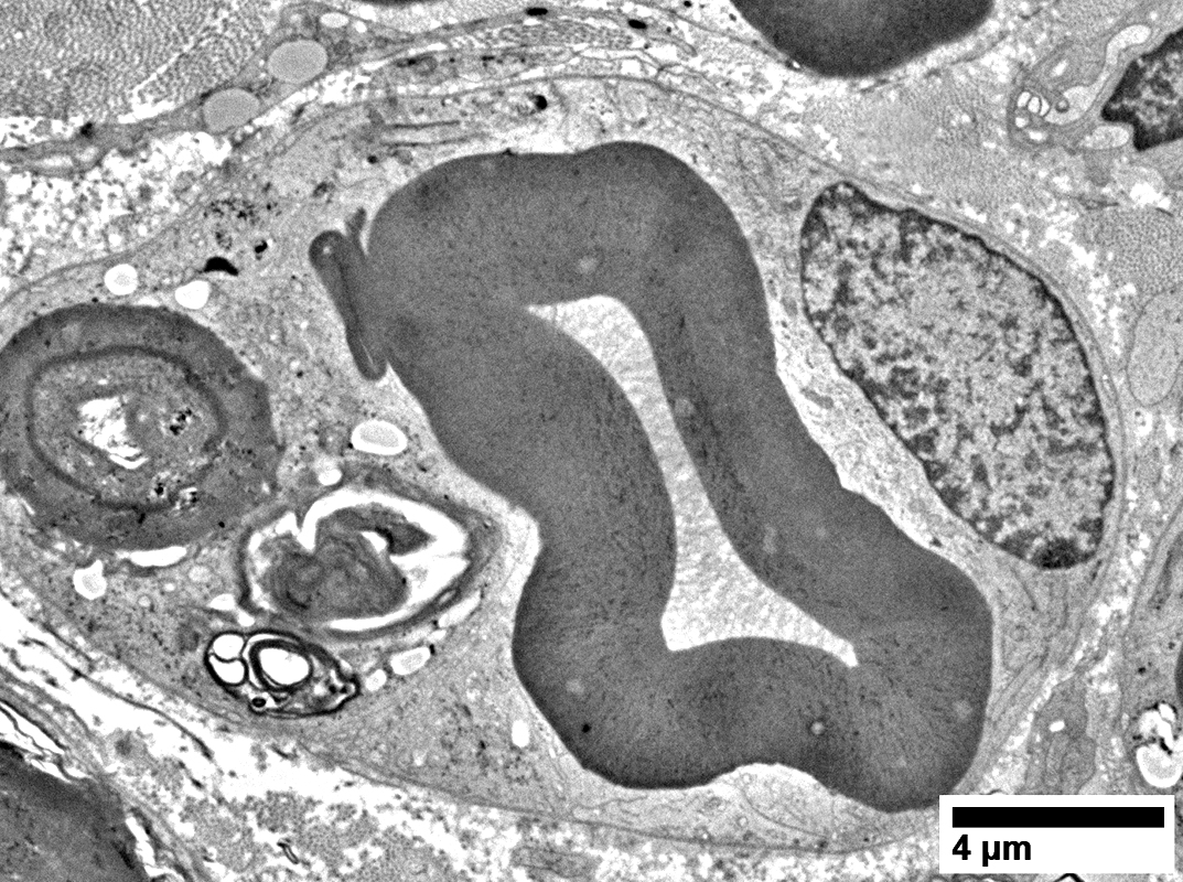
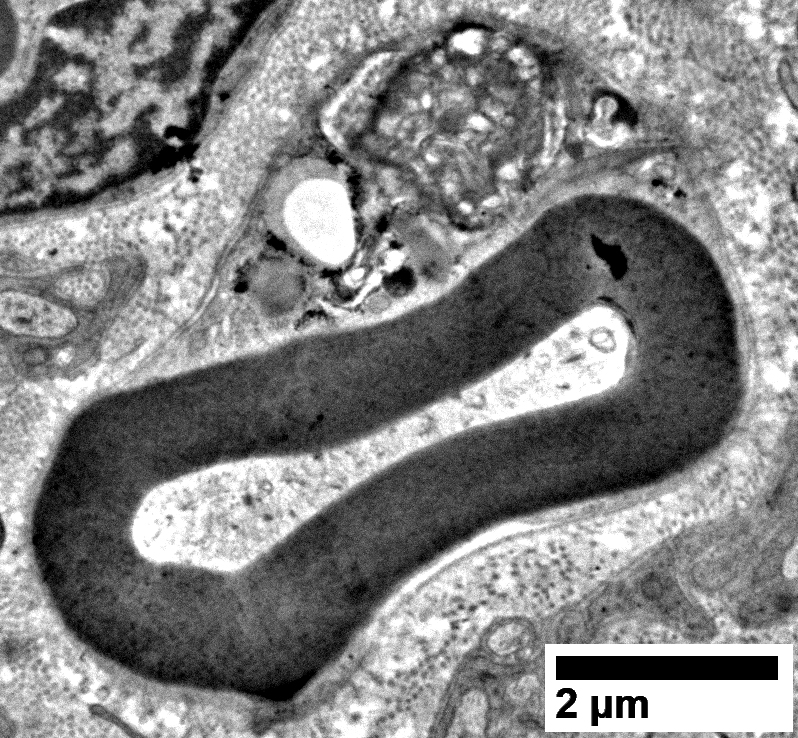
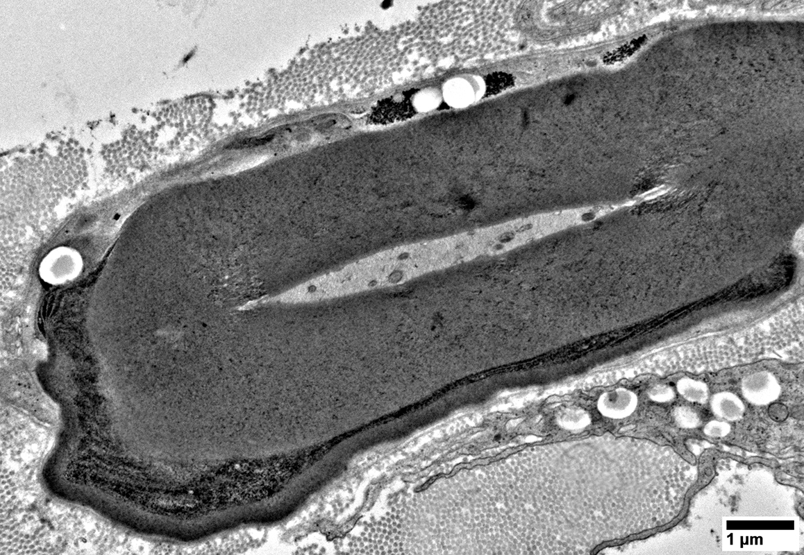
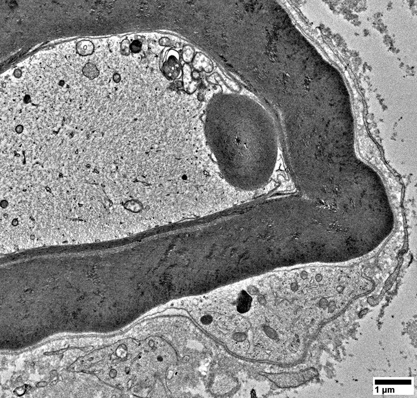
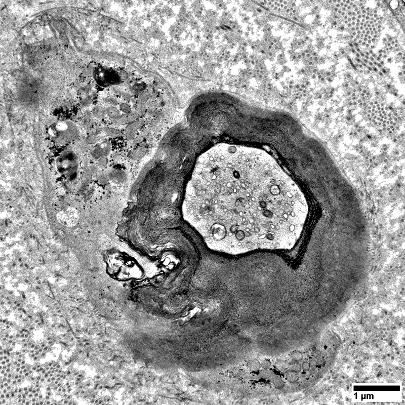
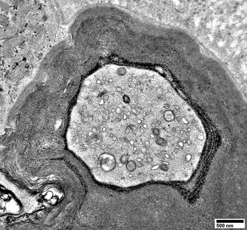
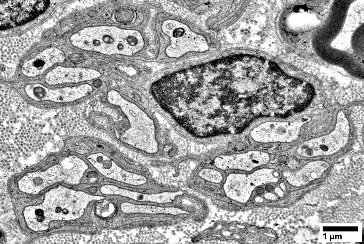
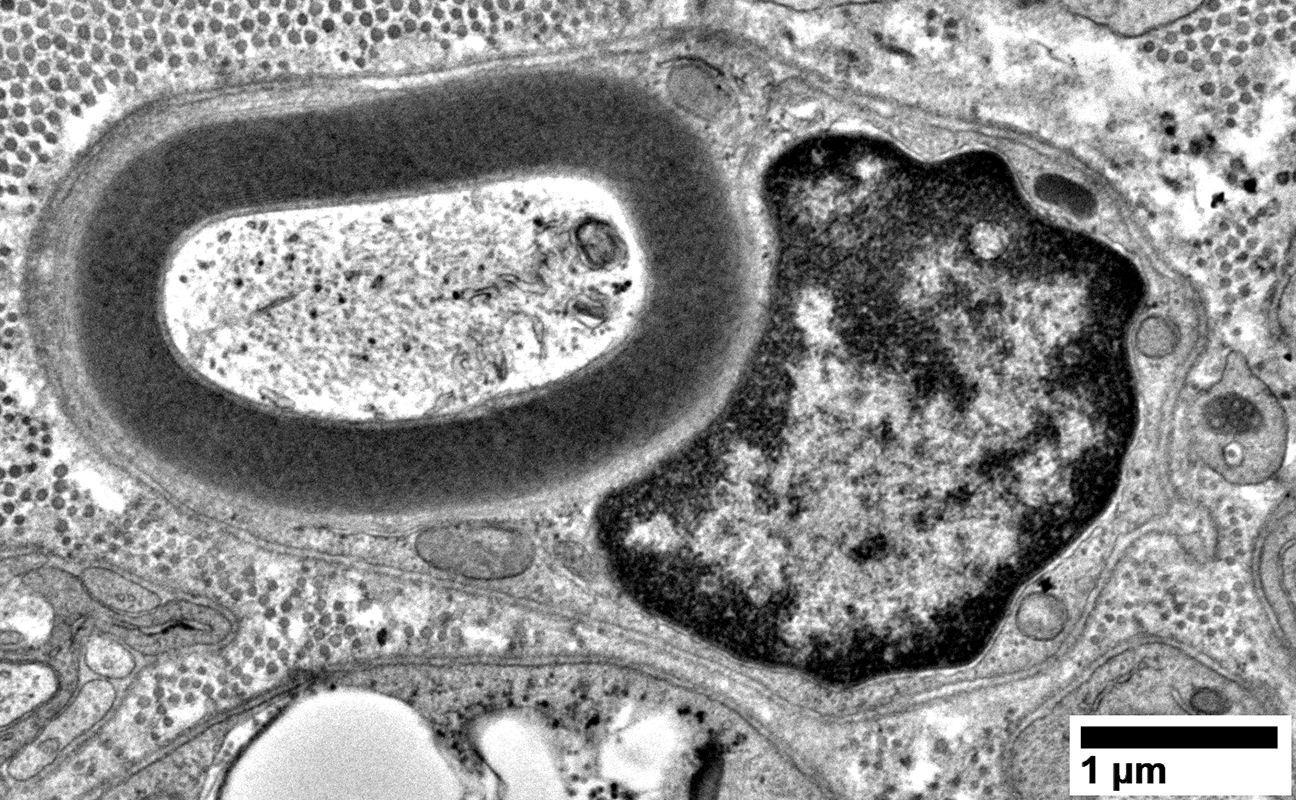
Axons: Large & Abnormal Structure with no surrounding Myelin
From: R Schmidt |
From: R Schmidt |
From: R Schmidt |
From: R Schmidt |
From: R Schmidt |
From: R Schmidt |
From: R Schmidt |
From: R Schmidt |
From: R Schmidt |
From: R Schmidt |
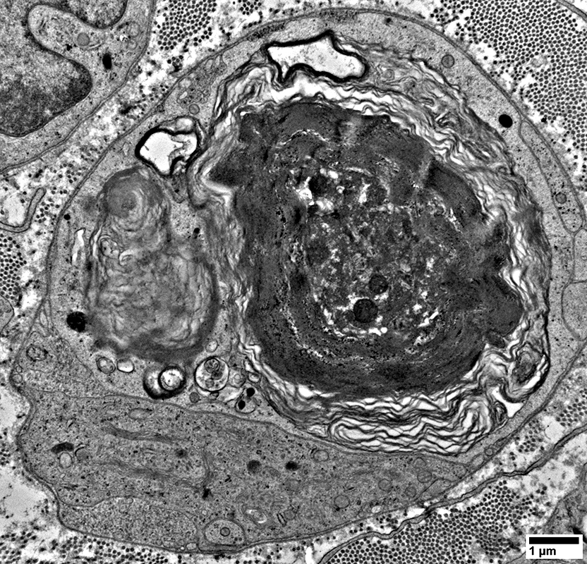
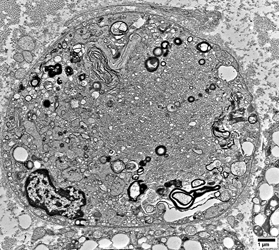
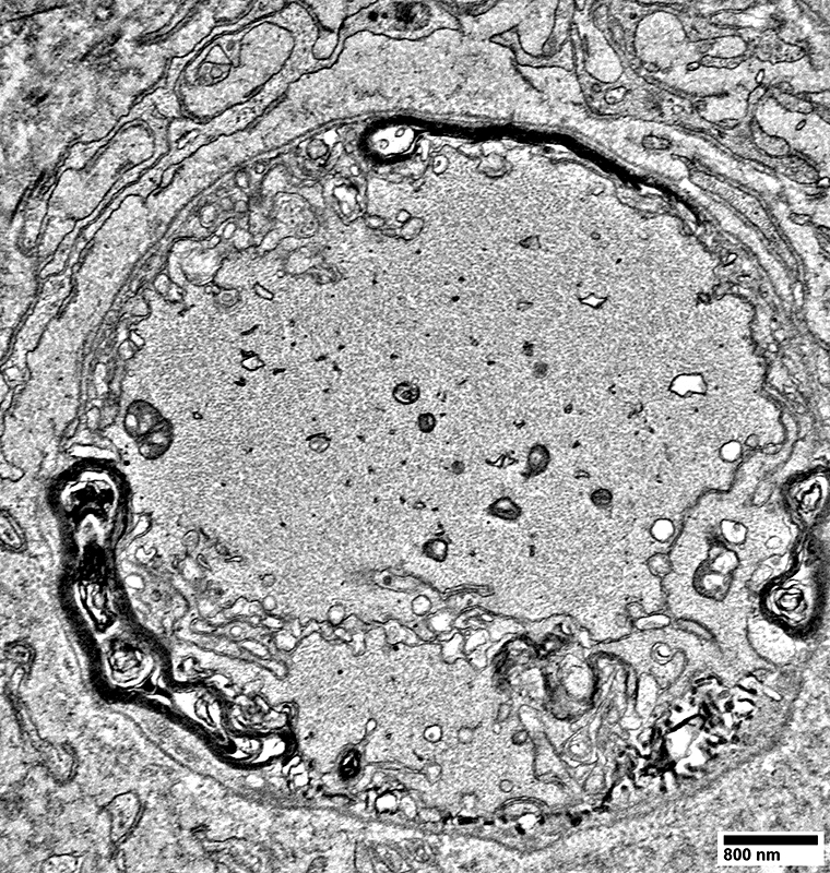
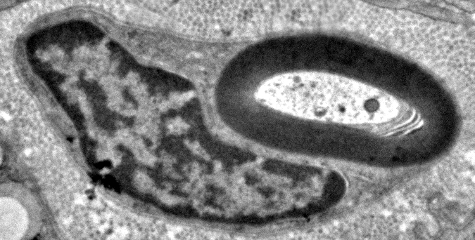
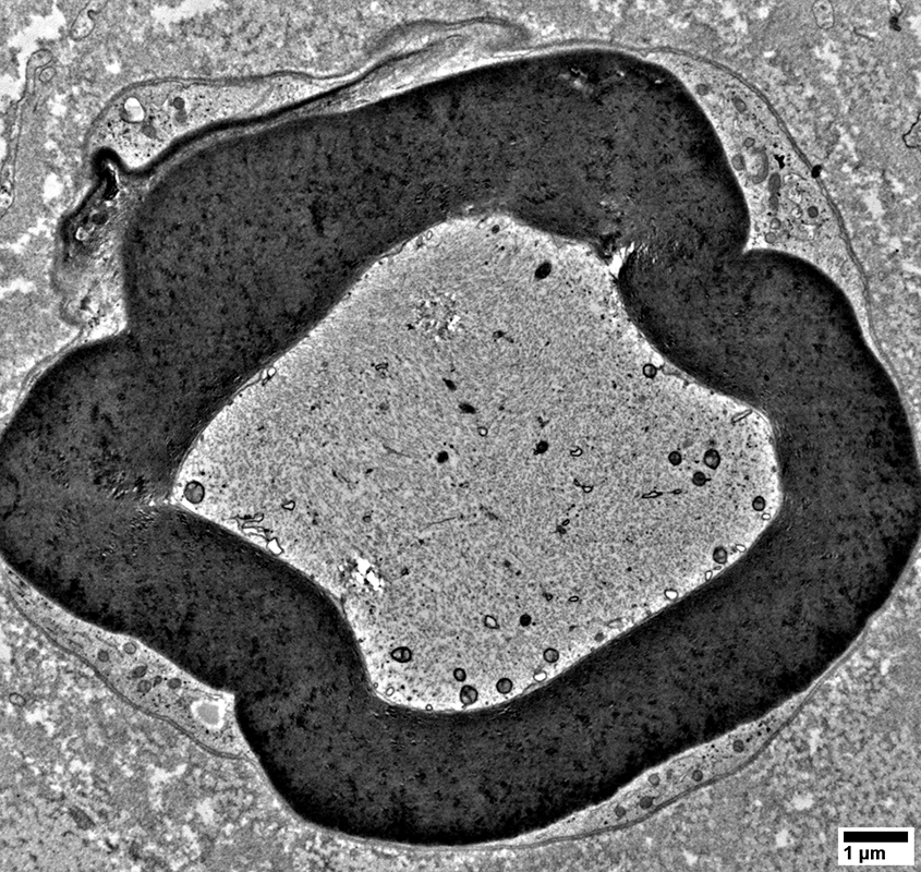
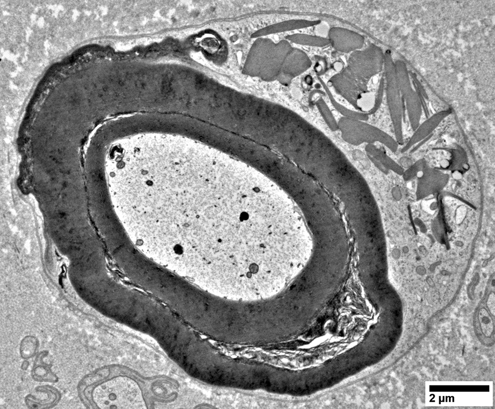
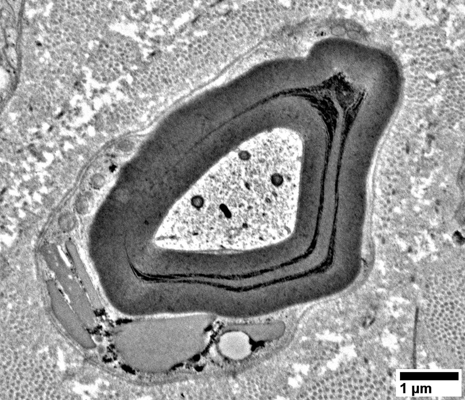
Schwann cells around Axons: Contain Myelin Degradation Products
From: R Schmidt |
From: R Schmidt |
From: R Schmidt |
From: R Schmidt |
From: R Schmidt |
From: R Schmidt |
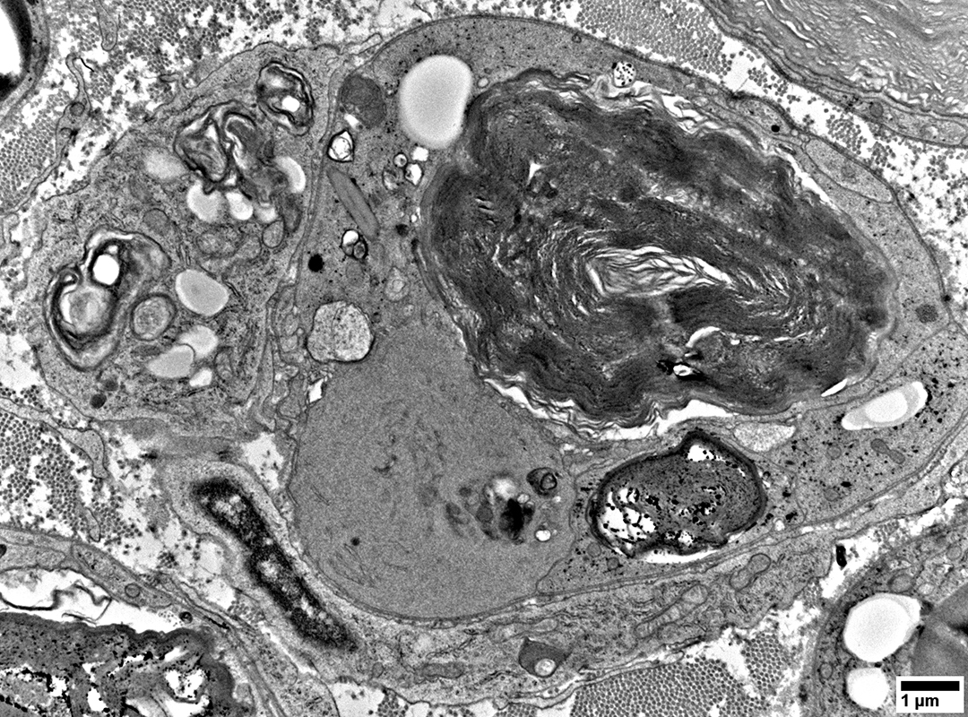
From: R Schmidt |
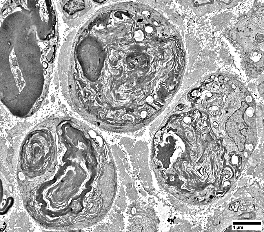
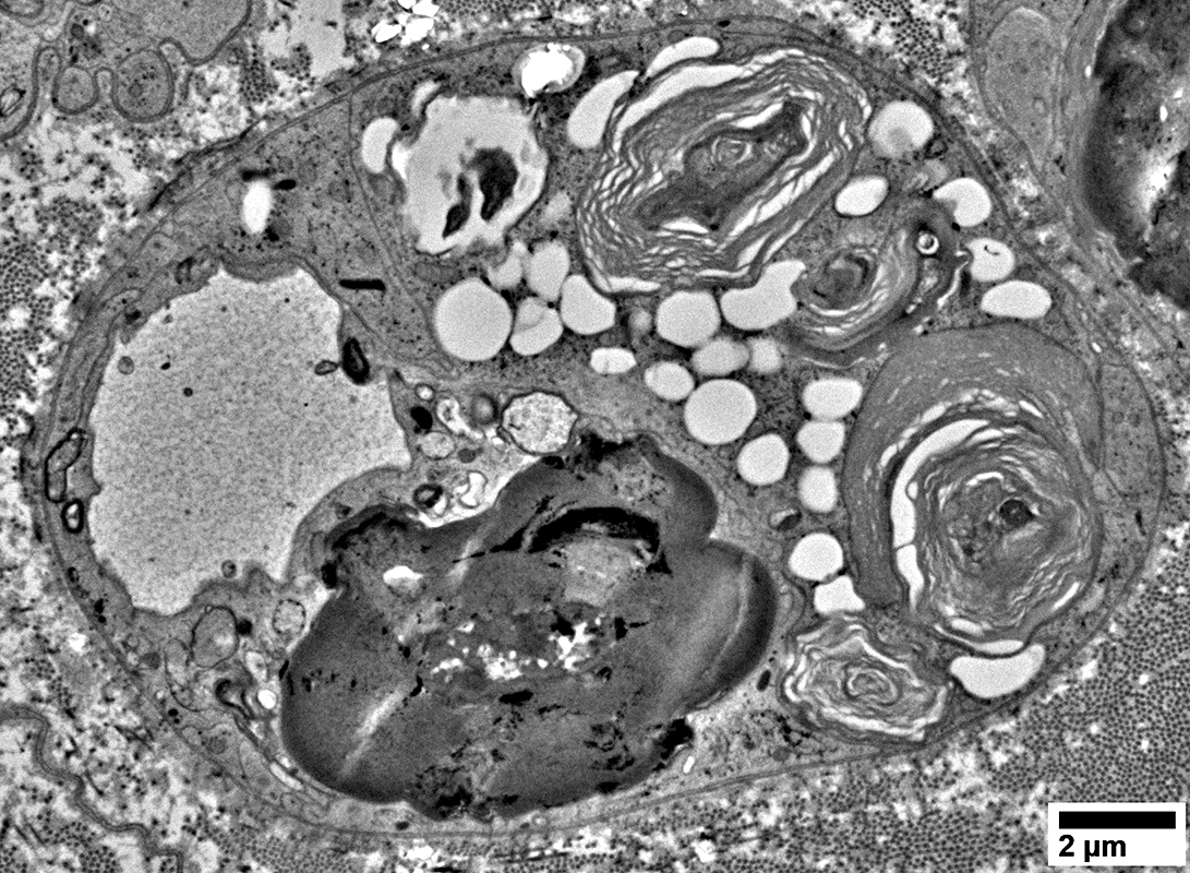
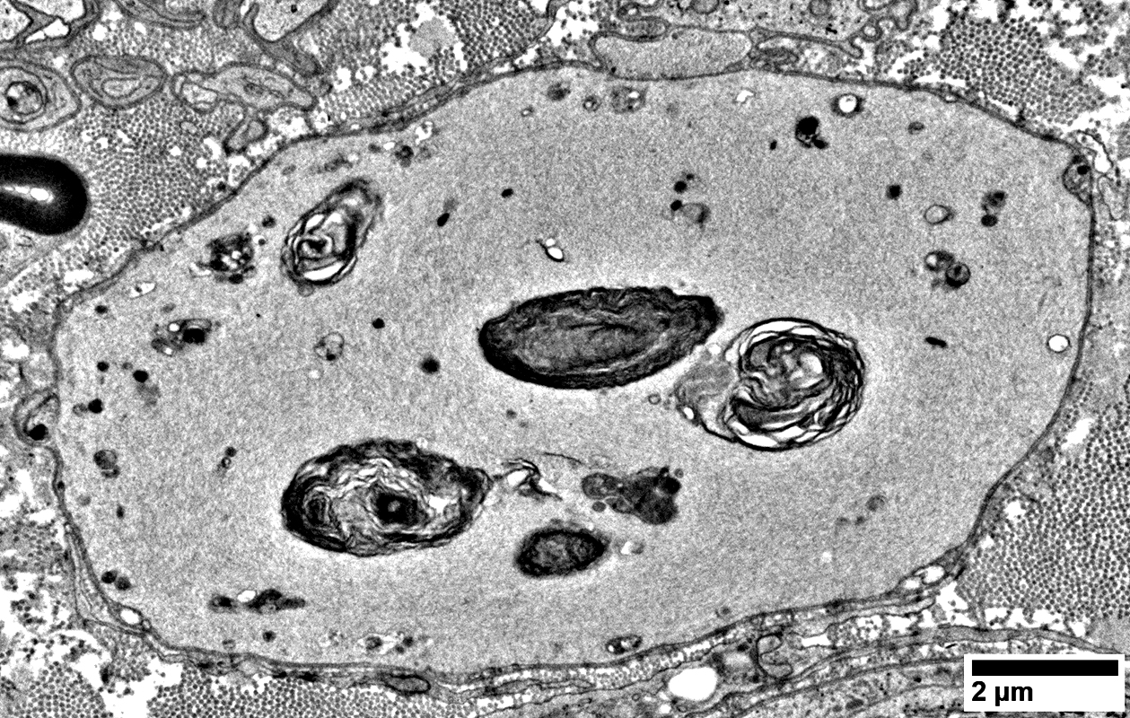
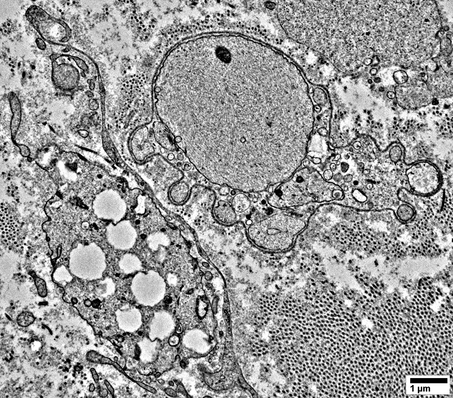
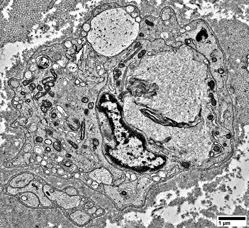
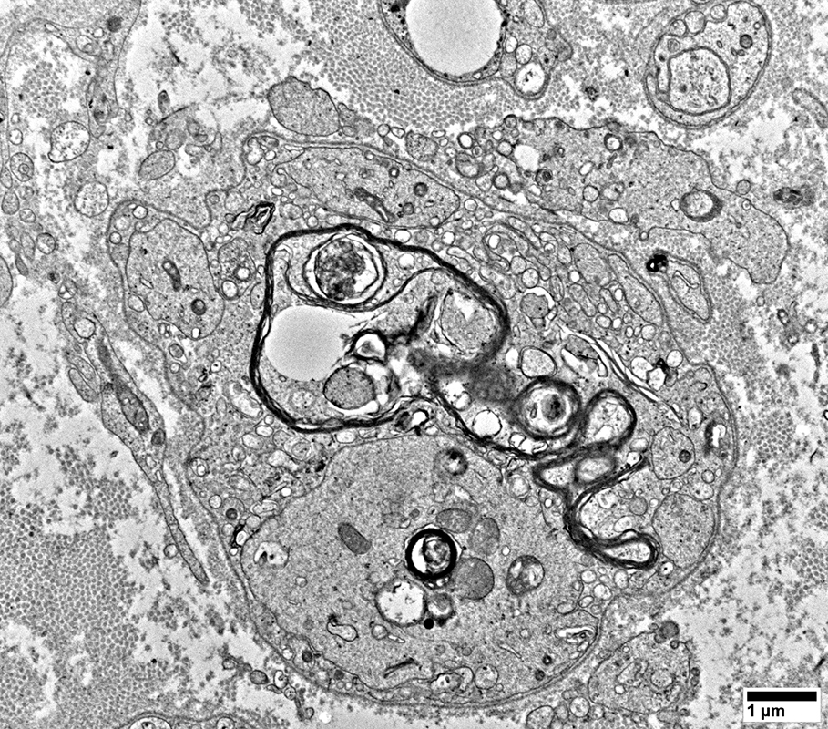
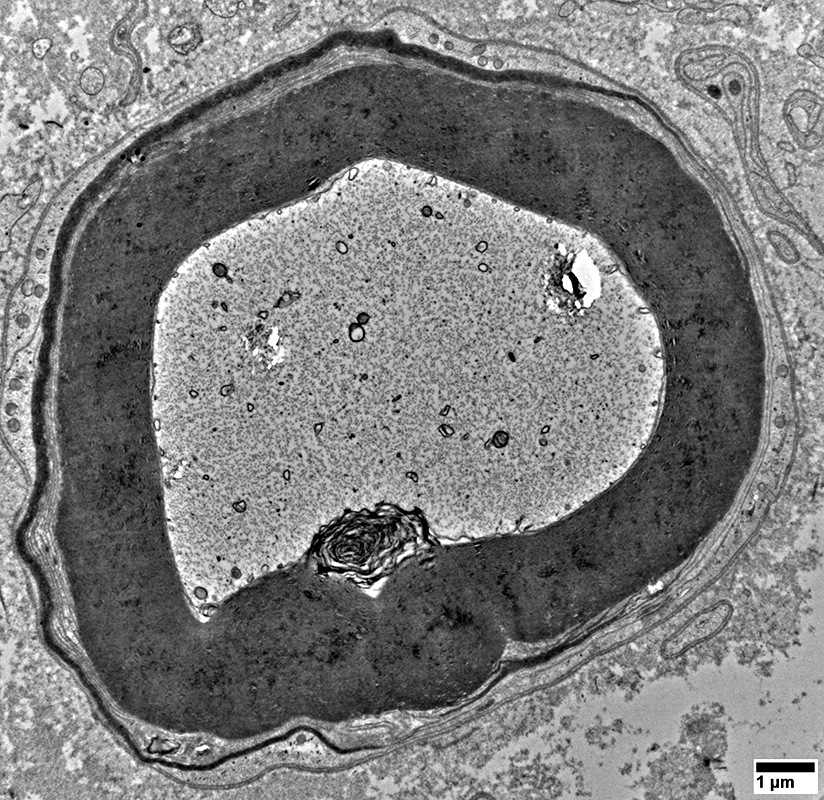
Myelin Pathology: Early
From: R Schmidt |
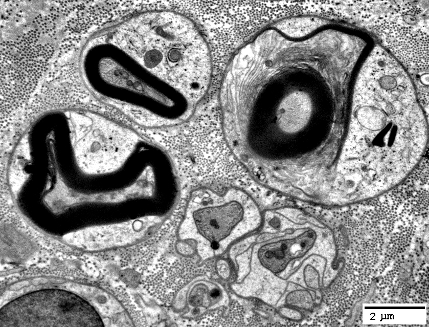
From: R Schmidt |
From: R Schmidt |
From: R Schmidt |
From: R Schmidt |
From: R Schmidt |
From: R Schmidt |
From: R Schmidt |
From: R Schmidt |
From: R Schmidt |
From: R Schmidt |
From: R Schmidt |
From: R Schmidt |
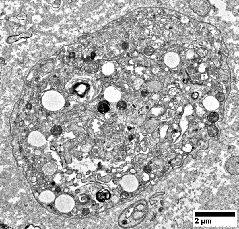
Non-Myelinating Schwann Cells & Unmyelinated Small Axons
NCAM stain |
NCAM stain |
NCAM(r)_09sm.jpg)
Neurofilament (Green), NCAM (Red) stain |
NCAM(r)_05sm.jpg)
Neurofilament (Green), NCAM (Red) stain |
)NCAM(r)_03sm2.jpg)
Neurofilament (Green), NCAM (Red) stain |
)NCAM(r)_07sm.jpg)
Neurofilament (Green), NCAM (Red) stain |
)NCAM(r)_09sm.jpg)
Neurofilament (Green), NCAM (Red) stain |
From: R Schmidt |
Numbers: Relatively preserved; Several per Schwann cell
Non-myeliinating Schwann cells
May have pale cytoplasm (Below)
From: R Schmidt |
Neurofilament (Green)stain |
May appear beaded
Numbers: Mildly reduced
Neurofilament (Green), NCAM (Red) stain |
From: R Schmidt |
A few have preserved structure
From: R Schmidt |
Return to: ANAN
Return to: Neuromuscular Home Page
1/13/2026