
Myopathy with Myonuclear & Vacuolar Pathology: Amiodarone-associated
 H&E stain |
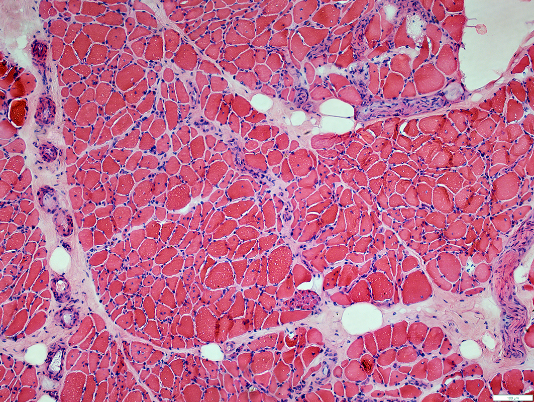
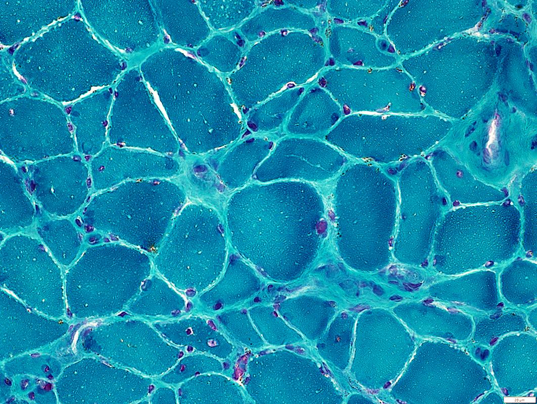
General: Atrophy
Size Distribution: Bimodal; Small, polygonal & Larger
Muscle Fiber Cytoplasm
Larger muscle fibers have multiple, small clear round regions
Nuclei
Internal in scattered muscle fibers
 Gomori trichrome stain |
 VvG stain |
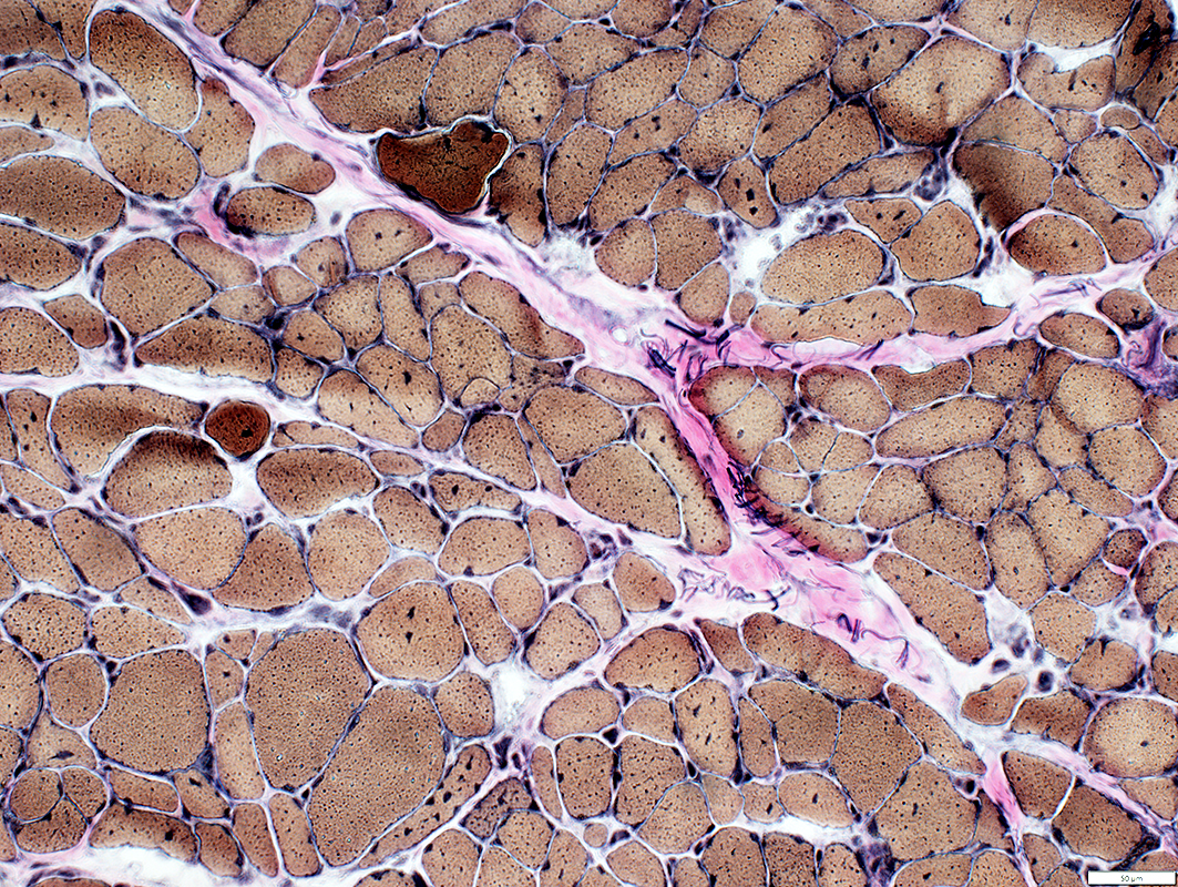
General: Atrophy
Size Distribution: Bimodal
Muscle Fiber Cytoplasm
Some muscle fibers have multiple, small dark round regions
Nuclei
Internal in scattered muscle fibers
Endomysial Connective Tissue
Mildly increased between muscle fibers
 SDH stain |
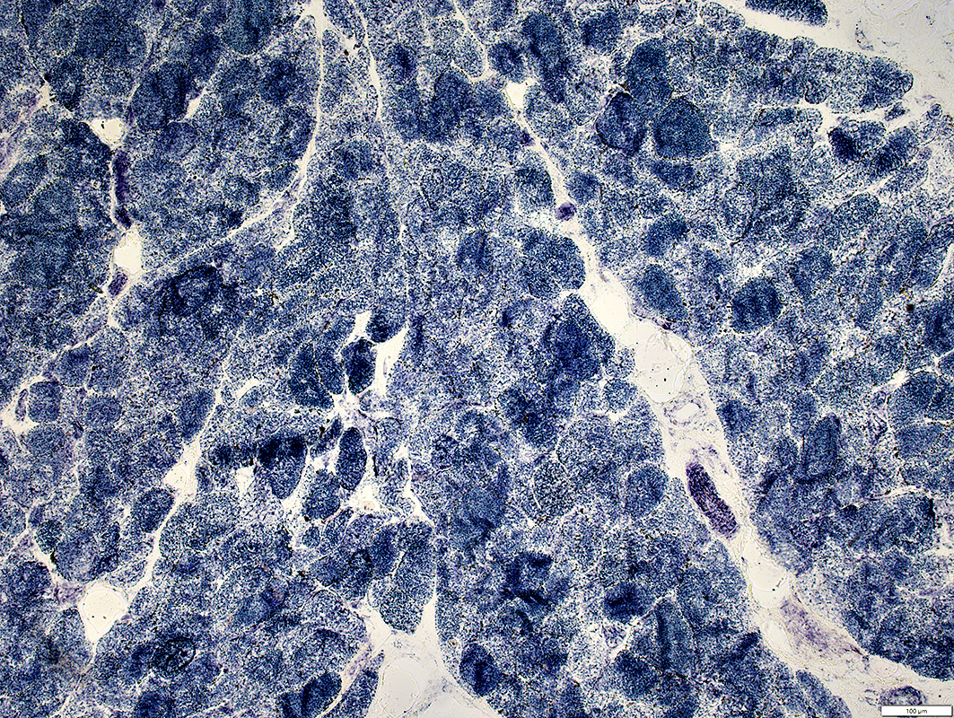
Few fibers are dark-stained on SDH
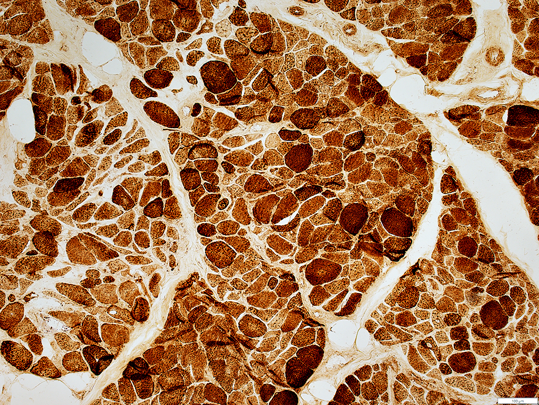 COX stain |
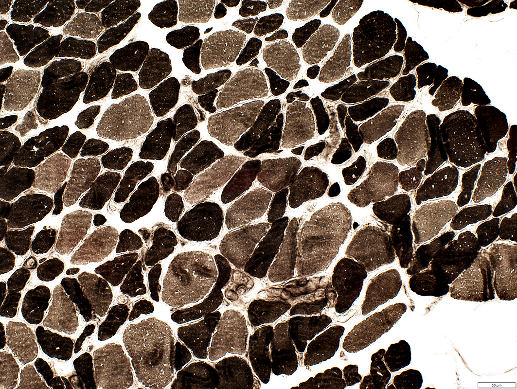 ATP pH 9.4 stain |
Largest fibers: Tend to be Type 1
Type 2C fibers (Intermediate stain; Below): Many scattered
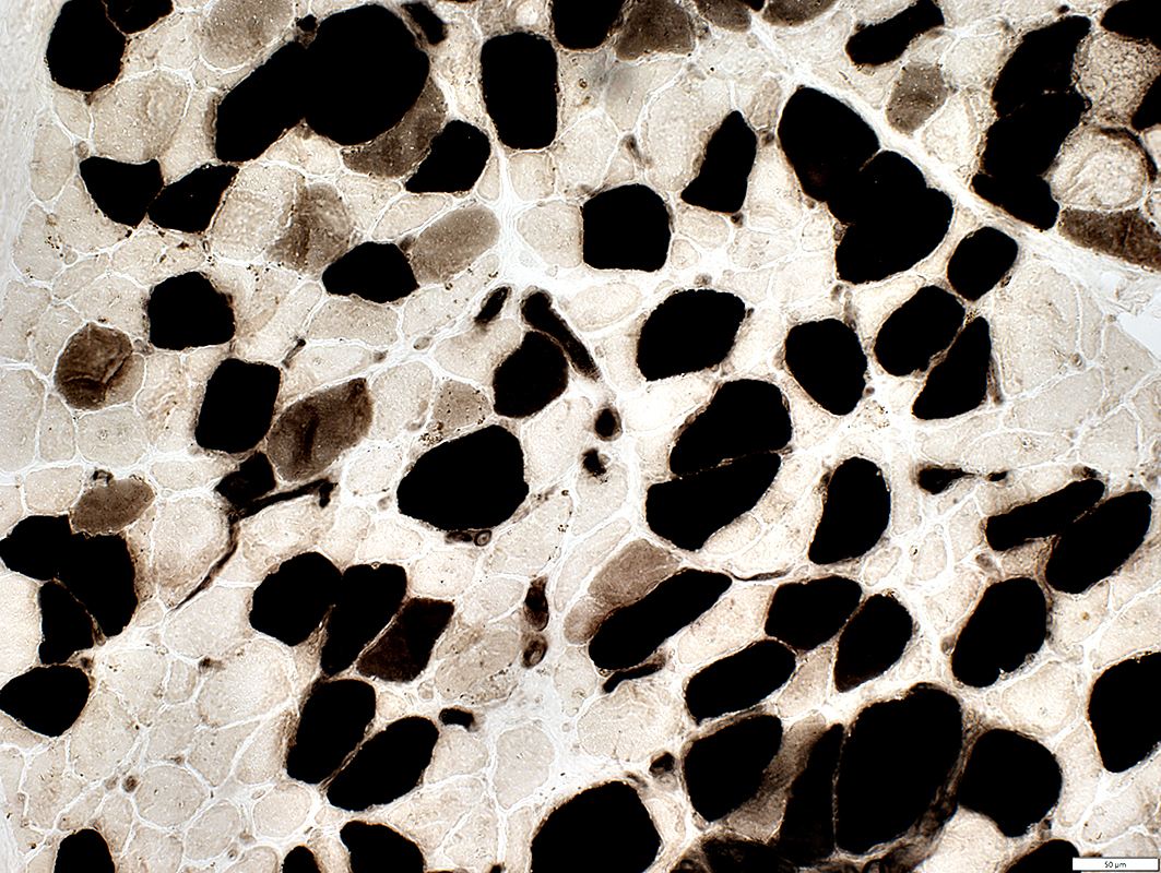 ATP pH 4.3 stain |
Muscle Fiber Internal Architecture
Coarse & Punctate
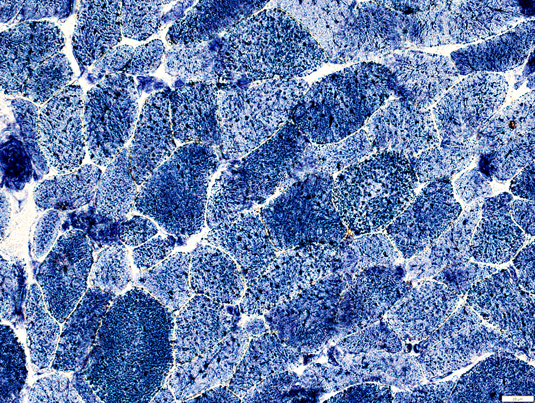 NADH stain |
Lipid Droplets
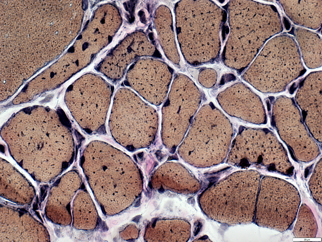 VvG stain |
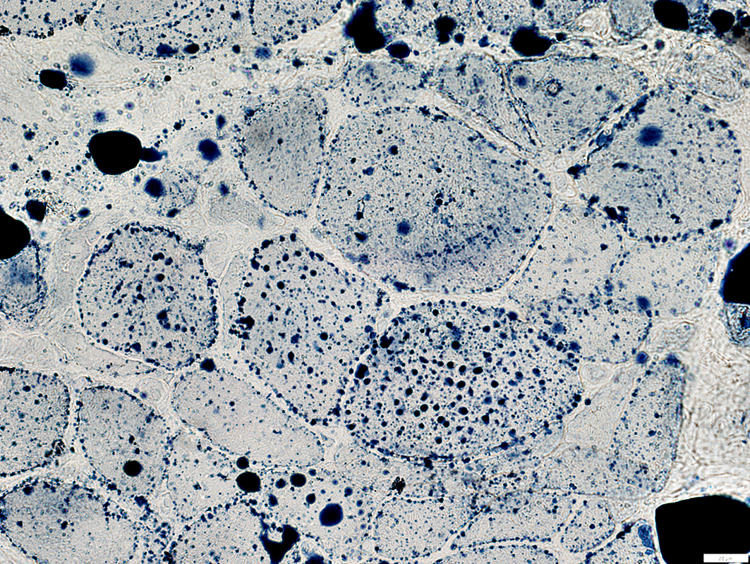 Sudan stain |
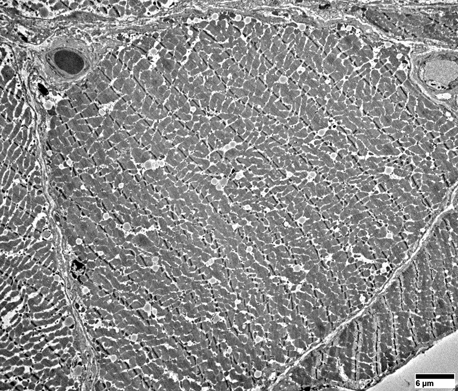 From: R Schmidt |
Distribution: Scattered in muscle fiber cytoplasm & subsarcolemmal
Structure: Homogenous gray regions surrounded by thin pale rim
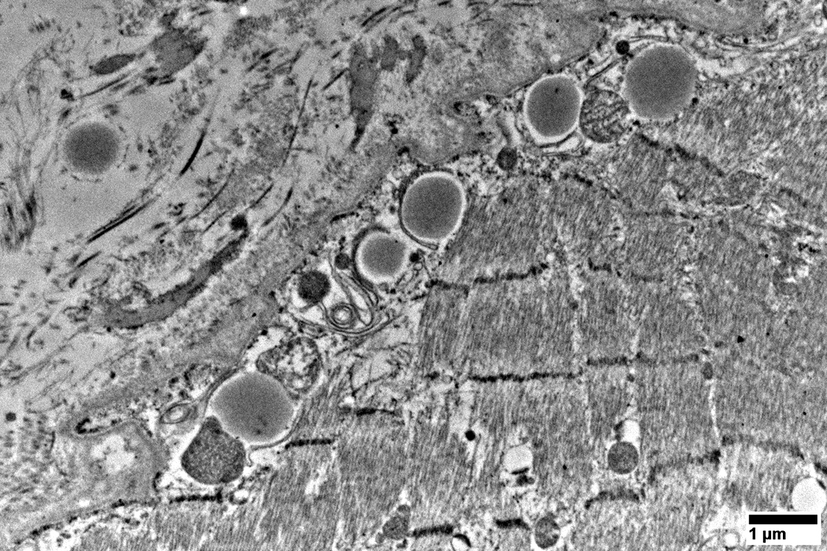 From: R Schmidt |
Distribution: Subsarcolemmal
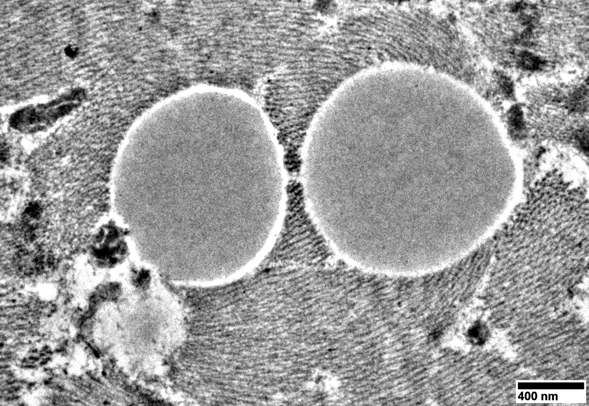 From: R Schmidt |
Structure: Homogenous gray regions surrounded by thin pale rim
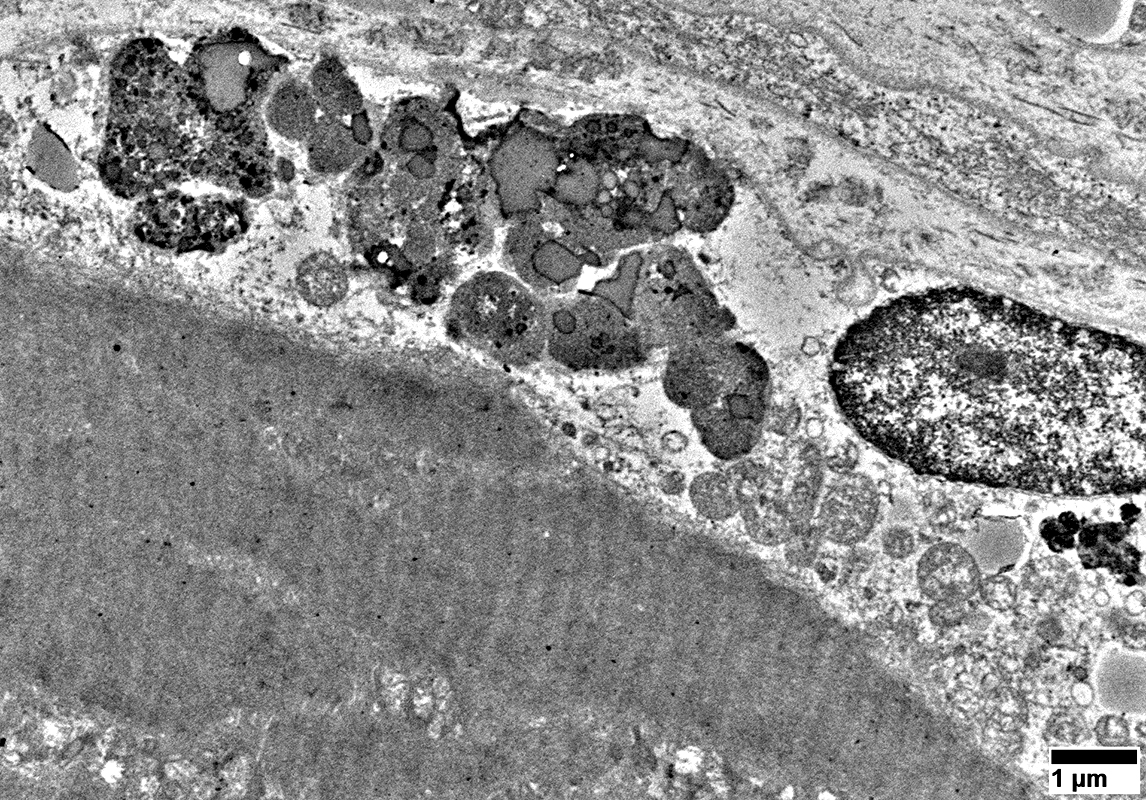
Lipopigment
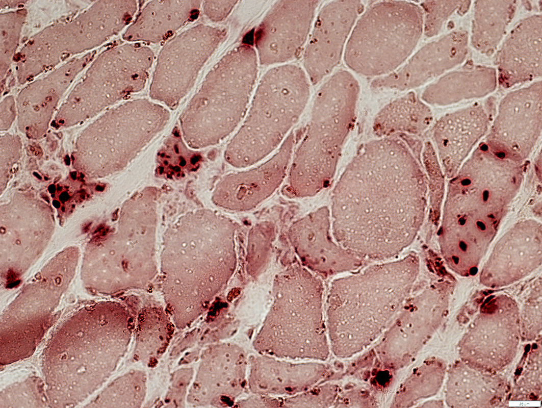
Acid phosphatase stain |
Clusters of small round brown material, often in subsarcolemmal locations
Acid phosphatase cytoplasmic granules are separate from the lipopigment
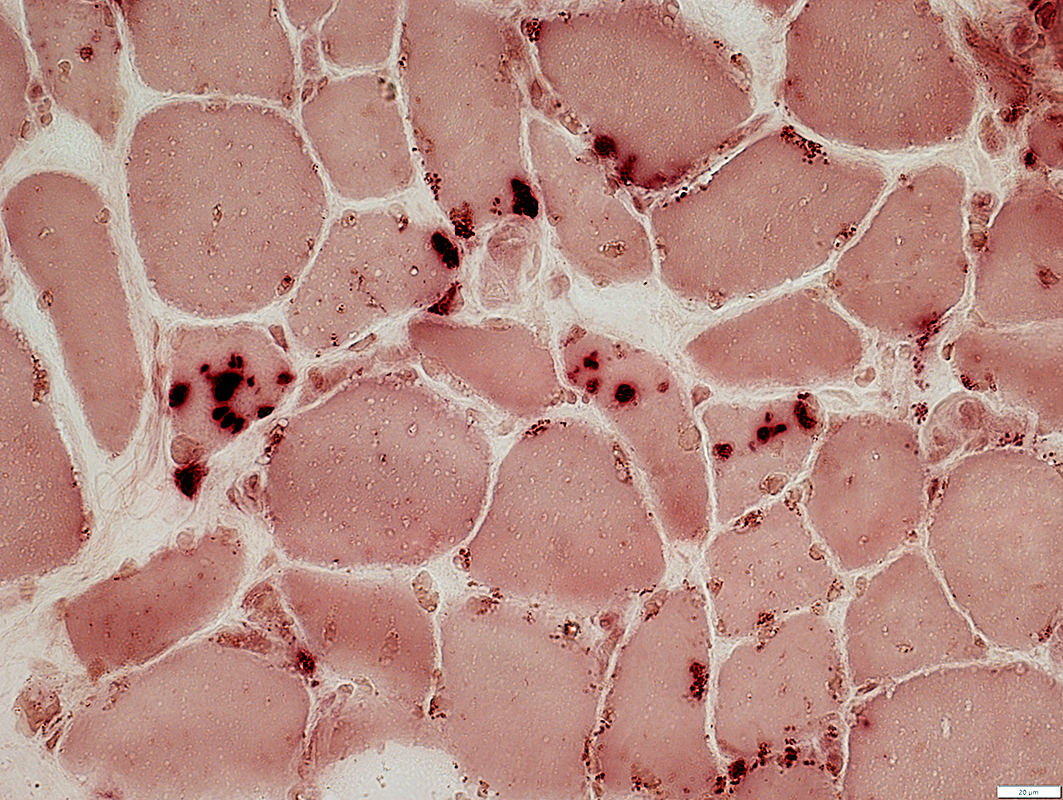 Acid phosphatase stain |
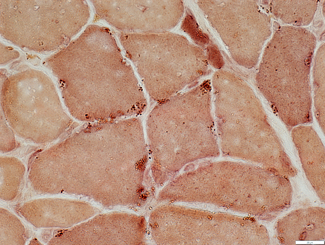 Esterase stain |
Clusters of small round brown material, most often in subsarcolemmal locations
 From: R Schmidt |
Clusters of irregularly shaped material, most often in subsarcolemmal locations
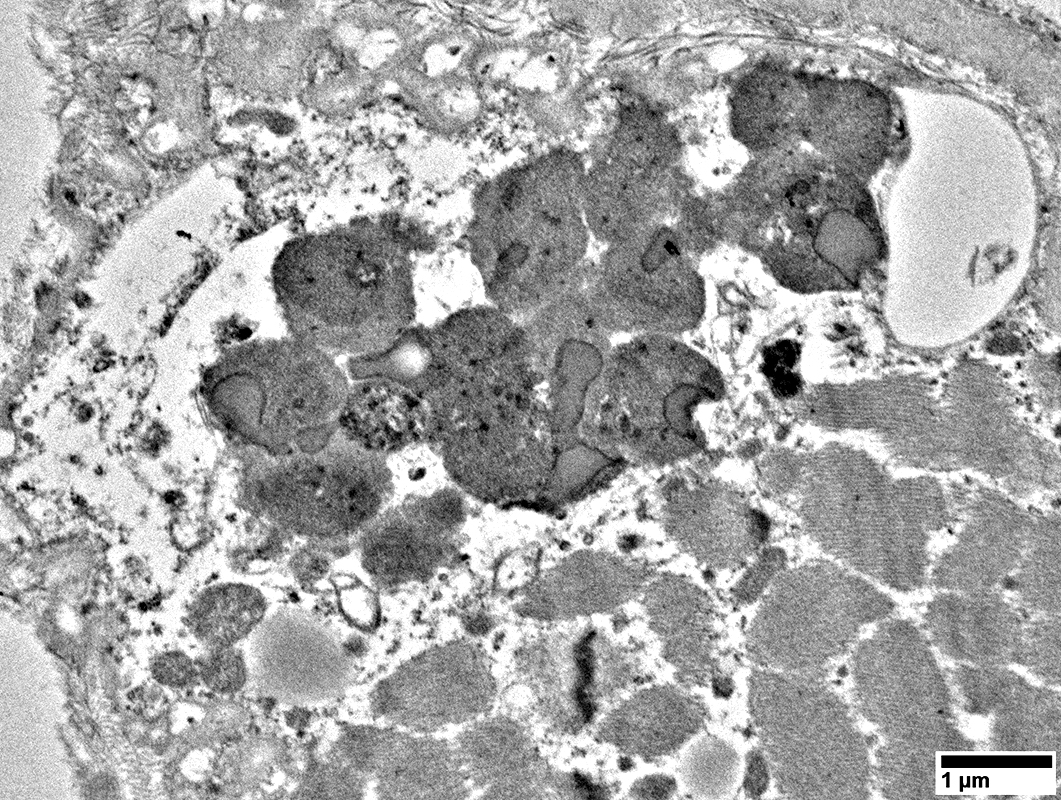
Individual lipopigment/ lipofuscin granules: Heterogeneous staining; May contain lipid-like droplets
 From: R Schmidt |
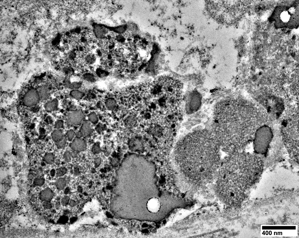
Individual lipopigment/lipofuscin granules
Heterogeneous structure
May contain lipid-like droplets
 From: R Schmidt |
 From: R Schmidt |
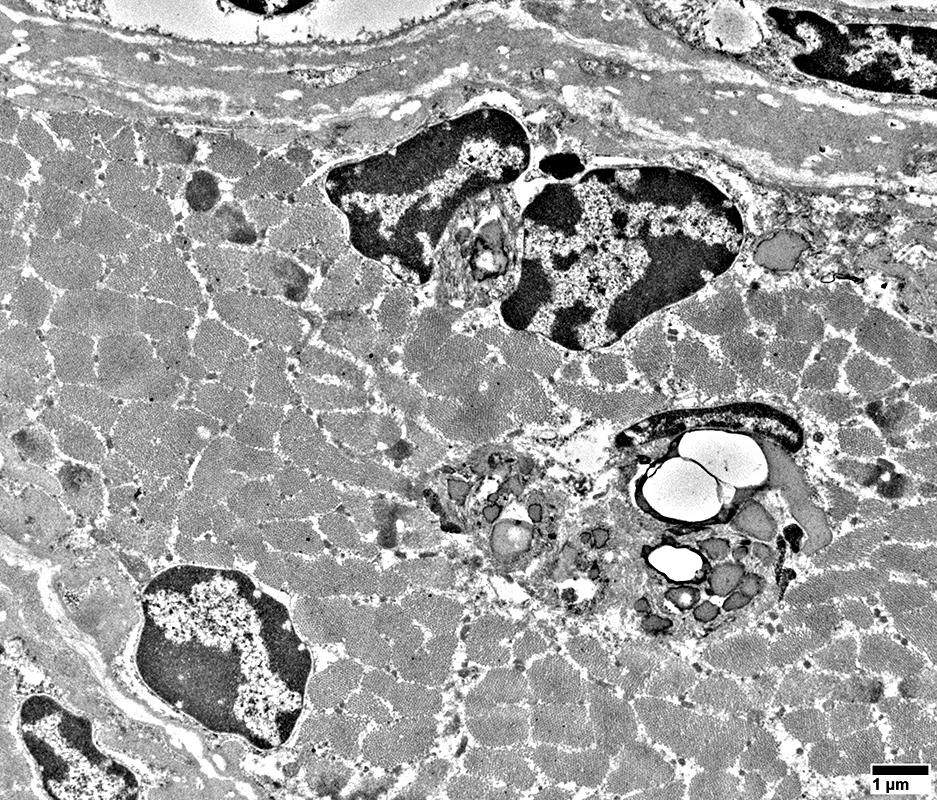
Lipopigment/lipofuscin clusters
May occur in cytoplasm
May contain pale lipid-like droplets
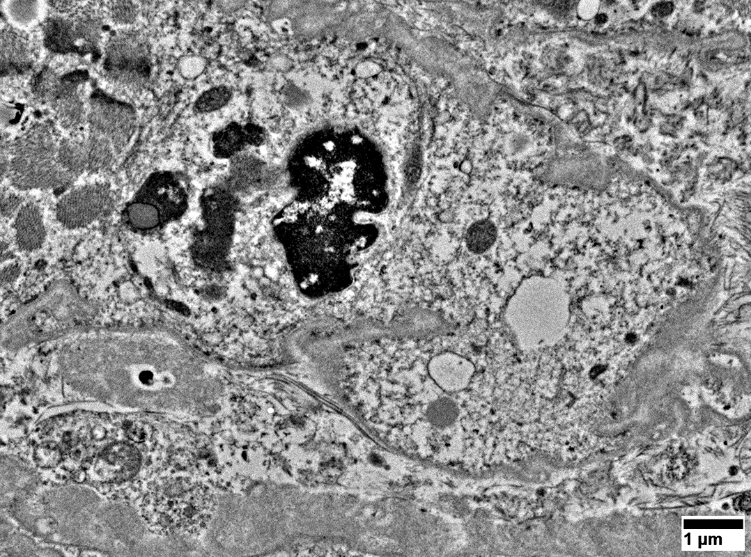
Myonuclei
Often have increased dark heterochromatin
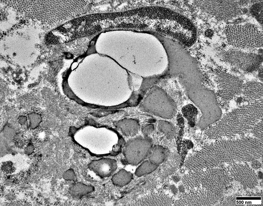 From: R Schmidt |
Vacuoles & Aggregates
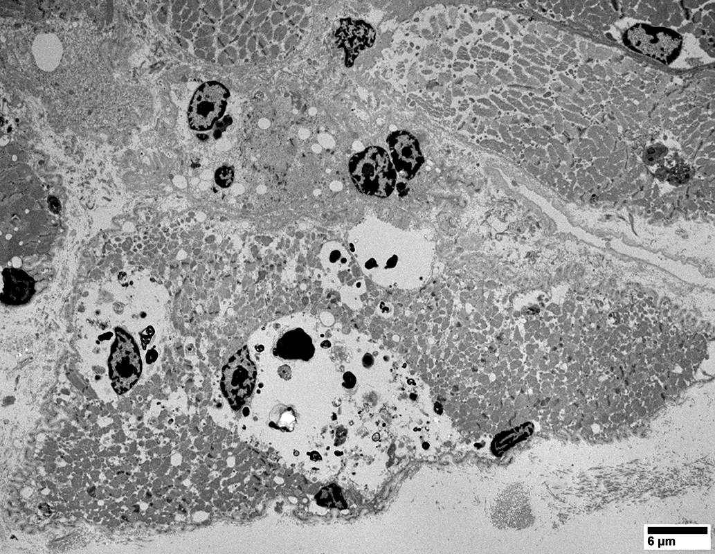 From: R Schmidt |
Rounded regions that may contain irregular debris & myonuclei distributed within a pale background
Membranes surrounding these cytoplasmic regions are often not prominent
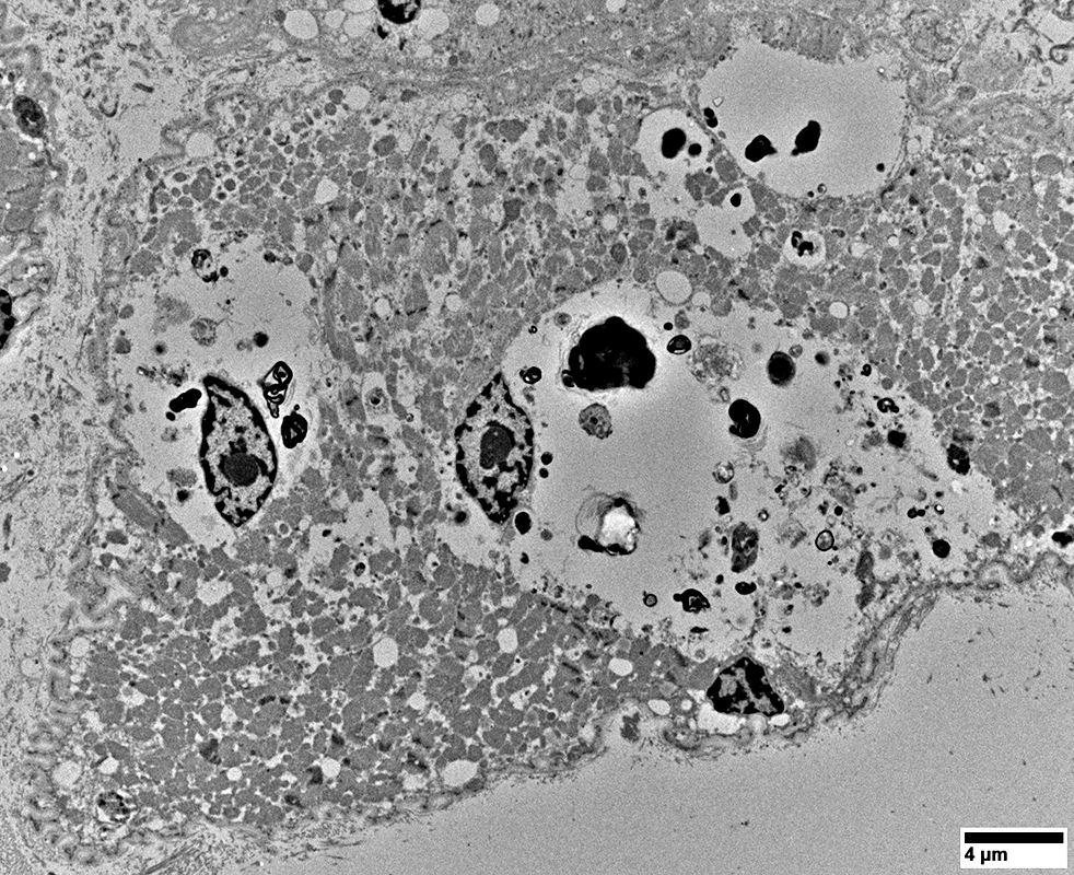 From: R Schmidt |
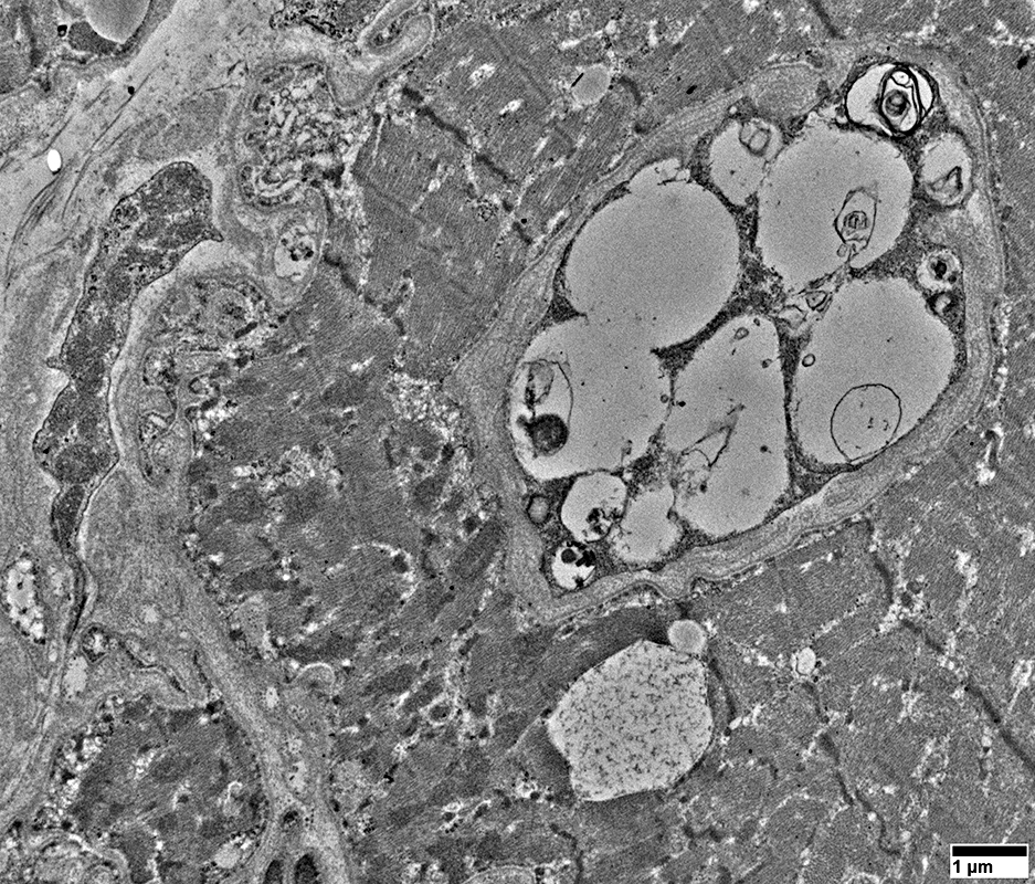 From: R Schmidt |
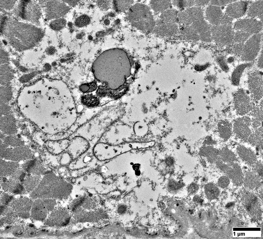 From: R Schmidt |
Rounded regions that may contain irregular debris, small vacuoles, lipopigment & myonuclei distributed within a pale background
Membranes surrounding these cytoplasmic regions are often not prominent
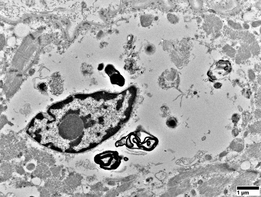 From: R Schmidt |
Cytoplasmic Aggregates
Scattered in the cytoplasm of some muscle fibers
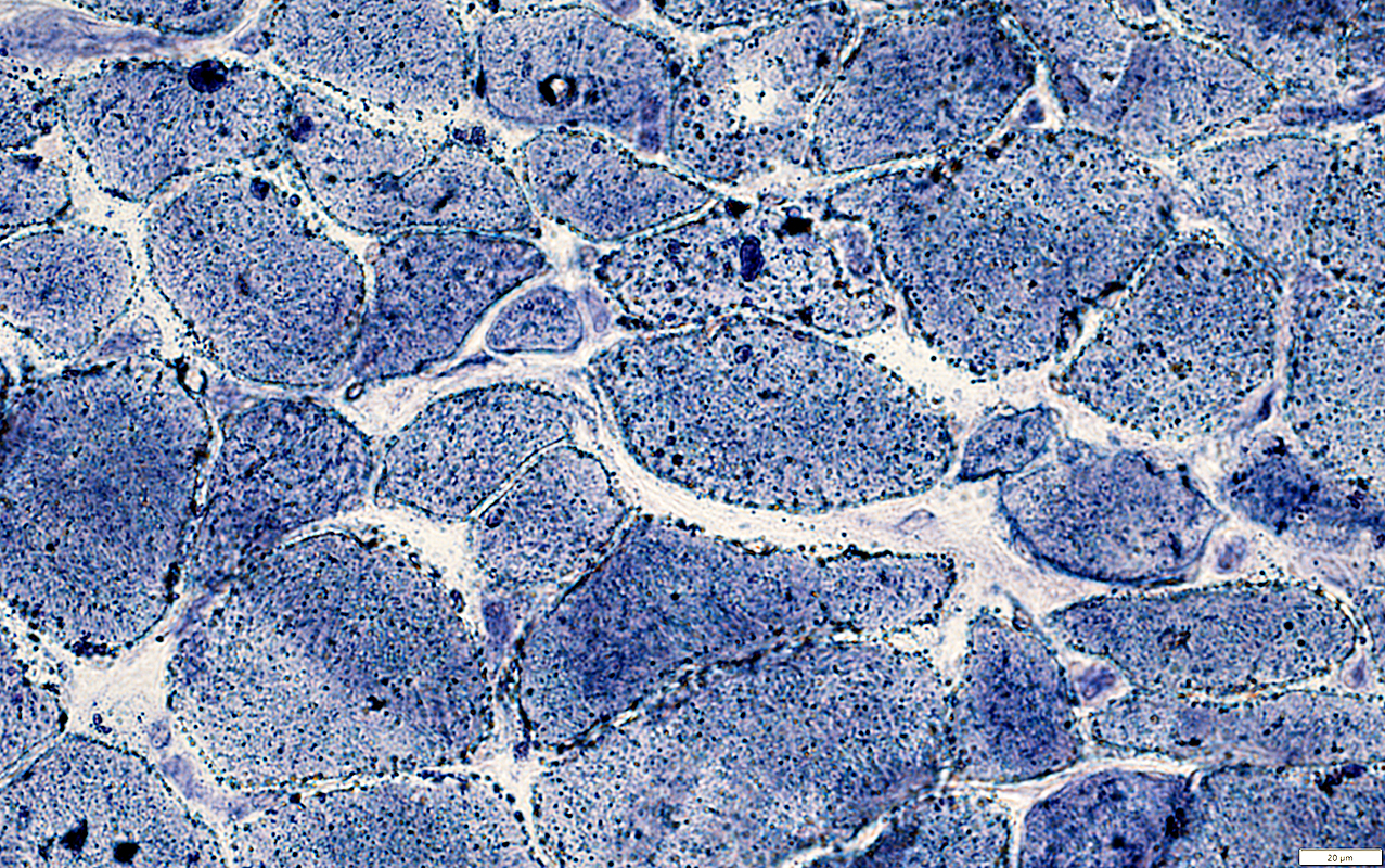 AMPDA stain |
Myonuclei
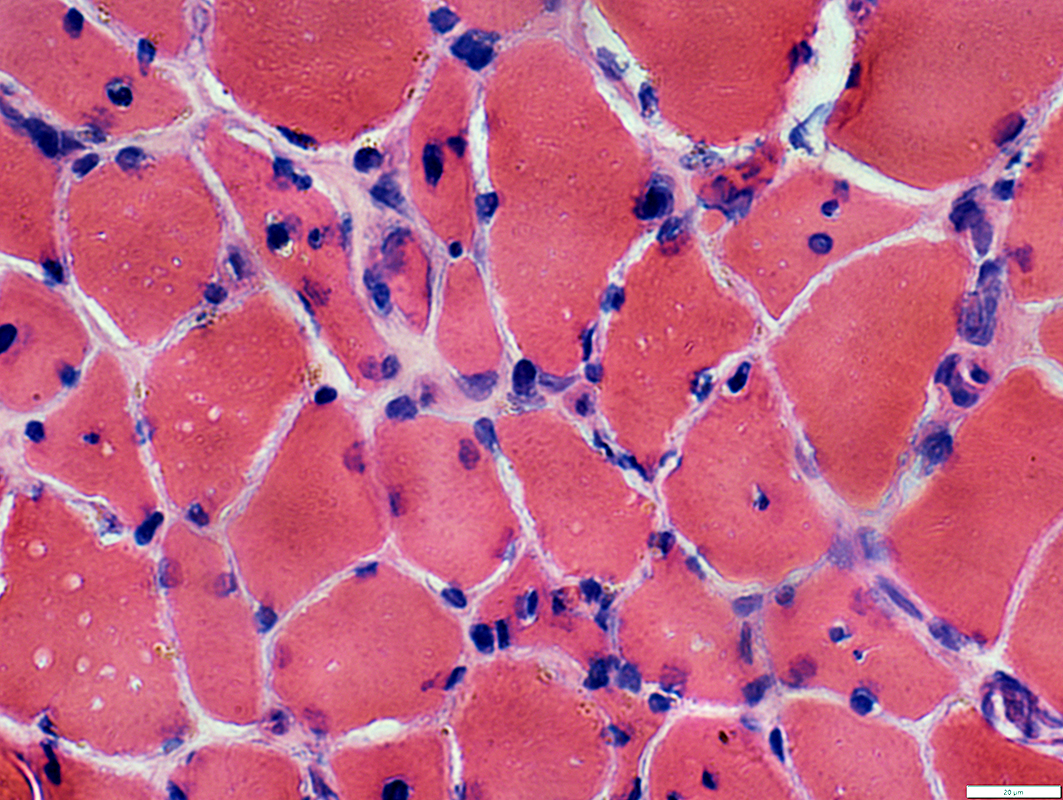 H&E stain |
Shapes: Irregular with blebs
Sizes: Often large
Distribution: some are clustered
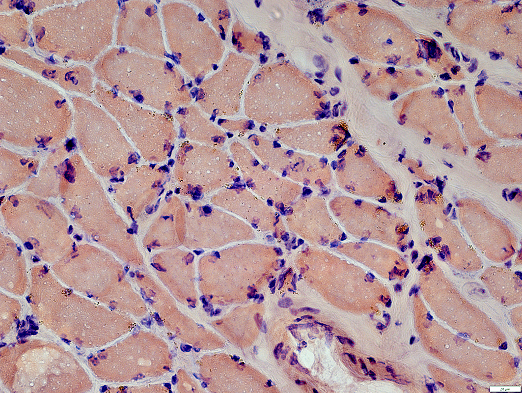 Congo red stain |
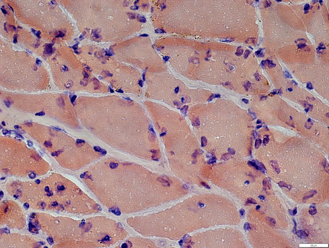 Congo red stain |
Shapes: Irregular with blebs
Sizes: Often large
Distribution: some are clustered
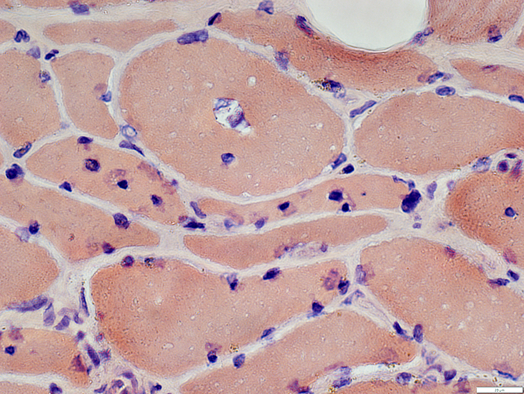 Congo red stain |
Myonuclei
Shapes: Emerin stain shows irregular nuclear shapes
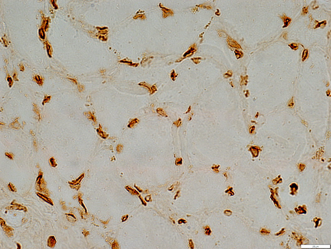 Emerin stain |
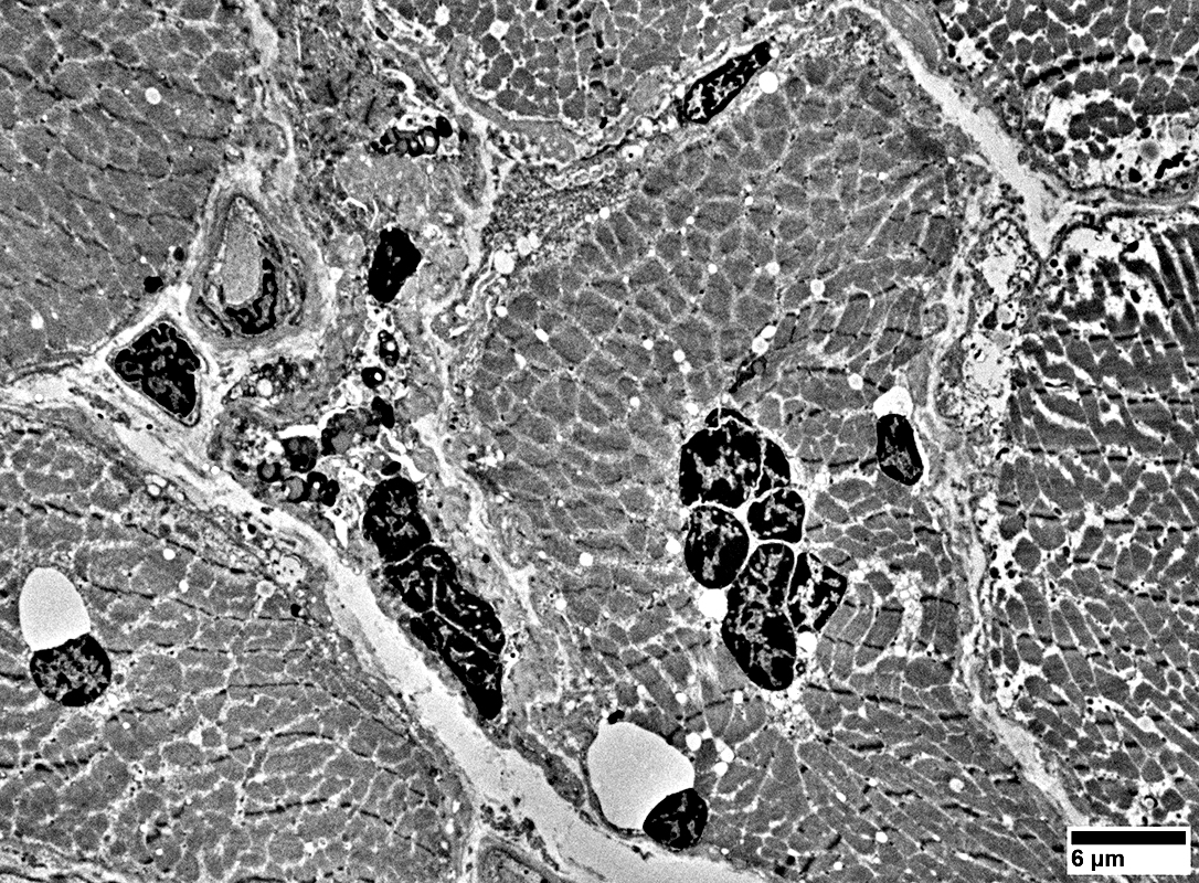 From: R Schmidt |
Heterochromatin: Increased
Distribution: Clustered, some together with lipopigment
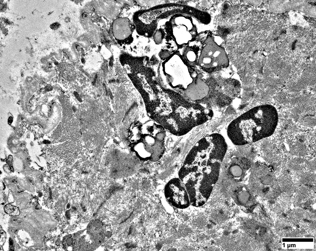 From: R Schmidt |
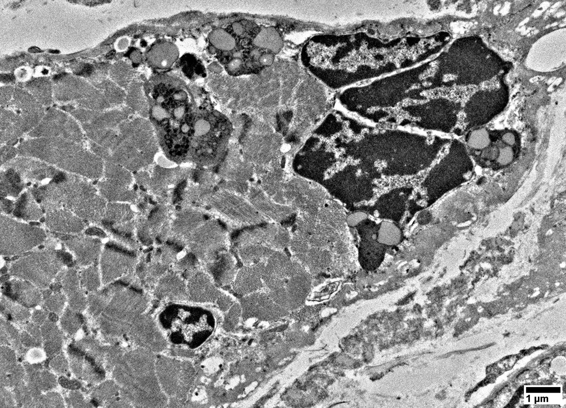 From: R Schmidt |
Heterochromatin: Increased
Distribution: Clustered, some together with lipopigment or vacuoles (Below)
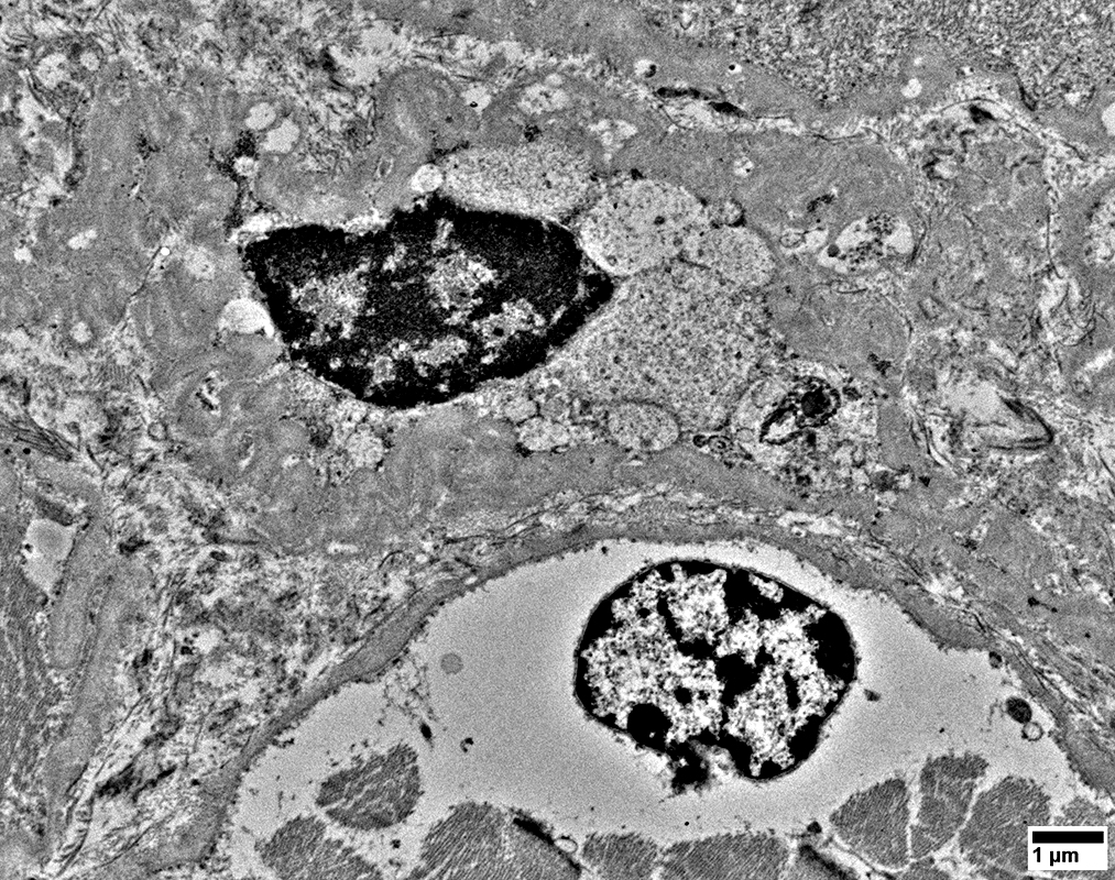 From: R Schmidt |
 From: R Schmidt |
Neuromuscular Junctions
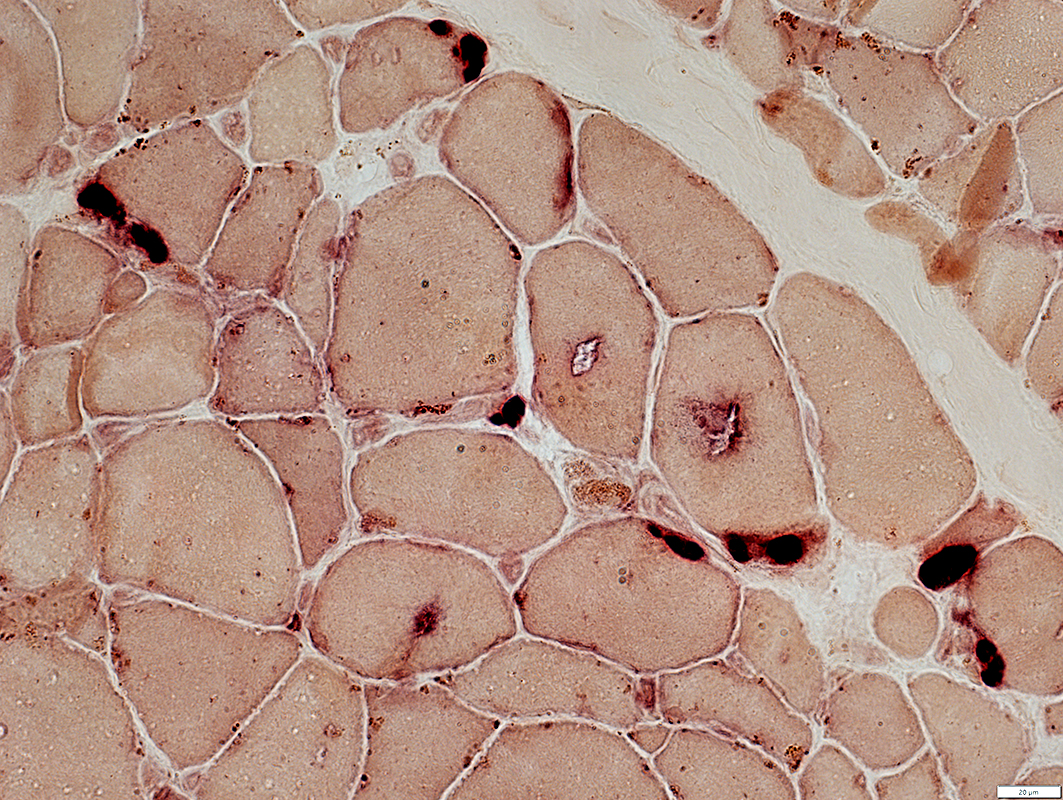 Esterase stain |
Some have several segments
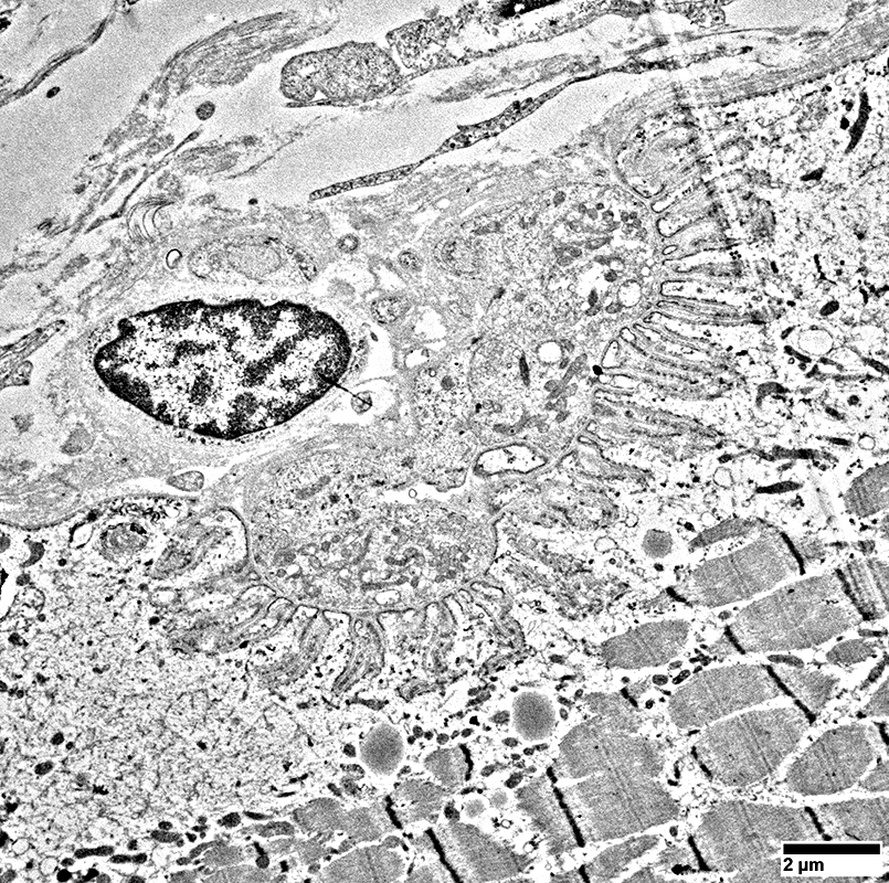 From: R Schmidt |
Increased cytoplasmic clear space below post-synaptic folds
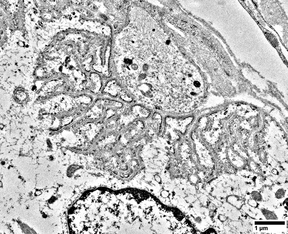 From: R Schmidt |
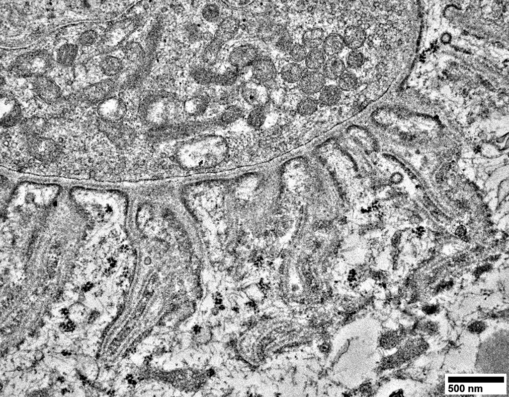 From: R Schmidt |
Some post-synaptic folds appear truncated
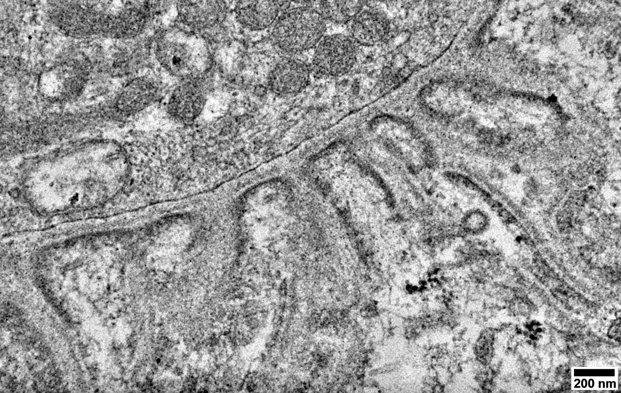 From: R Schmidt |
Capillaries
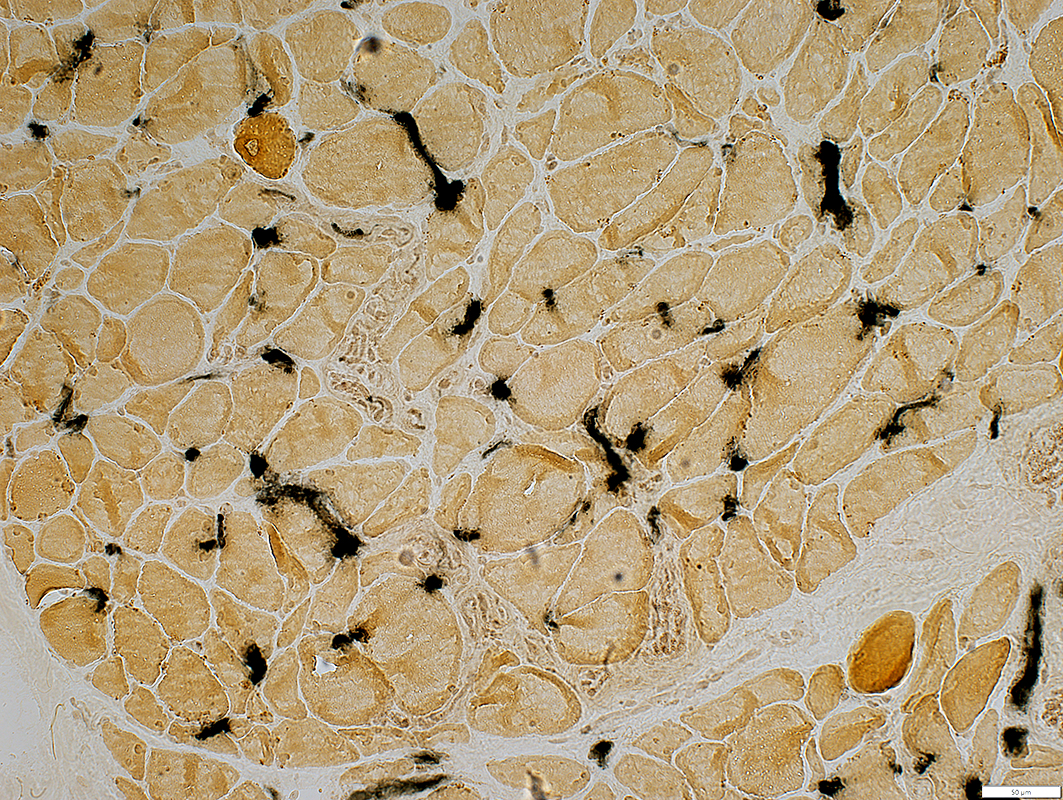 Alkaline phosphatase stain |
Increased numbers stain for alkaline phosphatase
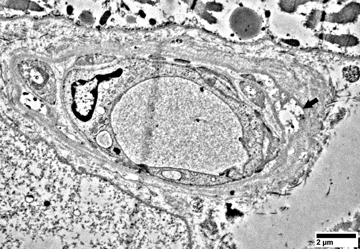 From: R Schmidt |
Vacuoles in some pericytes (Arrow; Above)
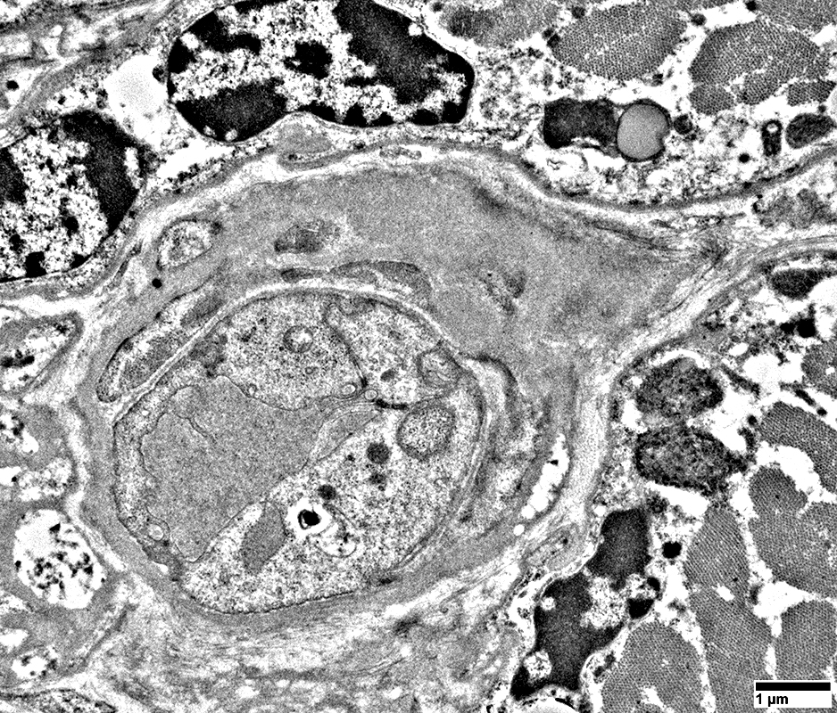 From: R Schmidt |
Basal lamina: Thick
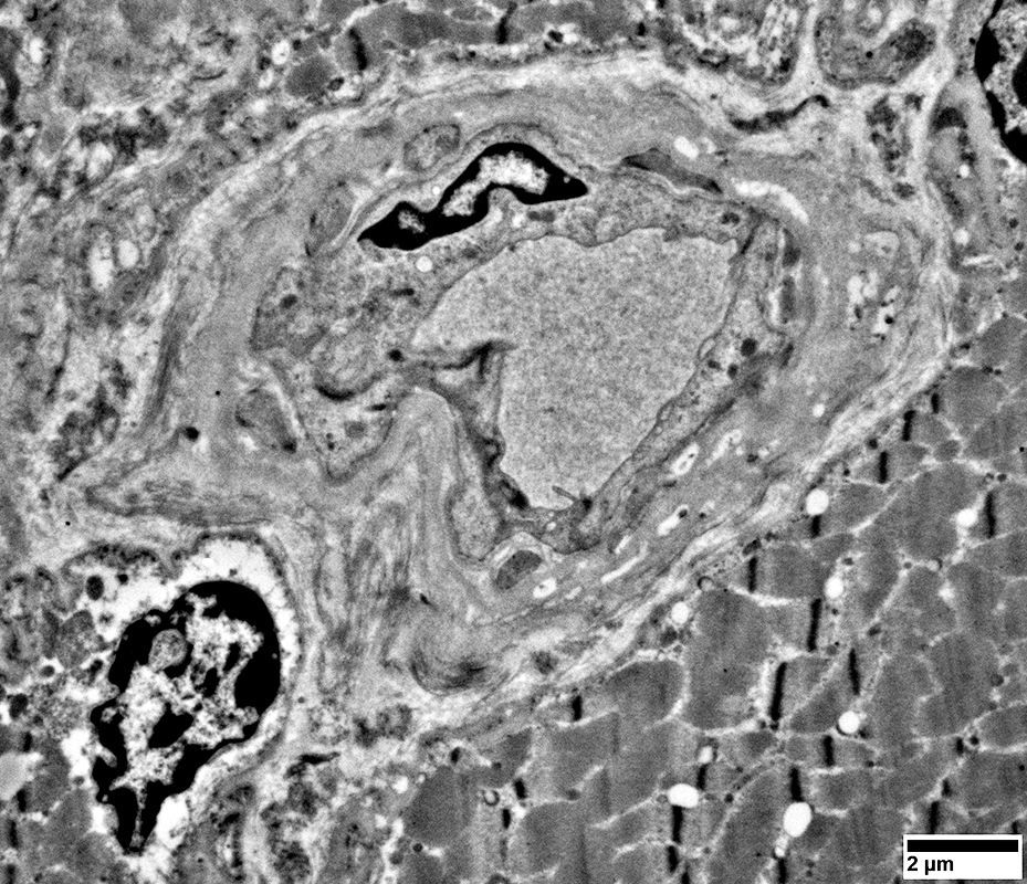 From: R Schmidt |
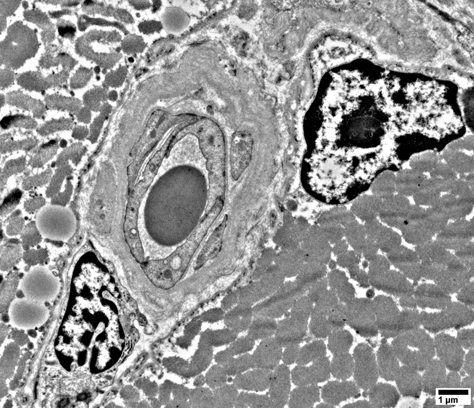 From: R Schmidt |
Return to: Amiodarone
Return to: Neuromuscular Home Page
9/17/2026