ACOX1: Mitchell syndrome
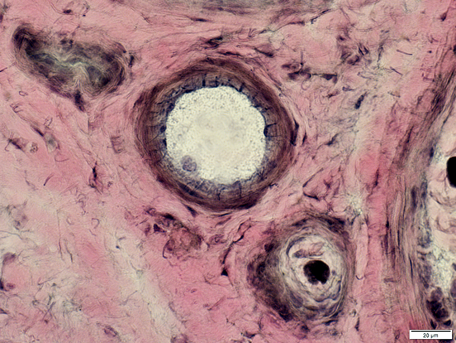
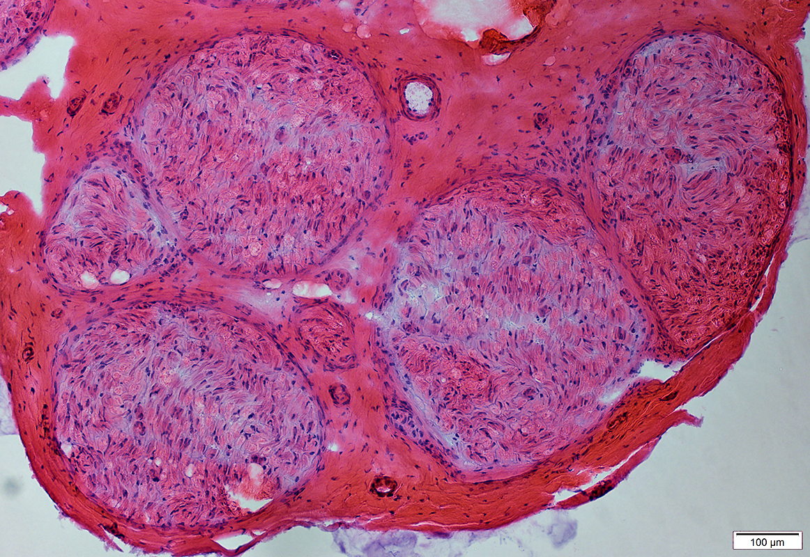 H&E stain |
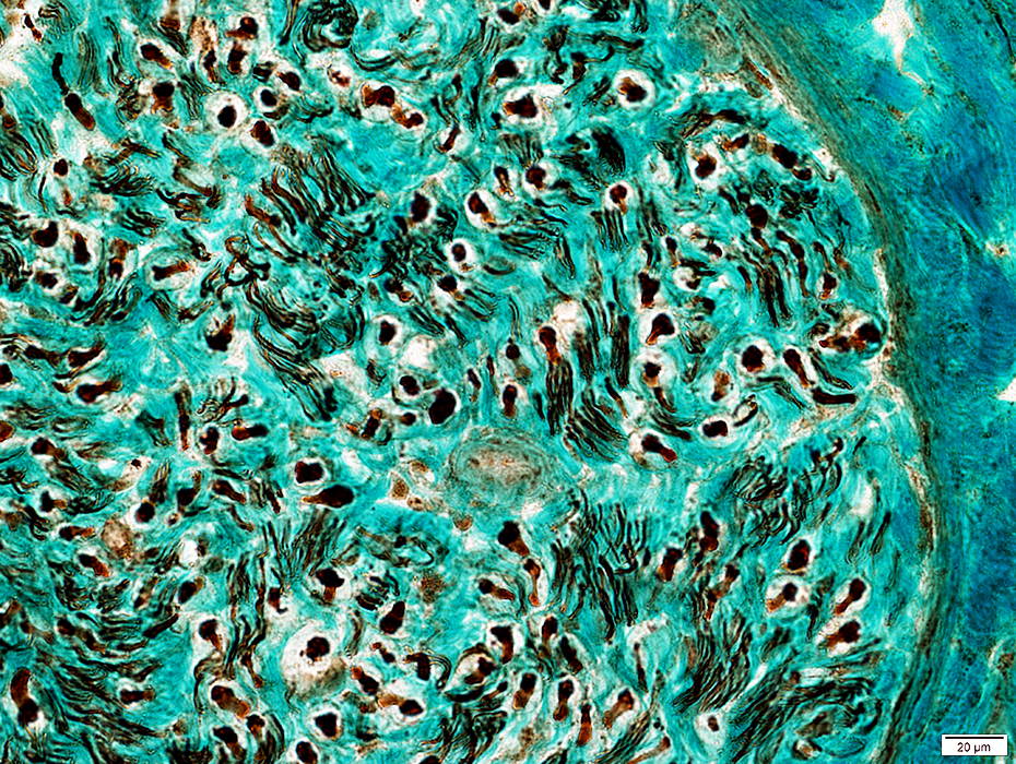
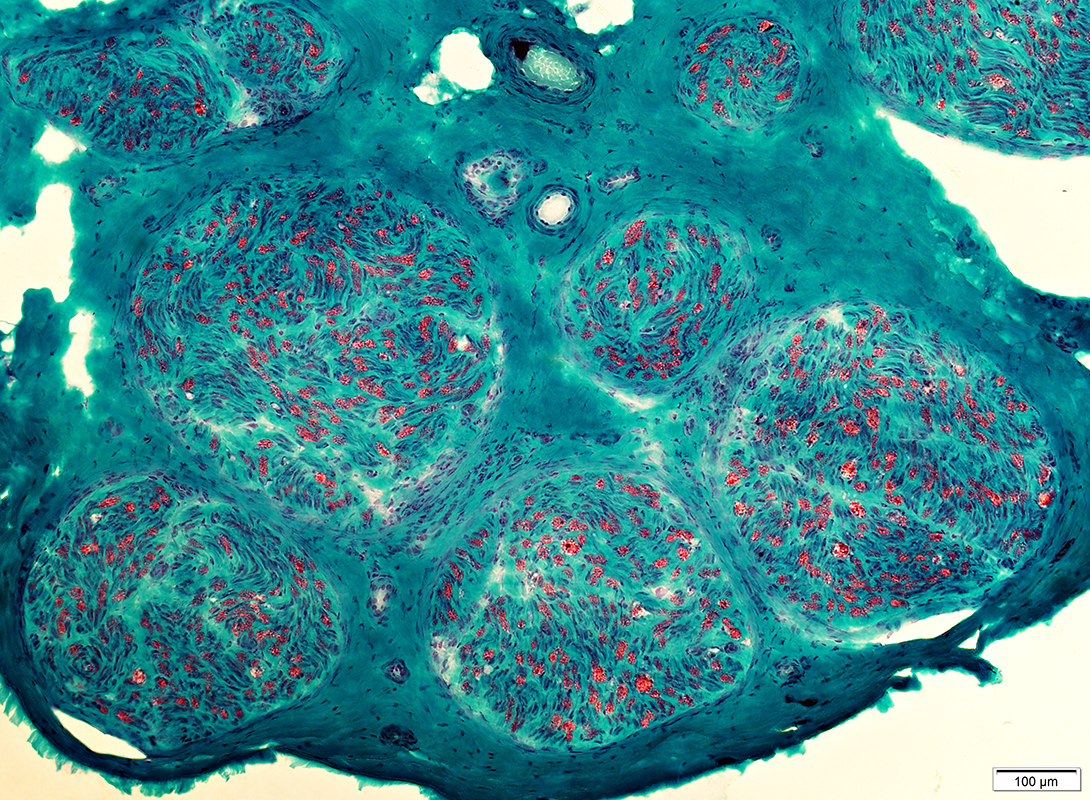 Gomori trichrome stain |
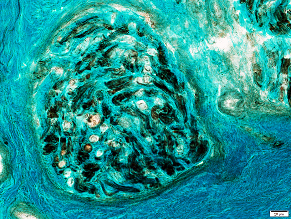
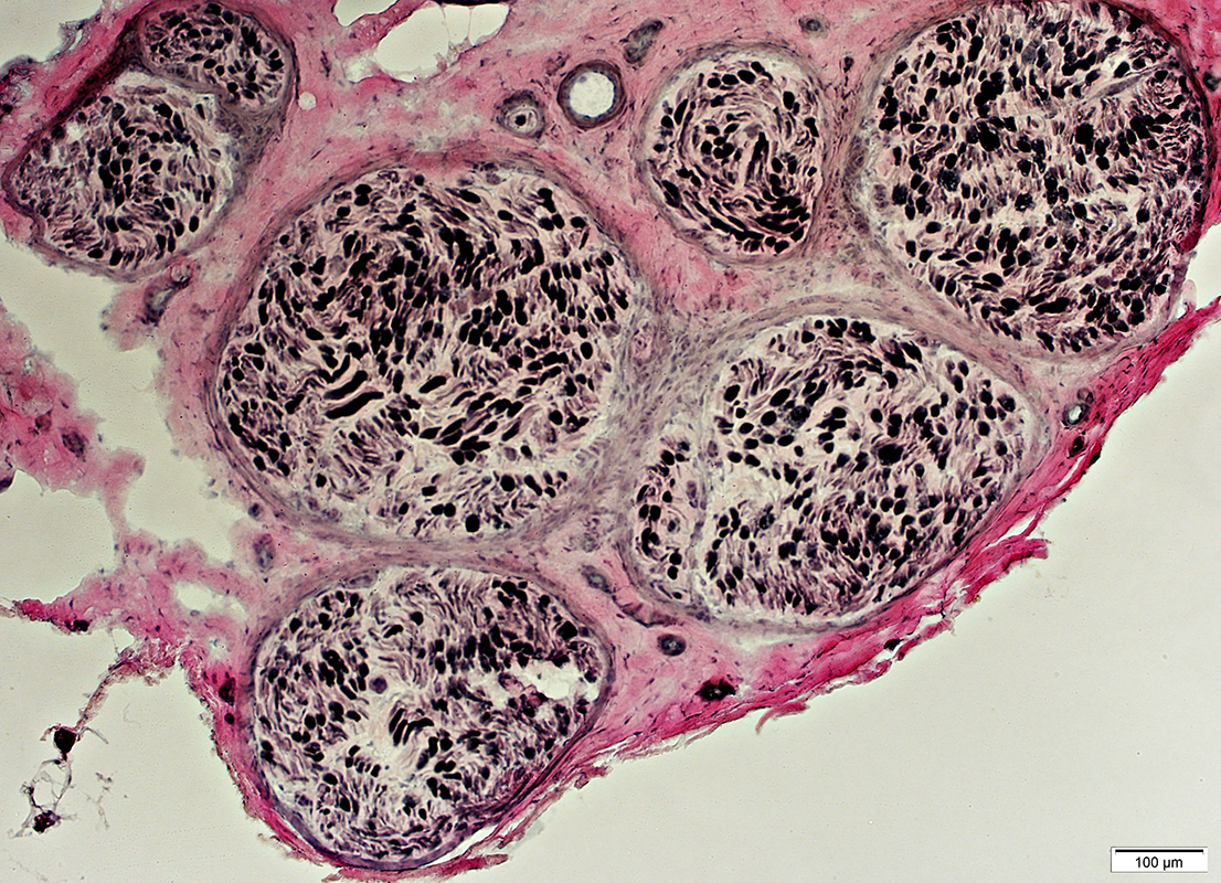 VvG stain |
Neurofilament stain |
Neurofilament stain |
 NCAM stain |
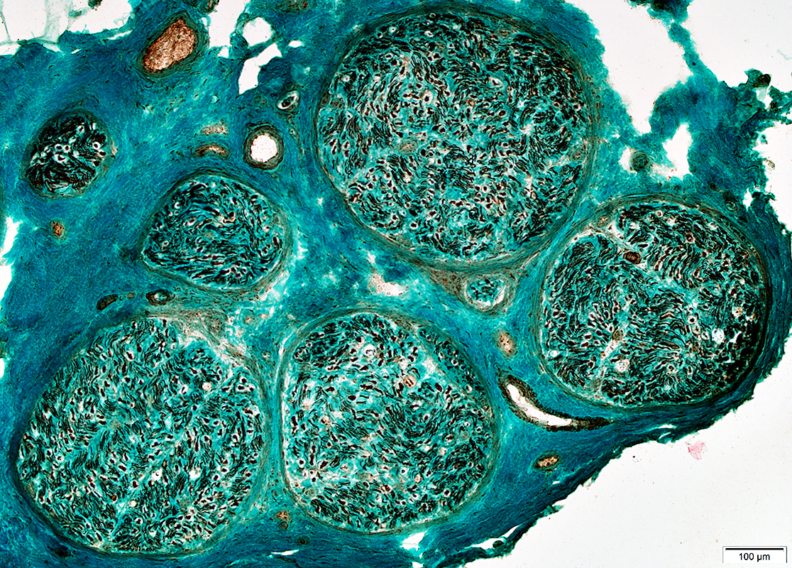
 VvG stain |
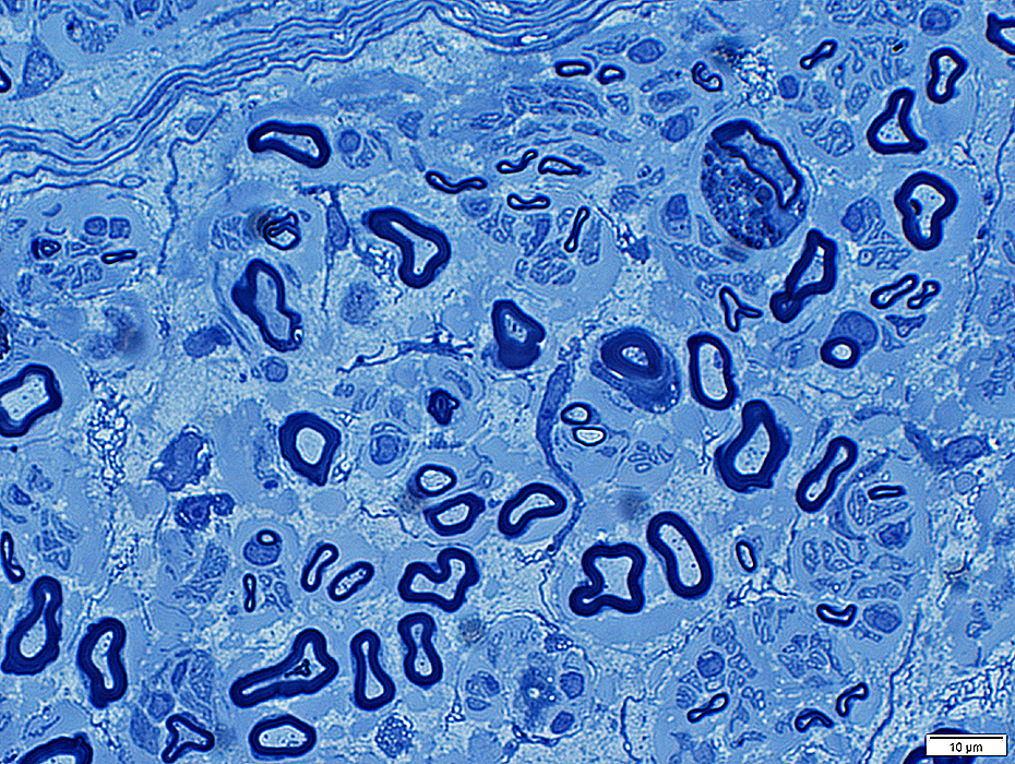
Myelinated axons: Morphology & Pathology
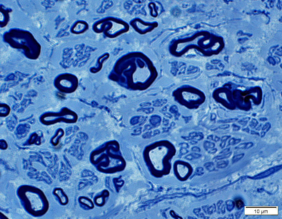
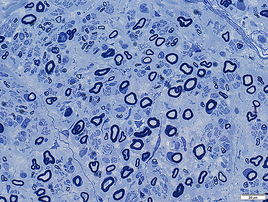 Toluidine blue stain |
Myelin sheaths: Normal thickness
Axon numbers: Loss of large & small myelinated axons
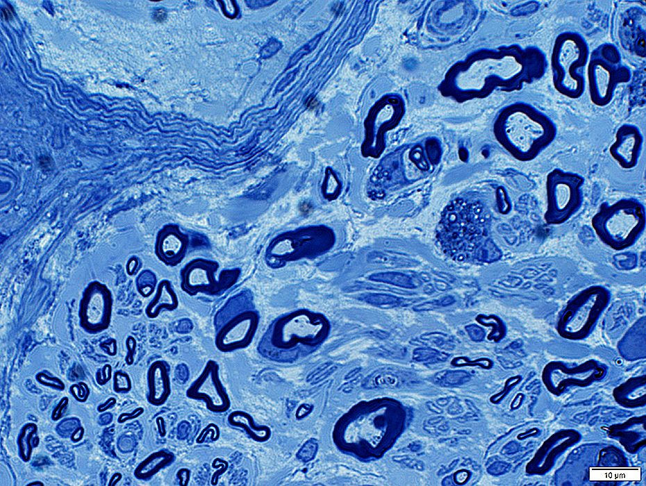
Wallerian degeneration
 Toluidine blue stain |
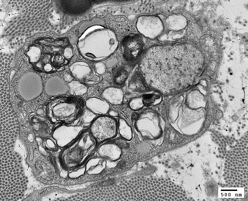
Scattered cells containing myelin debris & lipid droplets
 Toluidine blue stain |
|
 |
 |
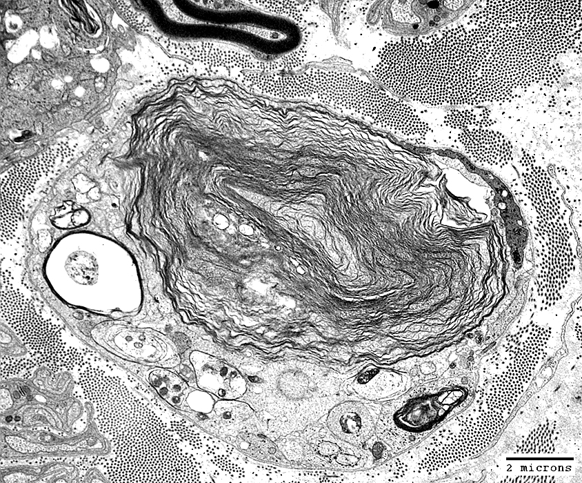
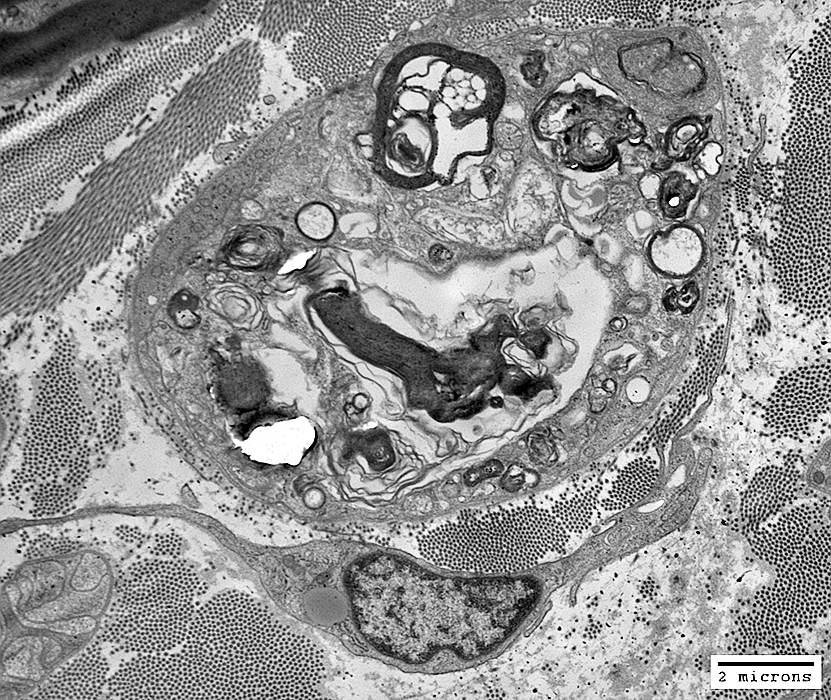
 Toluidine blue stain |
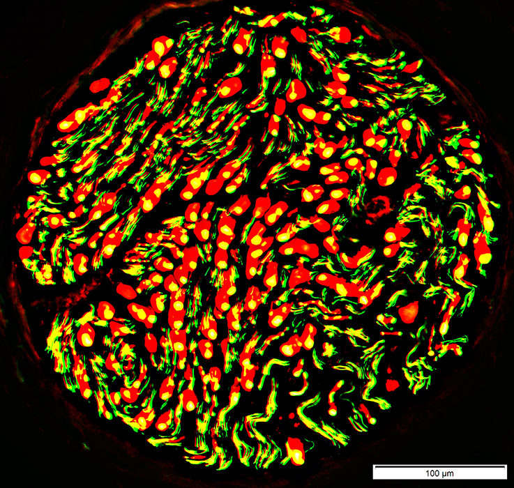
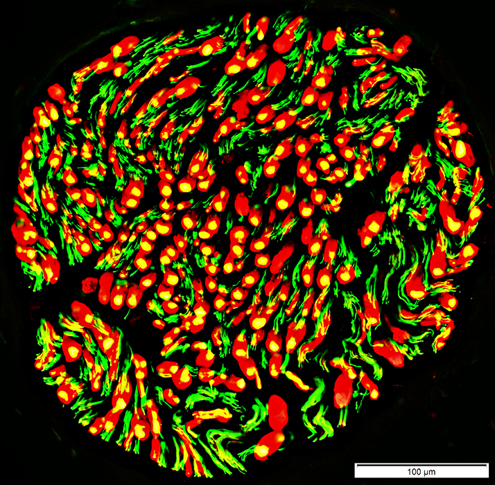 Neurofilament (Green) + P0 (Red) stains |
Acute axon loss: Some P0 regions (Red) with no axons
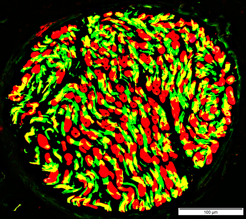
 Neurofilament (Green) + MBP (Red) stains Axon loss: Acute MBP regions (Red) with no associated axon |
 |
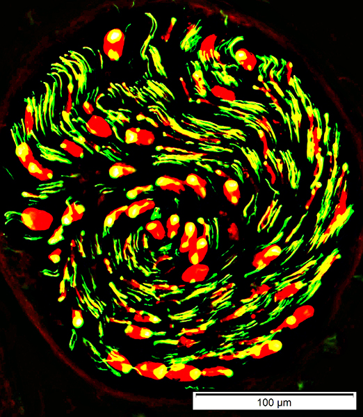
 NCAM (Green) + P0 (Red) stains |
Schwann cells, scattered, (Yellow) that co-stain for both NCAM & P0
Return to Neuromuscular Home Page
7/25/2020