U1RNP antibodies: Muscle Pathology
|
Capillary Pathology + Minimal Myopathy Active Myopathy |
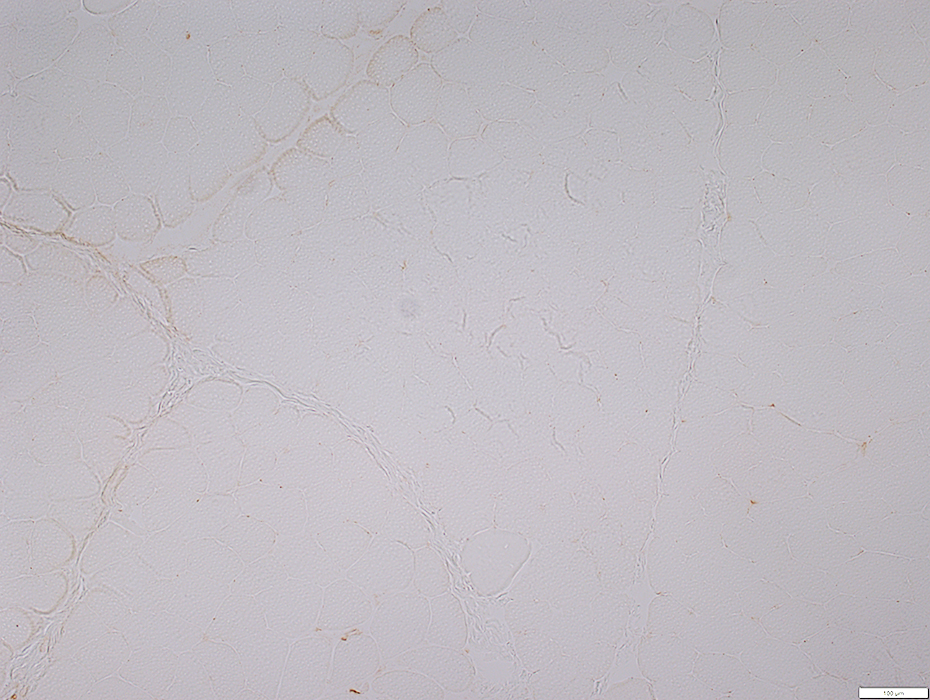
Endomysial Capillary Pathology with Minimal Myopathy
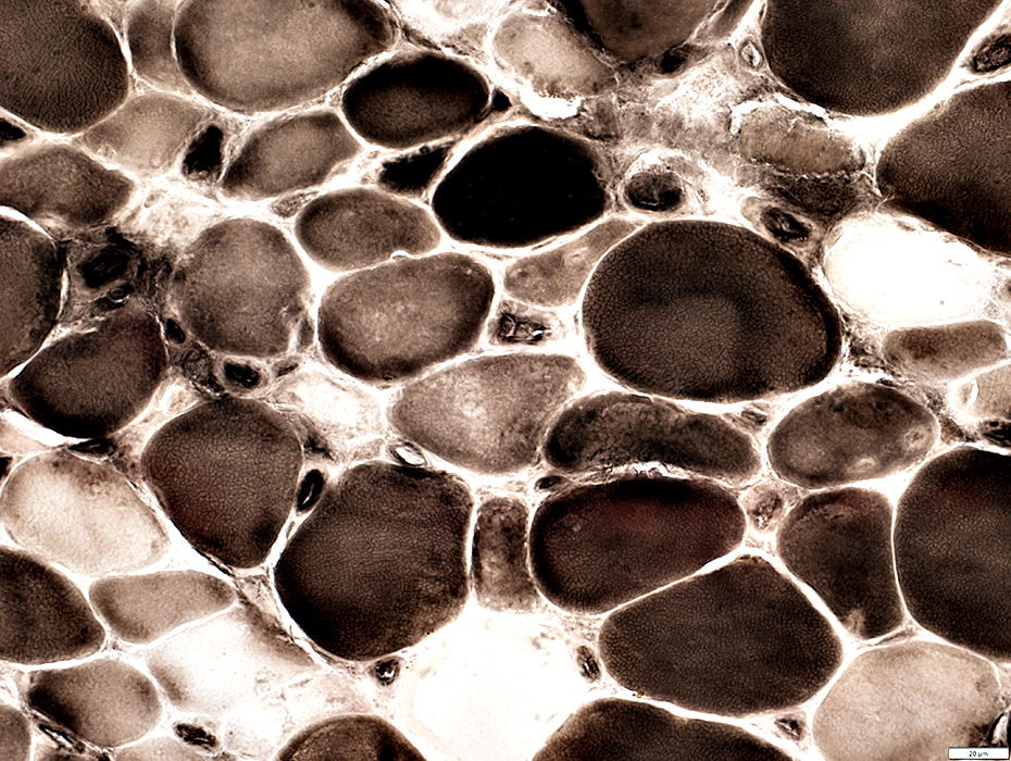
Endomysial Capillaries Morphology: Large; Moderately thick wallsEndothelium
Acid phosphatase
ATPase
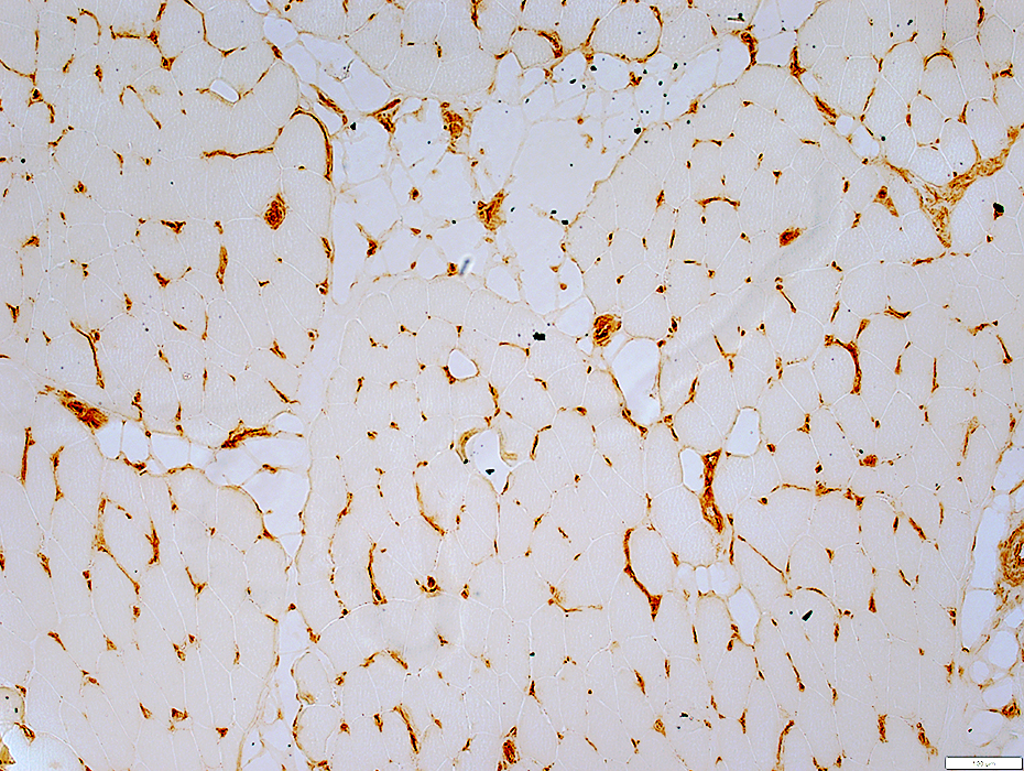
Ulex stain: Capillaries Large & Reduced numbers
MxA stain: Increased
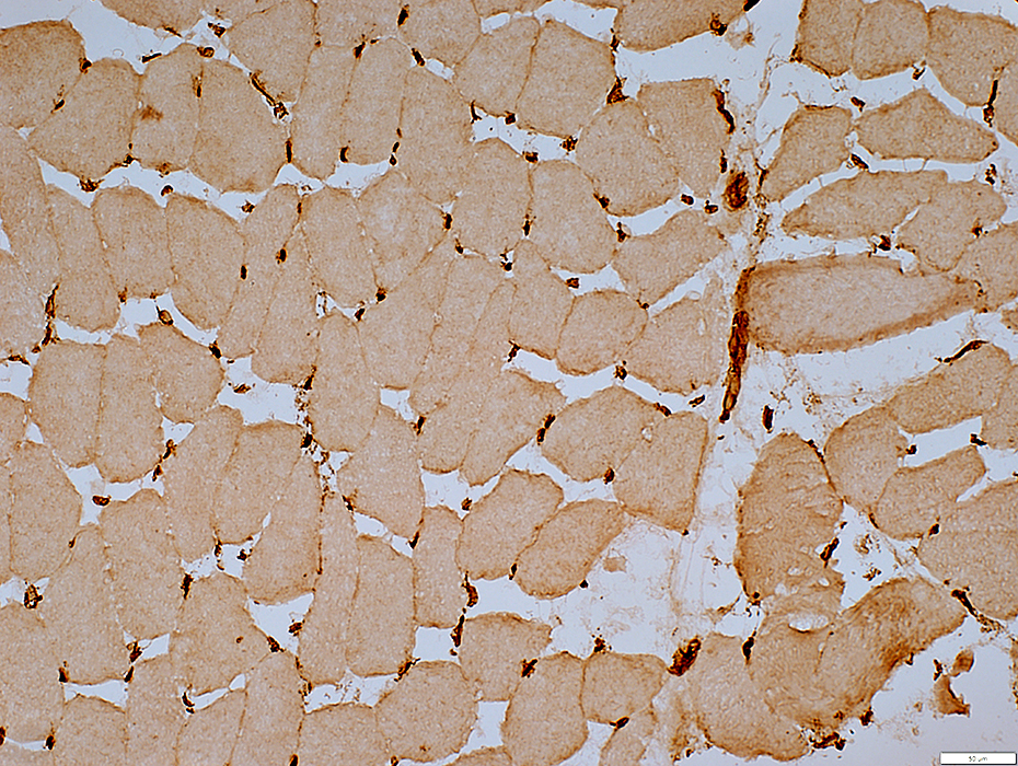
C5b-9 deposition
Muscle Fibers
Morphology: Normal
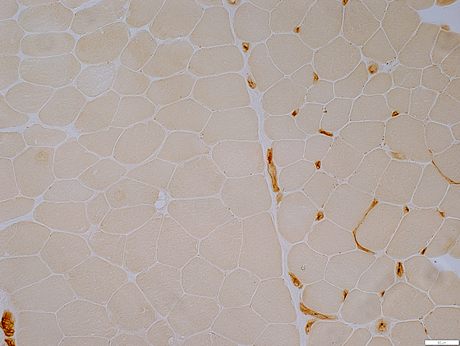
MHC Class I: Increased expression
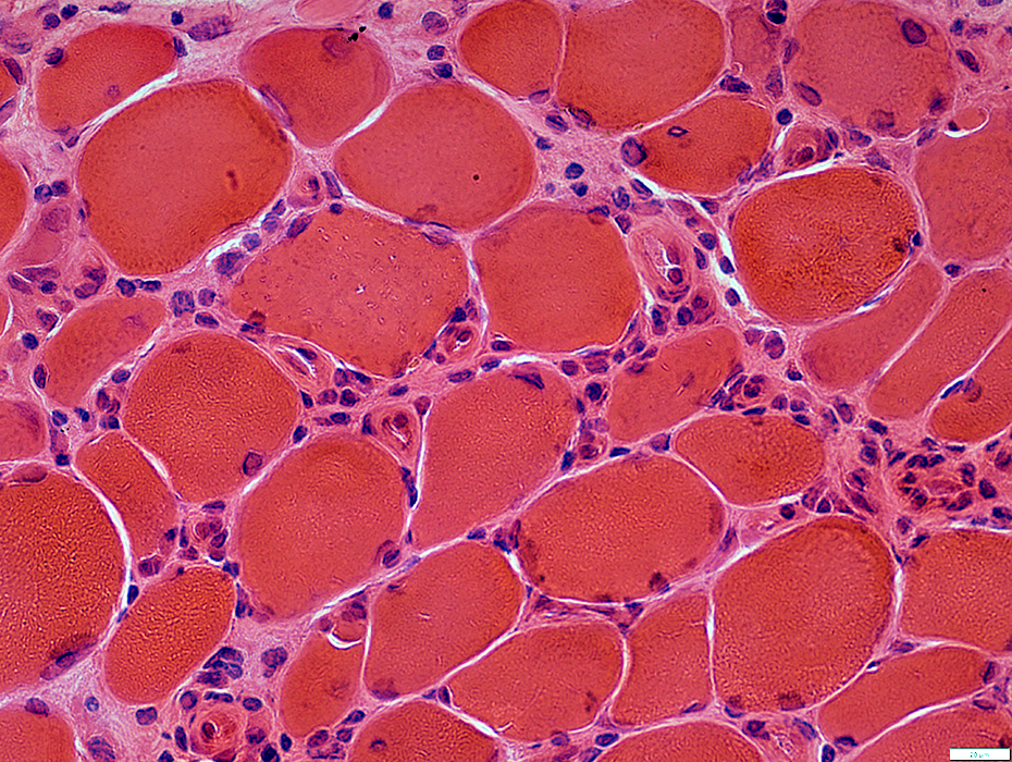
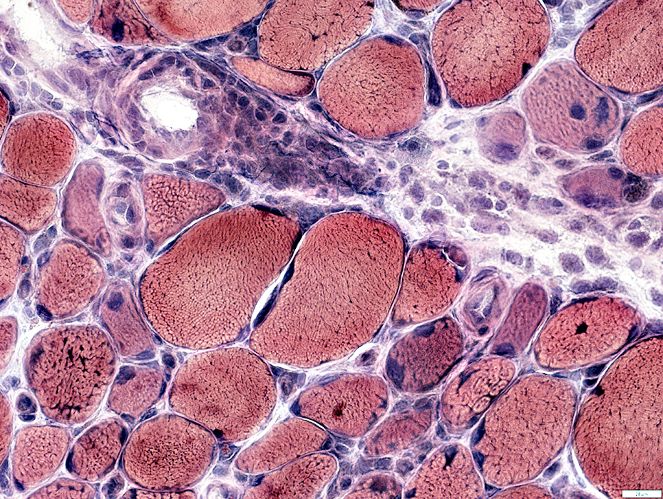
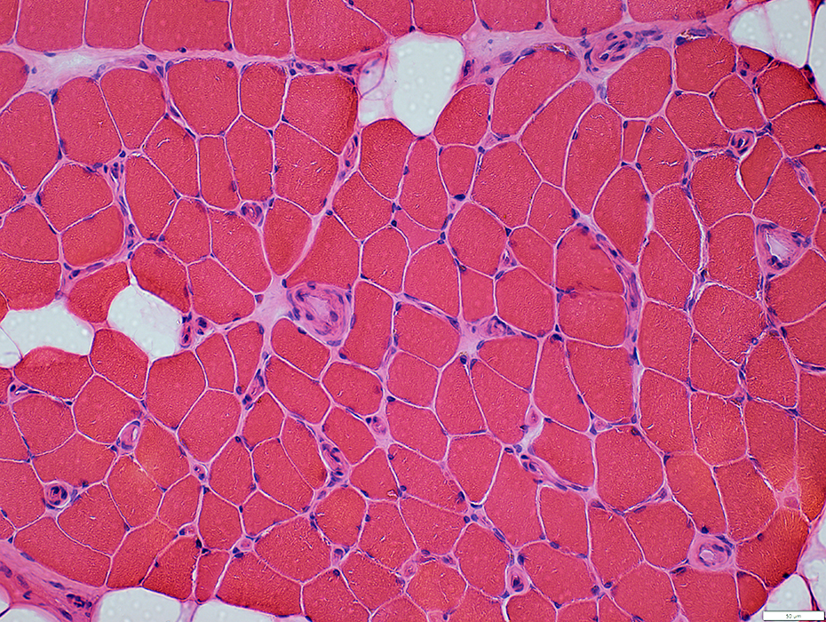 H&E stain |
Large
Basal lamina & Endothelial cells: Prominent
Scattered through muscle
Muscle fibers
Size variation: Moderate
Smaller fibers: Intermediate size
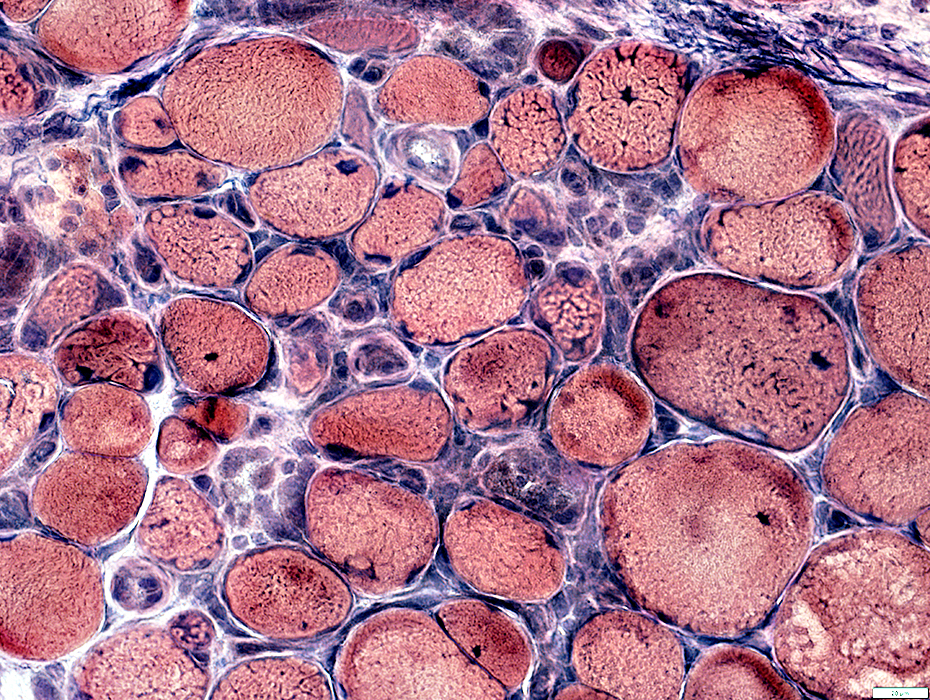
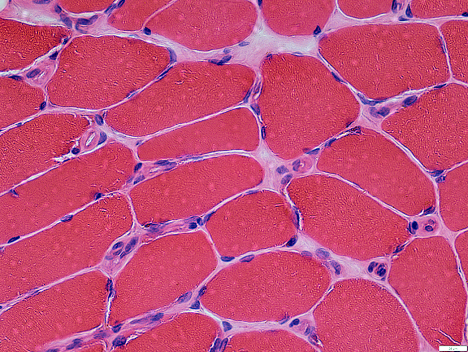 H&E stain |
 Gomori trichrome stain |
Large
Basal lamina & Endothelial cells: Prominent
Scattered through muscle
Muscle fibers
Size variation: Moderate
Smaller fibers: Intermediate size
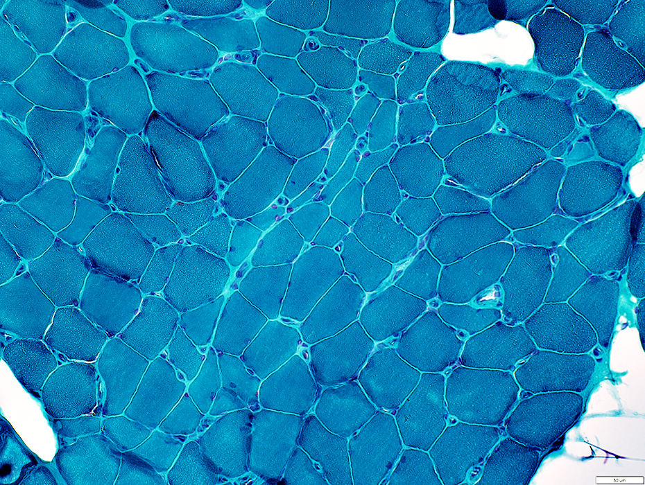
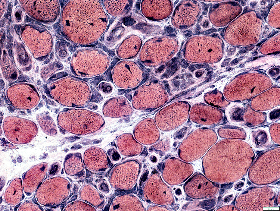
 VvG stain |
 Acid Phosphatase stain |
Endothelial cells: Acid phosphatase staining
 Acid Phosphatase stain |
 Ulex stain |
Numbers: Reduced
Stain intensity: Abnormally pale
Size: Large
 Ulex stain |
 Ulex stain |
Numbers: Reduced
Stain intensity: Abnormally pale
Size: Large
 Ulex stain |
Numbers: Reduced
Stain intensity: Abnormally pale
Size: Large
Control muscle (Below): Dark staining of small capillaries
 Ulex stain |
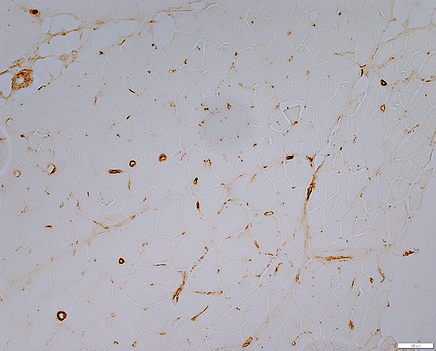
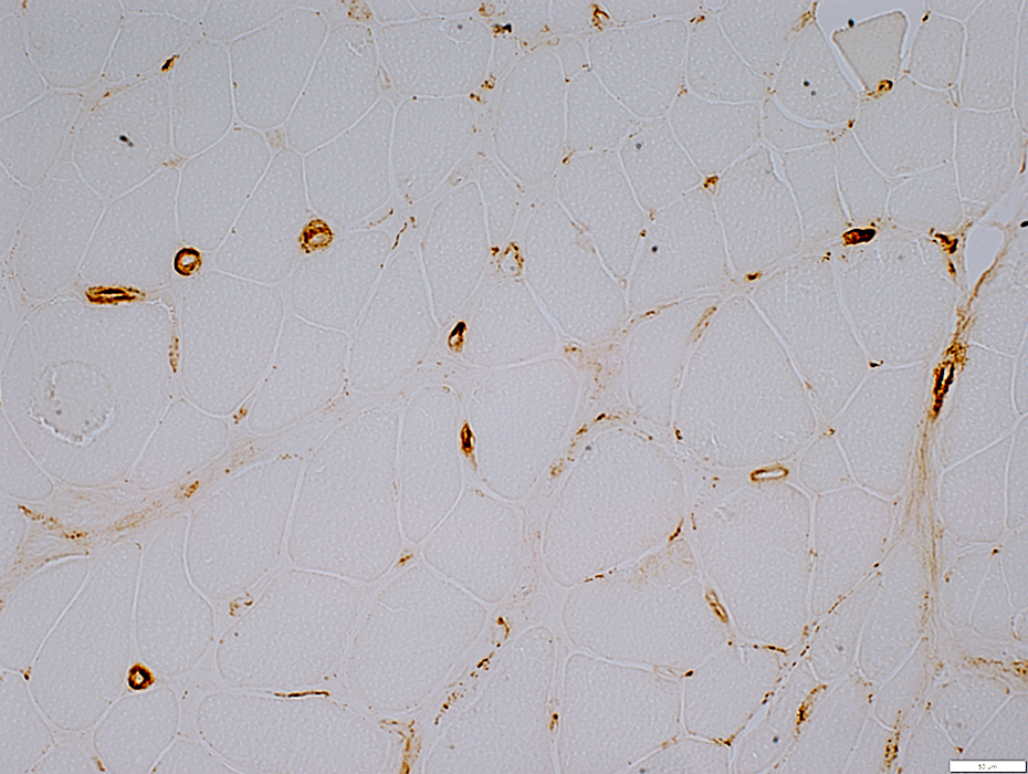
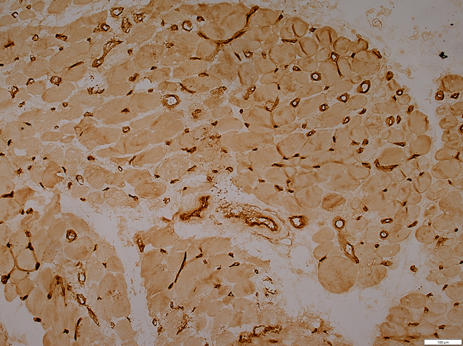
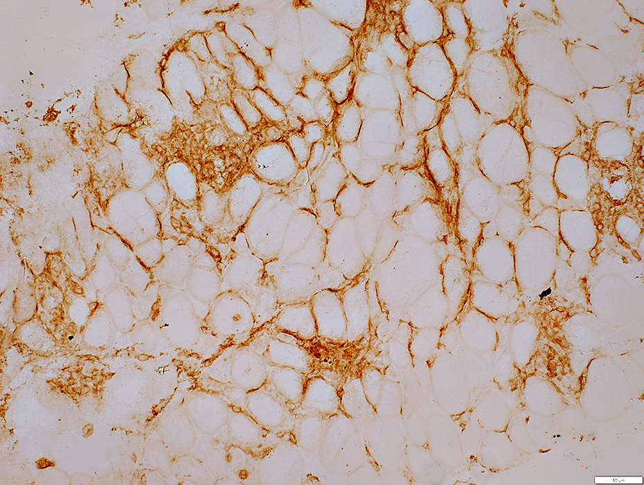
 MxA stain |
Capillary-associated cells: Dark stain
Muscle fibers: Mild cytoplasm stain
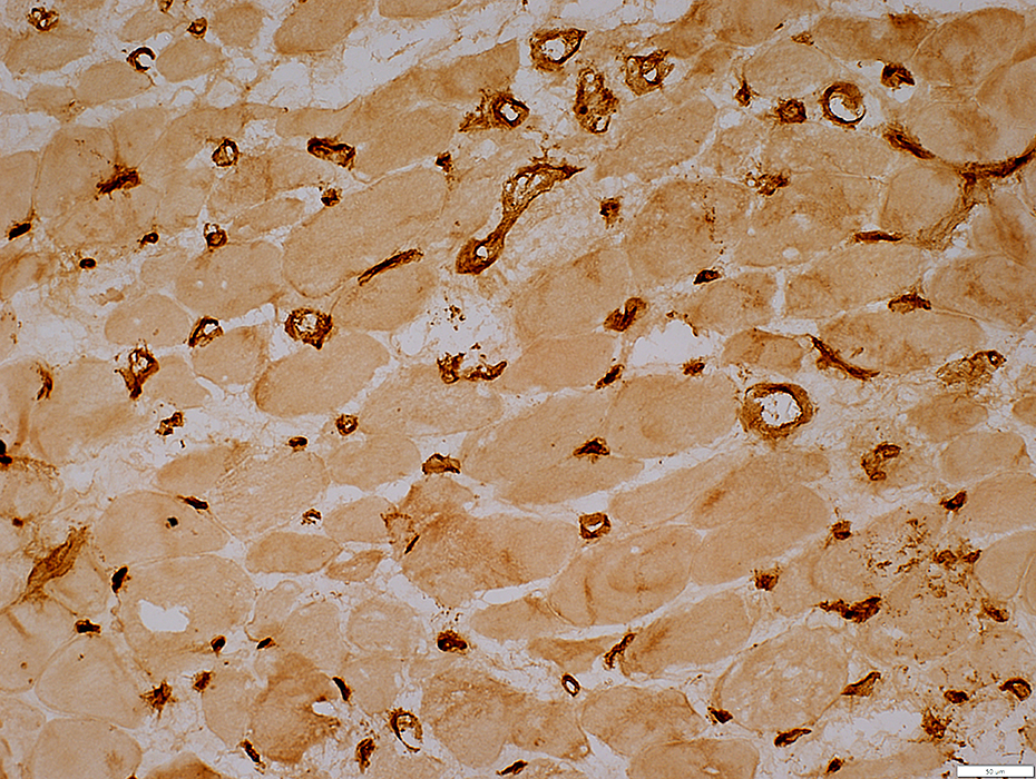
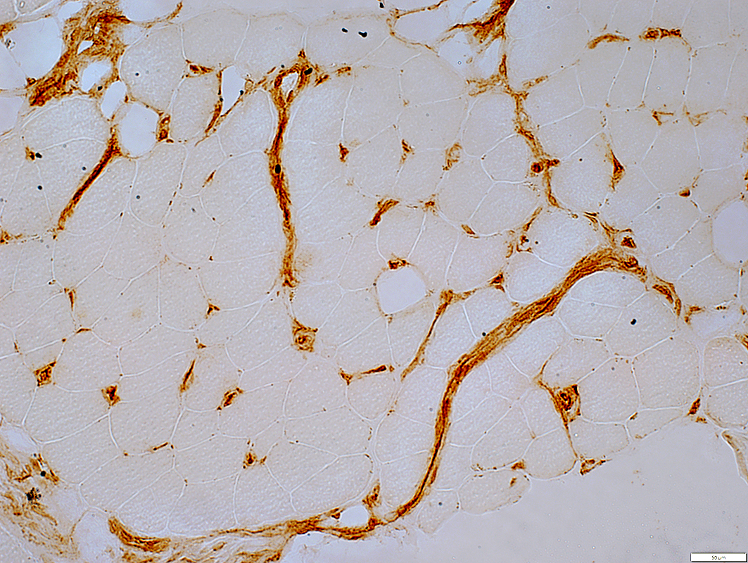 MxA stain |
Capillary-associated cells: Dark stain
Muscle fibers: Mild cytoplasm stain
Control muscle (Below): No staining of capillaries or muscle fibers
 MxA stain |
 C5b-9 stain |
Endomysial Capillary-associated
Muscle fibers: No staining
 C5b-9 stain |
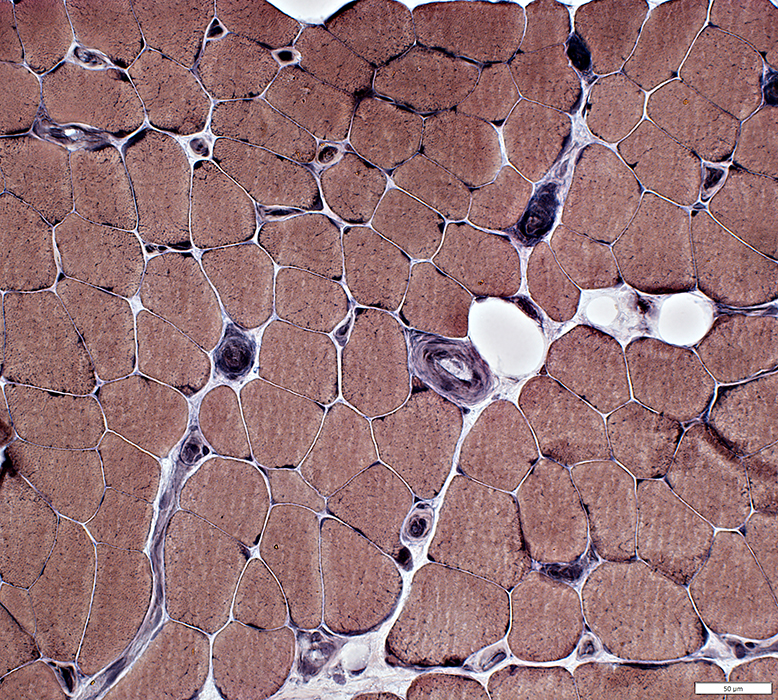
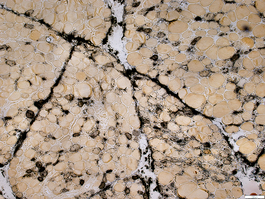
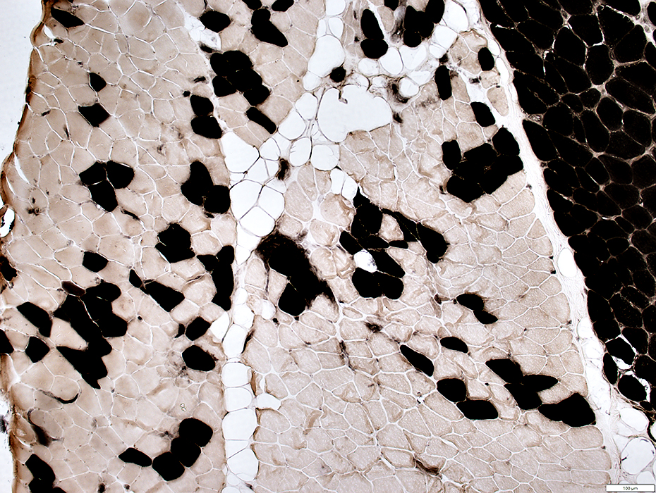 ATPase pH 4.3 stain |
Perimysial Connective Tissue: Replaced by fat (Above)
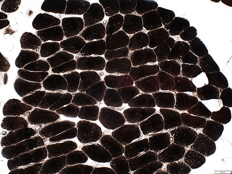 ATPase pH 4.3 stain |
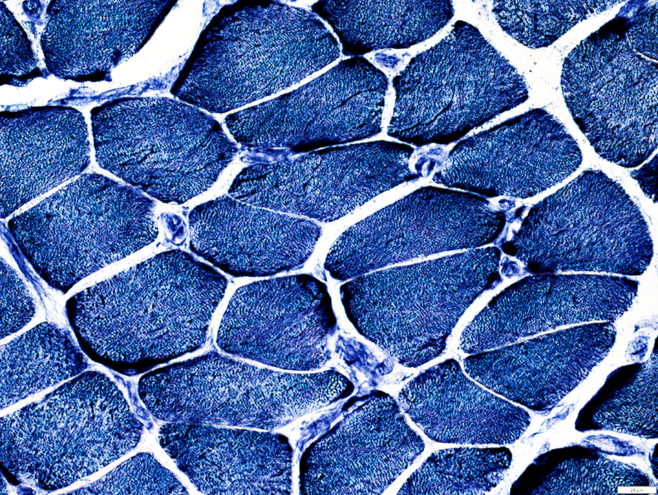 NADH stain |
Muscle fibers: Mildly coarse internal architecture
 NADH stain |
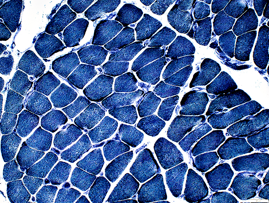
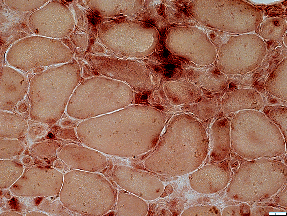
 Esterase stain |
Muscle fibers: MHC1 upregulation
 MHC I stain |
Myopathy with Capillary Pathology
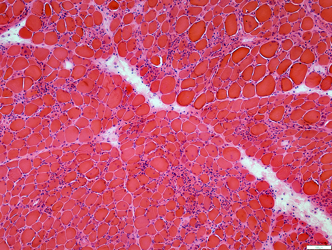 H&E stain |
Capillaries
Muscle fibers
Connective tissue
Inflammation
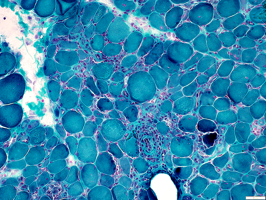 Gomori trichrome stain |
 VvG stain |
Capillary Pathology
 H&E stain |
 VvG stain |
 VvG stain |
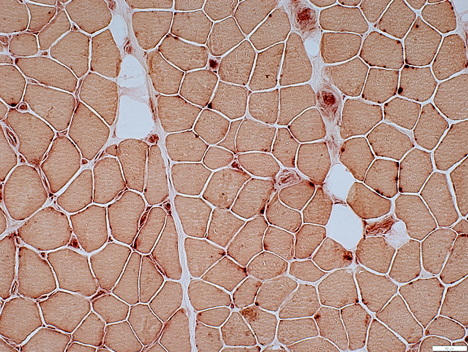
Endomysial Capillaries: Acid phosphatase stains endothelium
 Acid phosphatase stain |
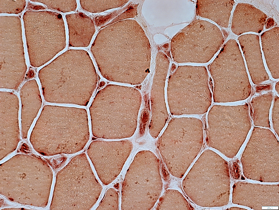
Endomysial Capillaries: ATPase stains endothelium
 ATPase pH 4.3 stain |
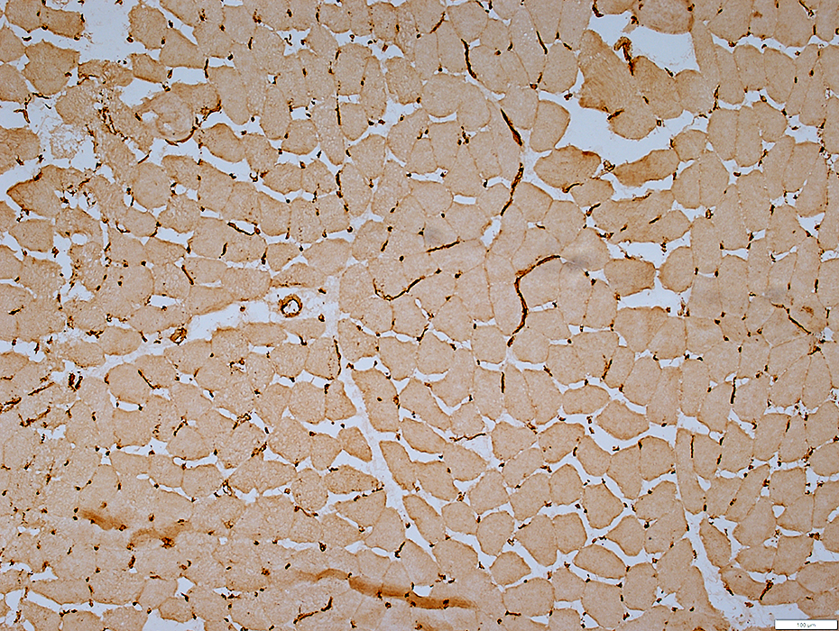
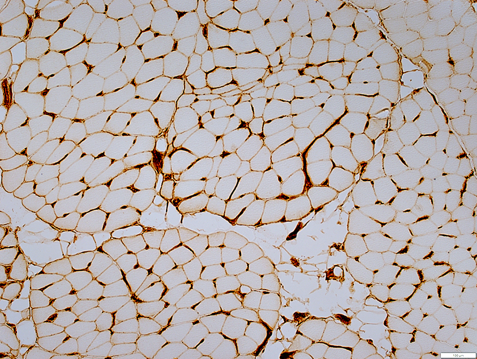
Endomysial Capillaries: Ulex lectin shows large size & Reduced numbers
 UEA-I stain |
 UEA-I stain |
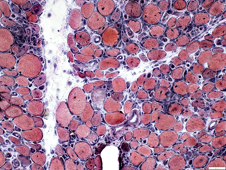
Muscle Fiber Pathology
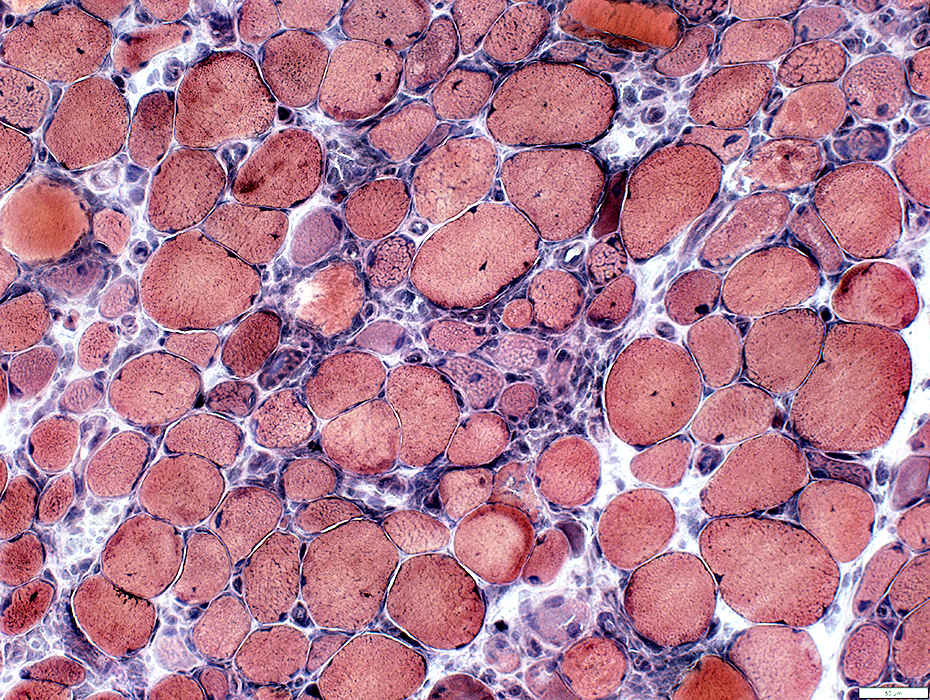 VvG stain |
Sizes: Varied
Many immature fibers
Necrosis: Scattered fibers
Myonuclei: Large
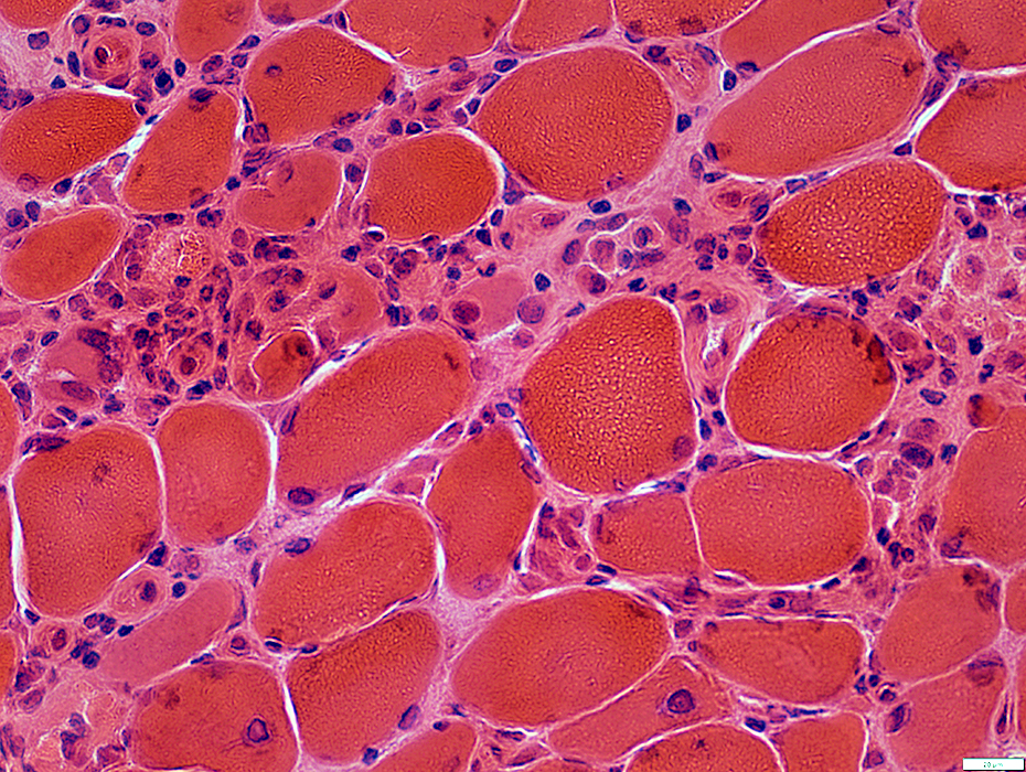 H&E stain |
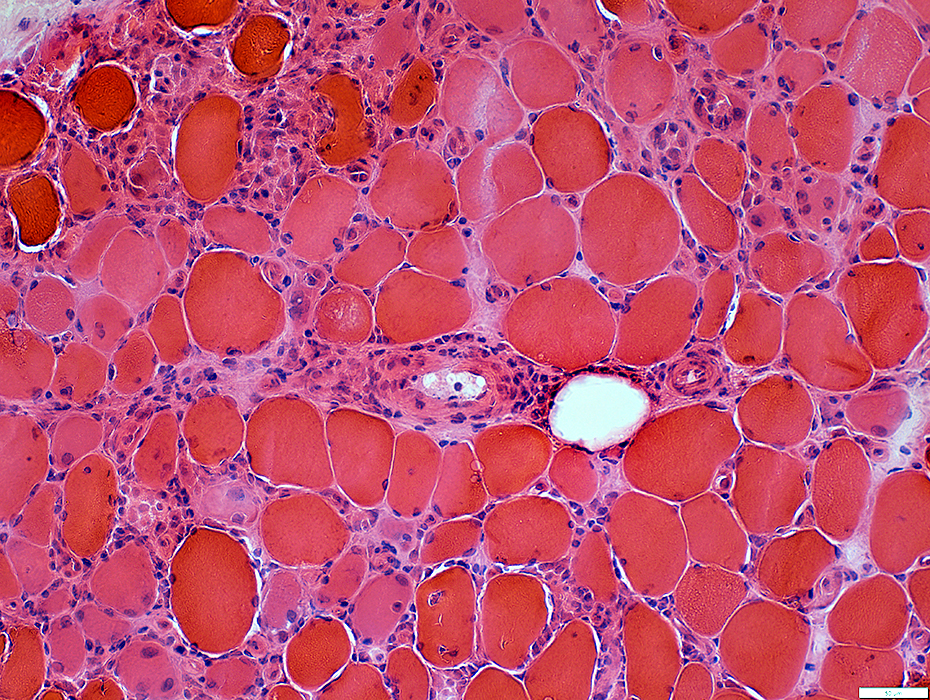
Muscle Fiber Pathology
Necrotic fibers invaded & replaced by histiocytes
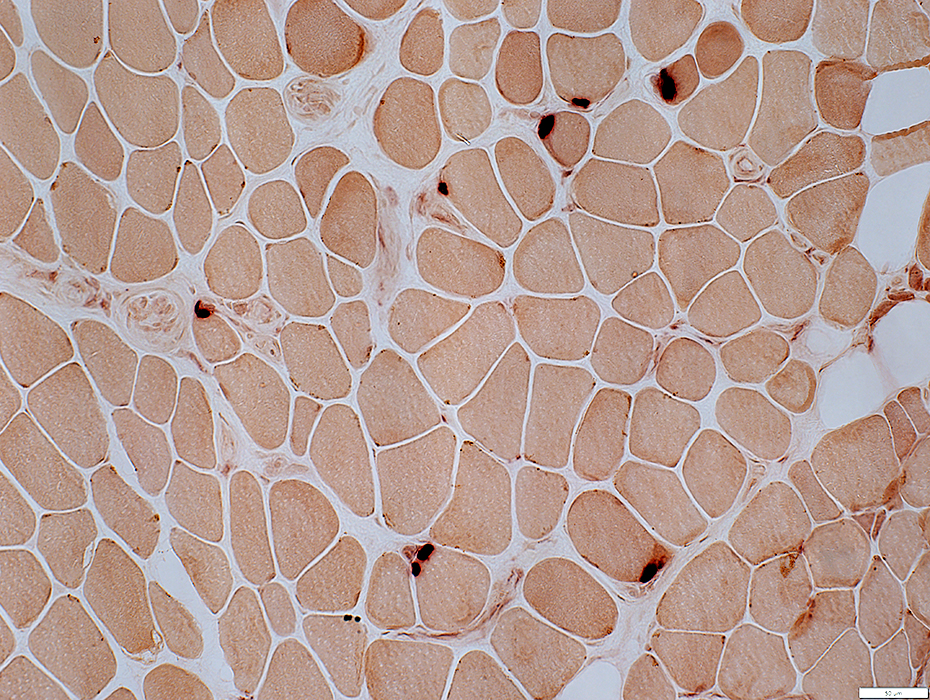
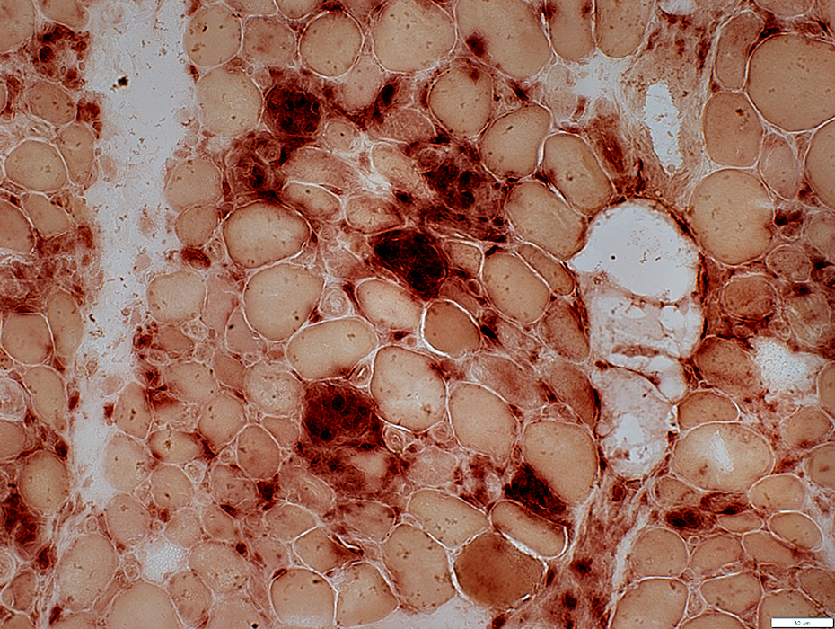 Acid phosphatase stain |
Muscle Fiber Pathology
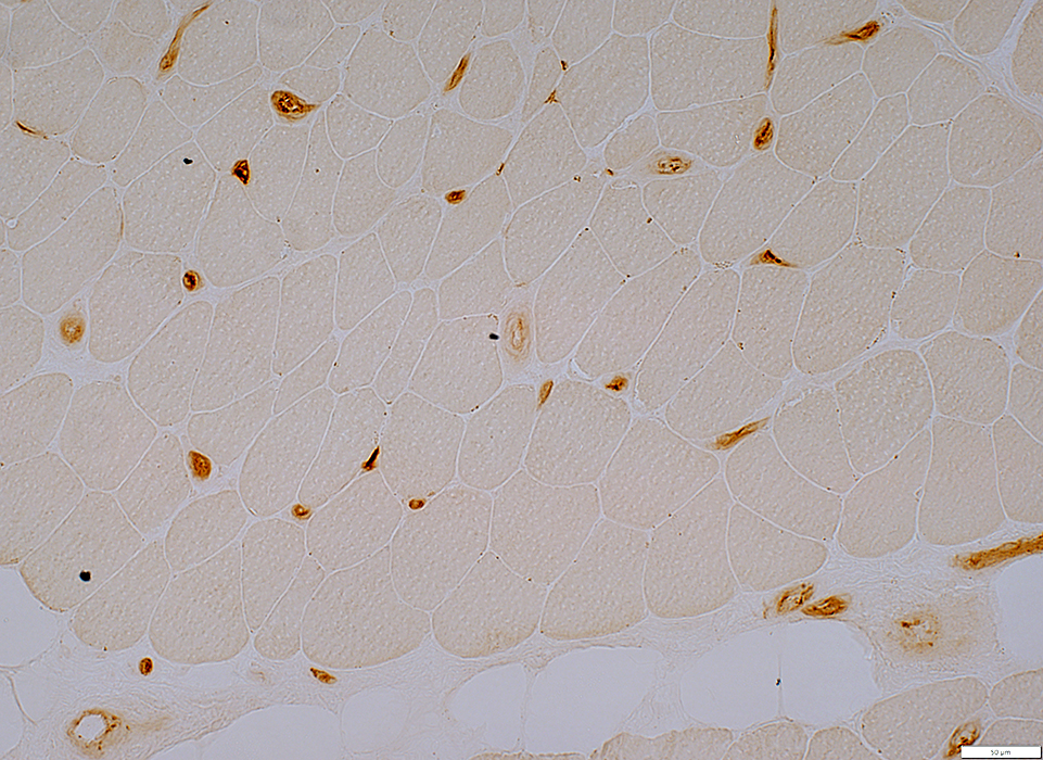
MHC Class I: Upregulated by muscle fibers
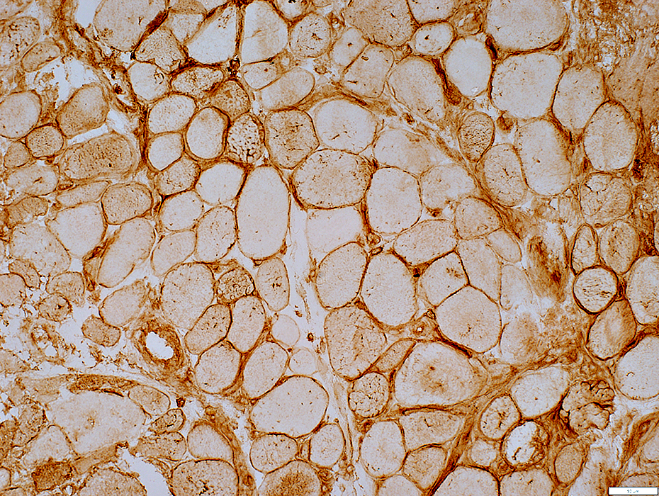 MHC Class I stain |
Muscle Pathology
C5b-9 stains
Cytoplasm of necrotic muscle fibers
Endomysial connective tissue
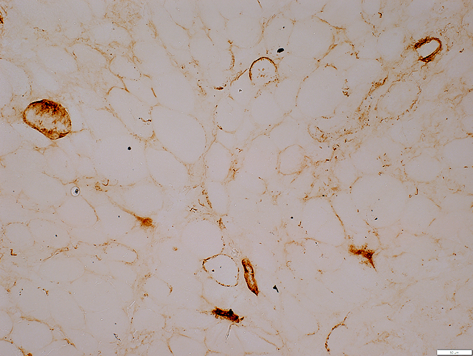 C5b-9 stain |
Perimysial Connective Tissue Pathology
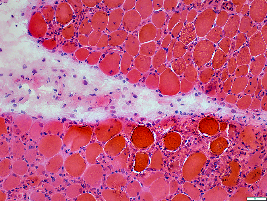 H&E stain |
Perimysial Connective Tissue
Structure: Damaged
Contains: Large, Scattered Histiocytes
Stains for: Alkaline phosphatase (Below)
 Alkaline phosphatase stain |
 H&E stain |
Lymphocytes: Surround perimysial vessel
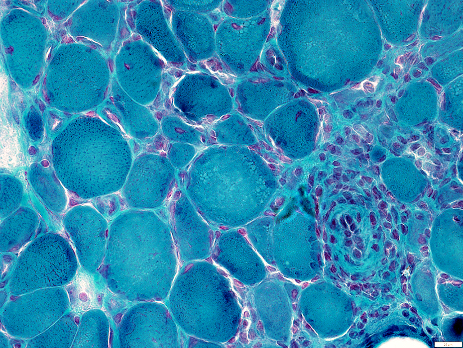 Gomori trichrome stain |
 VvG stain |
Lymphocytes: CD4 stains many endomysial cells
 CD4 stain |
Return to: U1RNP
Return to: Neuromuscular Home Page
12/17/2023