Regional Ischemic Immune Myopathy (RIIM)
|
Muscle fibers Regional pathology: Varied involvement in different areas Necrosis zone: In border regions between vessels Purlieu zone: Region between vessels & necrosis zone Regeneration zone: Clustered regeneration after necrosis Vessels Capillaries: Necrotic (In areas with necrotic muscle fibers) Veins with damaged walls NXP-2 antibody-related pathology MRI images |
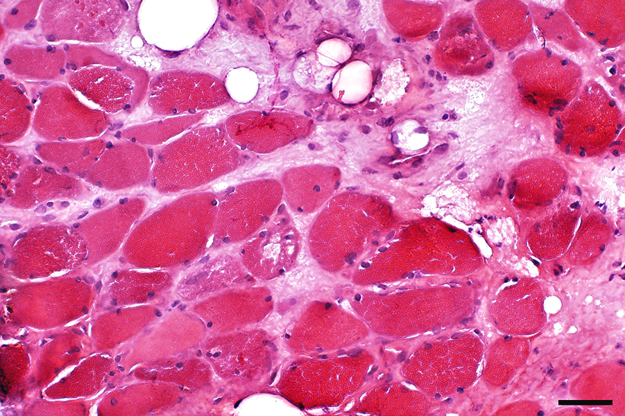
Regional pathology: Necrotic areas
All tissues are involvedMuscle fibers: Necrosis
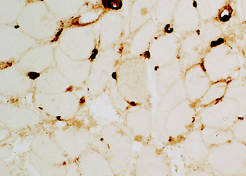
Capillaries: Necrosis
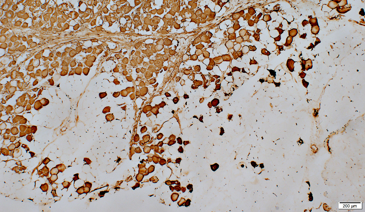
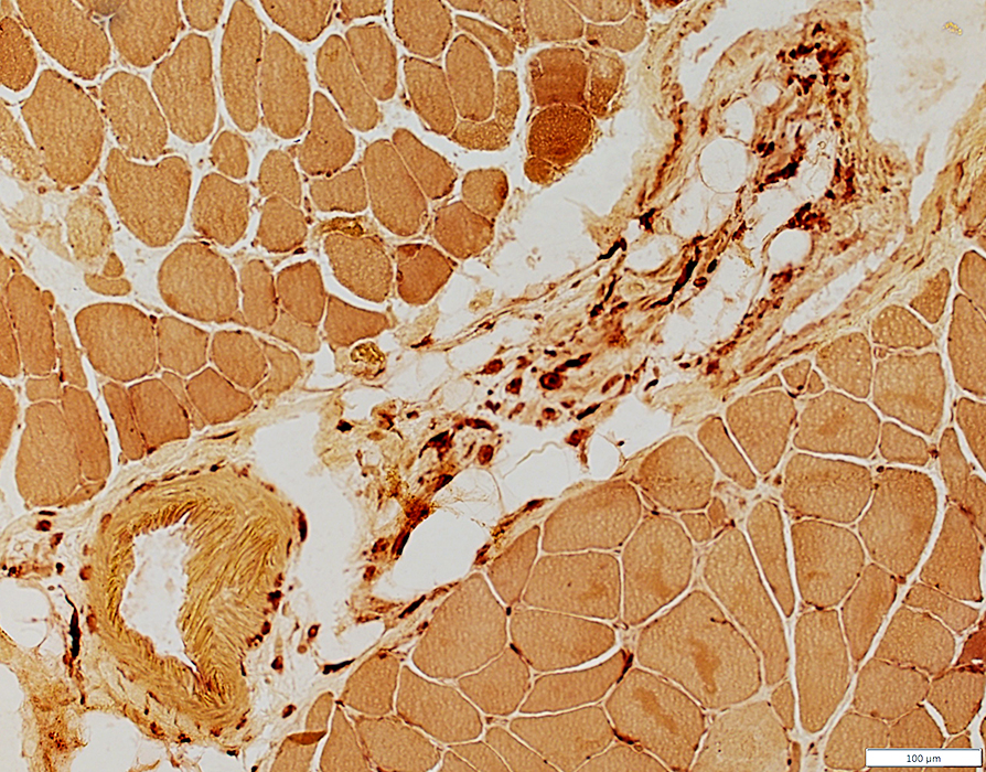
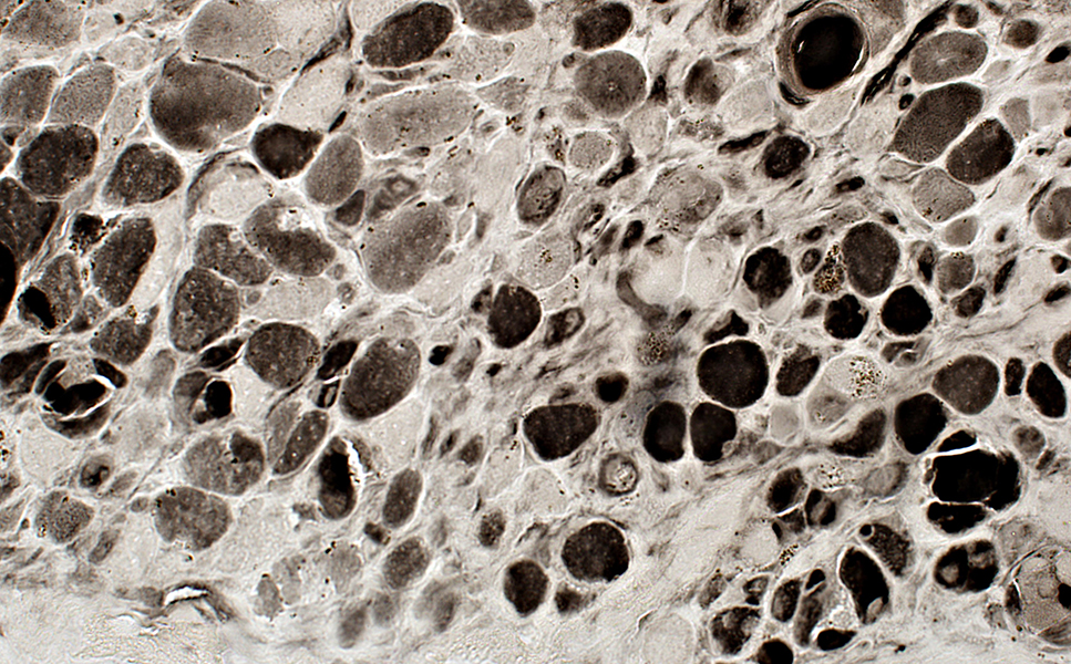
Perimysial & endomysial connective tissue: Abnormal structure & CA9 staining
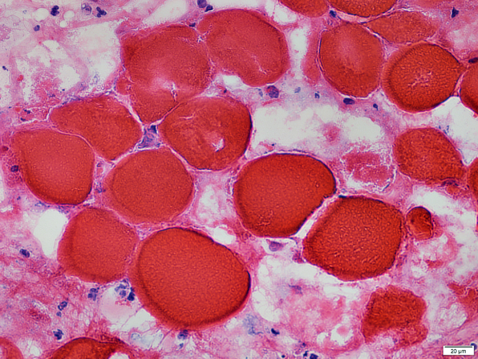
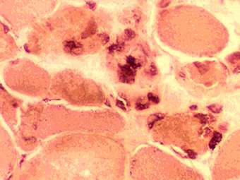
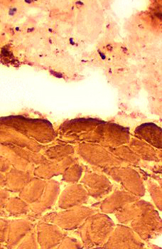
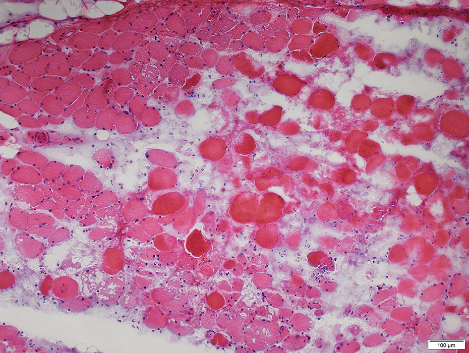
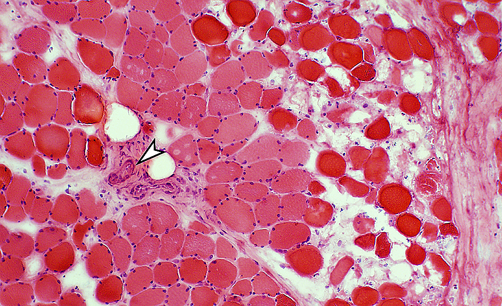 H&E stain |
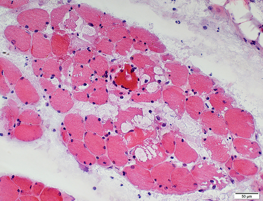
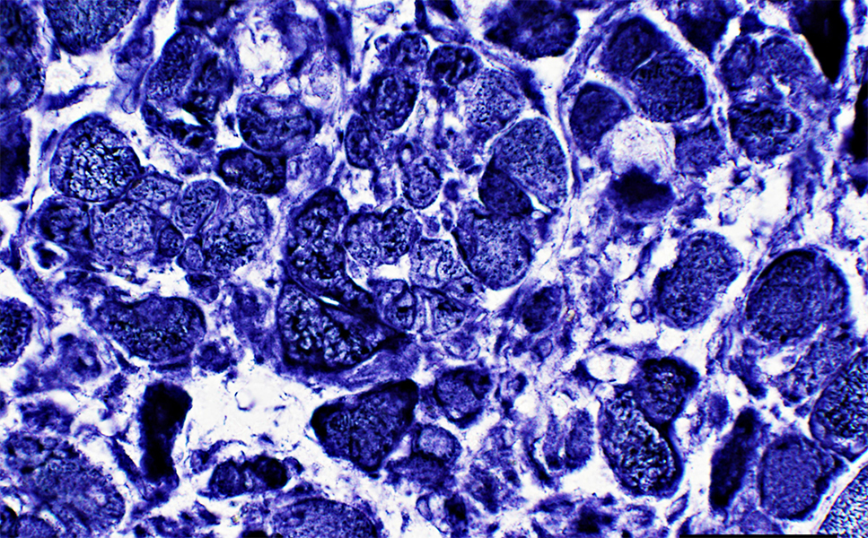
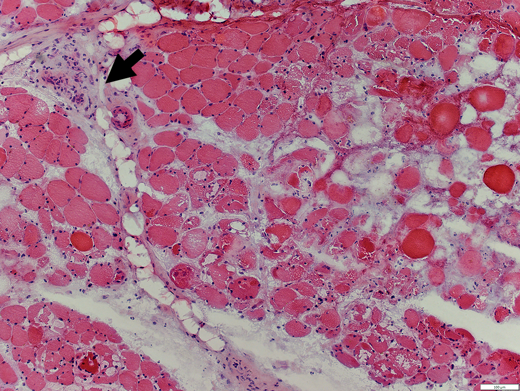
Clustered
Pale stained or Hypercontracted
No nuclear staining
None have macrophage infiltration
Perimysium (Far right): Pale stained
Vessel Bundles (Arteries & Veins; Arrows): Not contiguous with necrosis zone
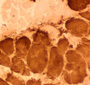
 H&E stain |
 H&E stain |
 H&E stain |
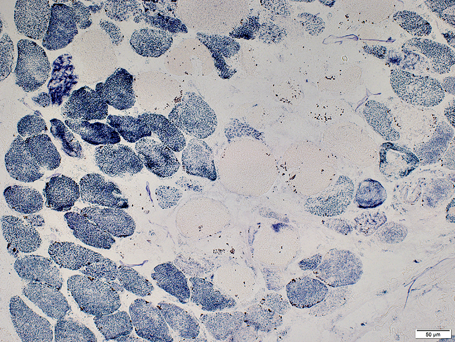
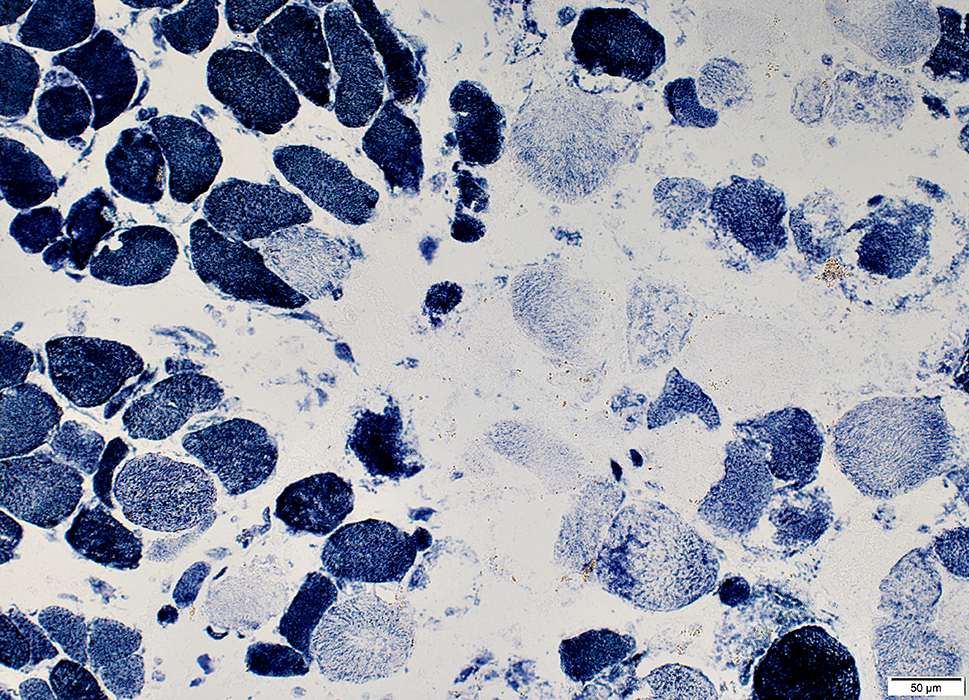
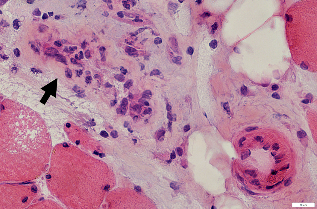
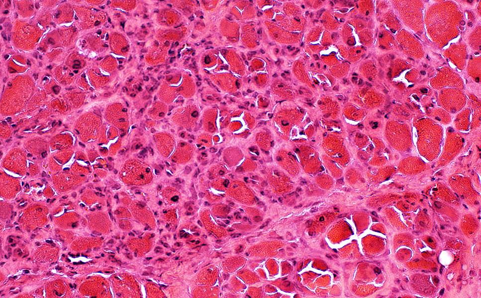
RIIM: Borderzone Necrosis
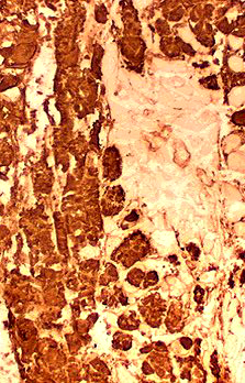
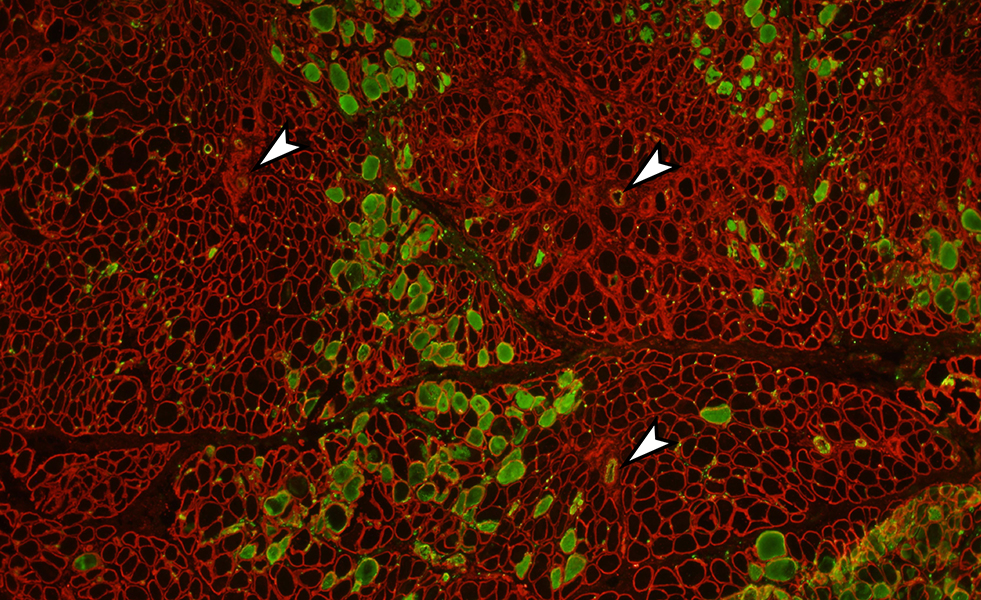
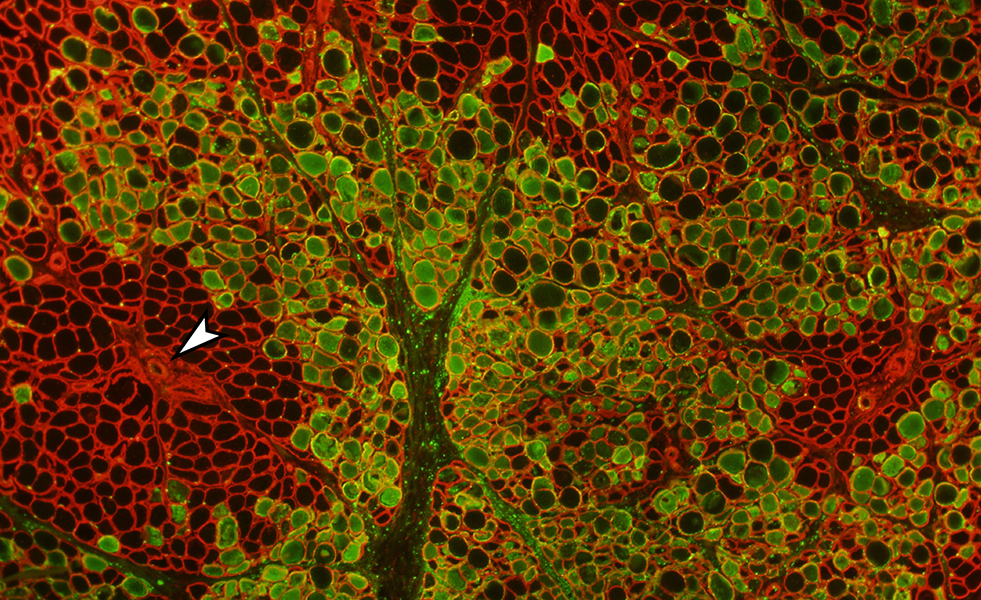 C5b-9 (Green) & Collagen IV (Red) stains Regional necrosis Zones of necrosis Surrounding but not neighboring vessels (Arrow) Do not respect fascicular borders Necrotic muscle fibers Clustered in borderzones between large arteries & veins (Arrows) Muscle fiber cytoplasm: Stains for C5b-9 complement (Green; Above) Connective tissue (Perimysium & endomysium) is also damaged CA9: Abnormal staining of perimysial & endomysial connective tissue & muscle fibers(Below) C5b-9 complement: Punctate deposition in perimysium (Above) Capillaries, endomysial: Necrotic & effaced Purlieu zone: Muscle fibers between vessels & necrotic zone have no C5b-9 deposition Vessels (Arrows): Inside purlieu zone & Distant from necrotic zone |
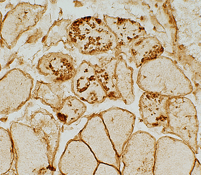
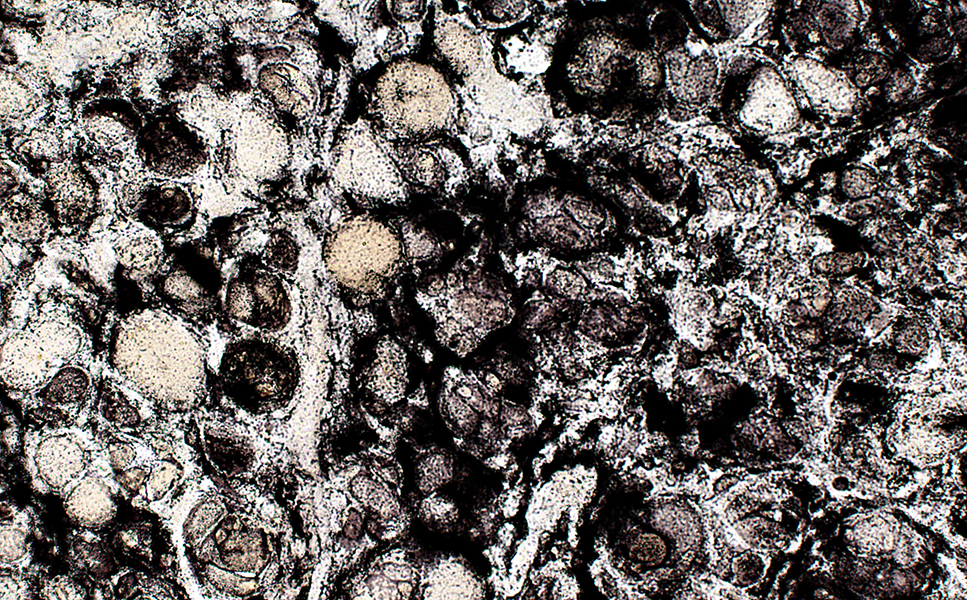
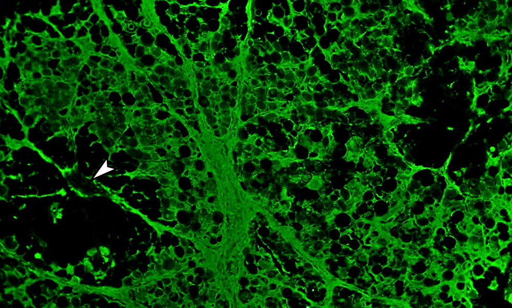 CA9 stain CA9 staining in necrotic zone: Abnormal on perimysium & endomysium |
 C5b-9 (Green) & Collagen IV (Red) stains Borderzone regions of necrosis: No relation to fascicular borders
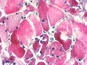
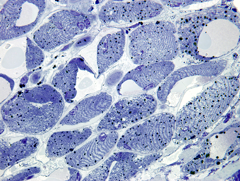
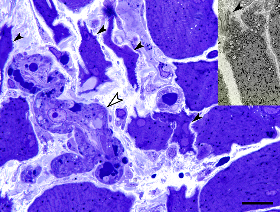
General: Most in similar, early stage, of necrosis Pale staining or Hypercontracted No nuclear staining Edges: Some are invaded by phagocytic cells
Necrotic muscle fibers: Absent SDH staining
Necrotic muscle fibers: Reduced or Absent NADH staining
Muscle fibers in Necrotic zones Terminal components of complement ((C5b-9; Membrane attack complex) C5b-9: Deposited in cytoplasm of clusters of necrotic muscle fibers
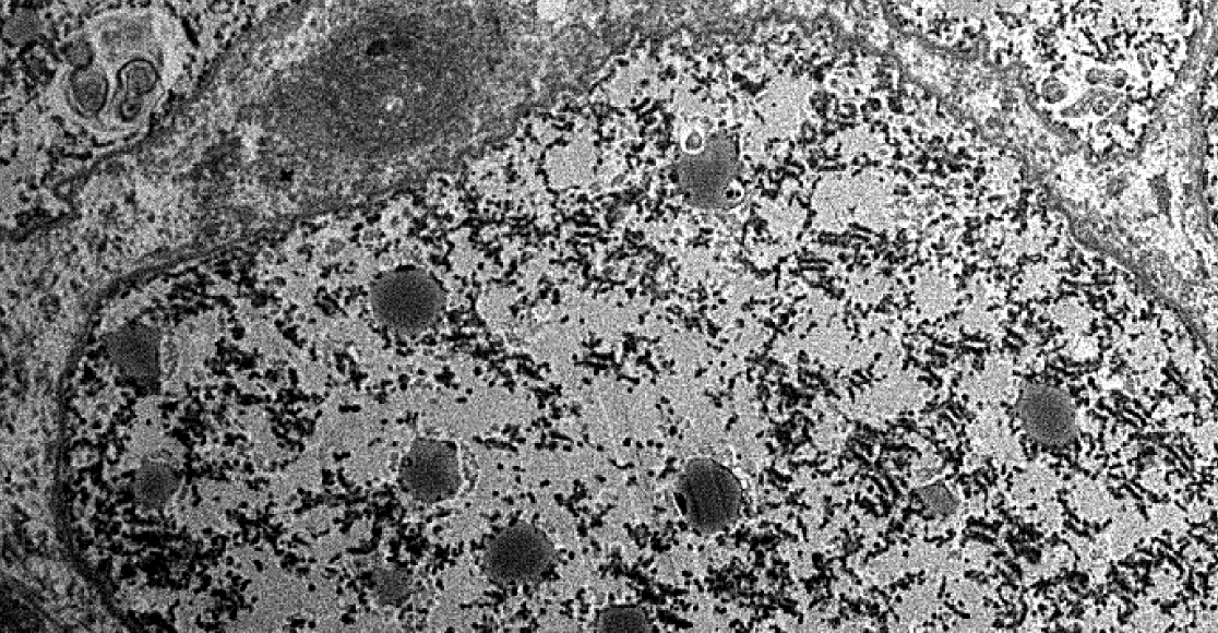
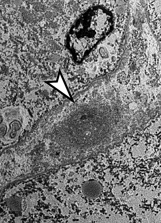
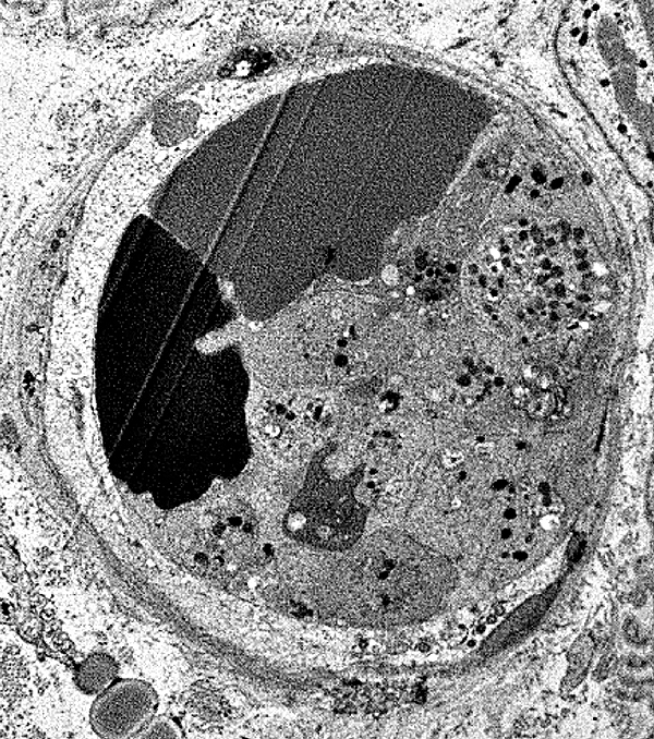
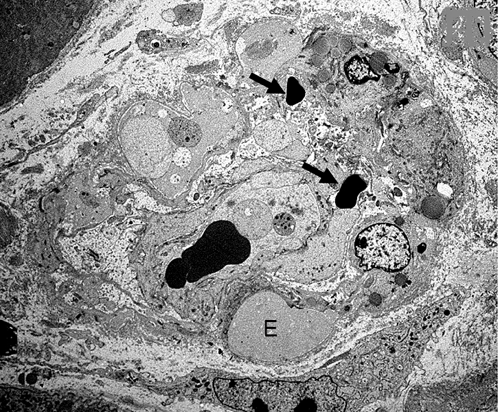
RIIM: Capillaries
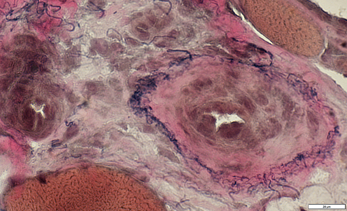
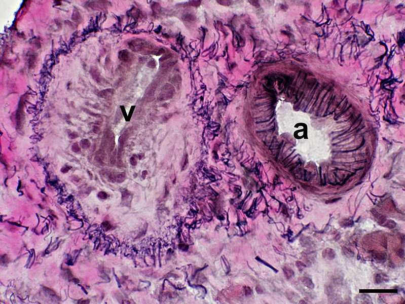
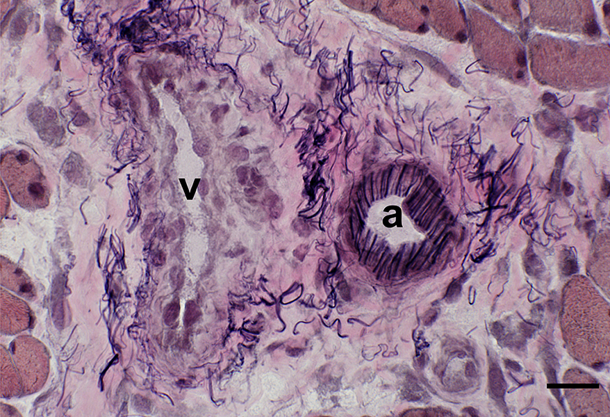
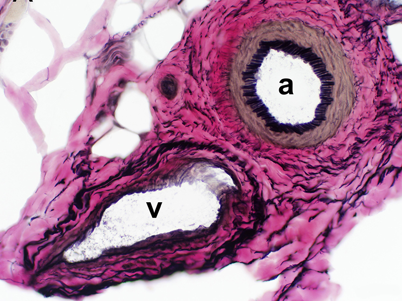
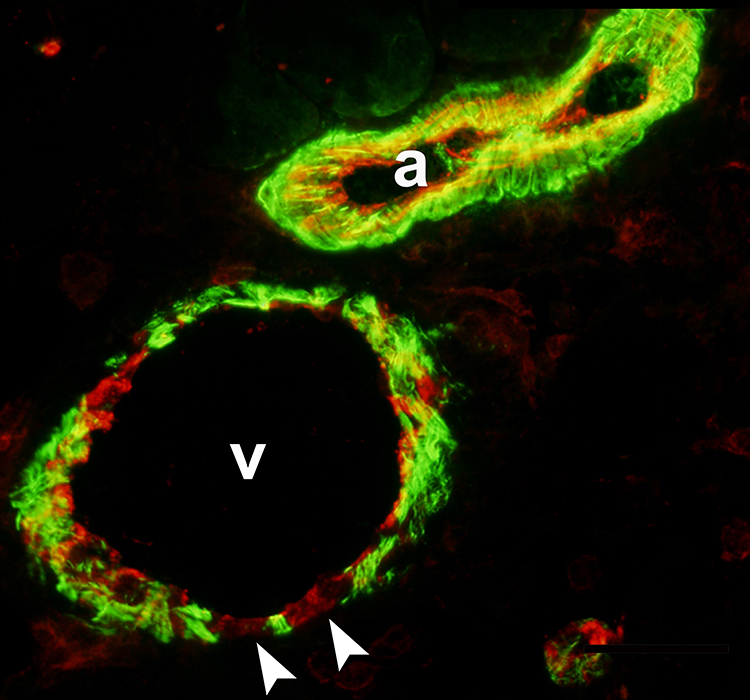
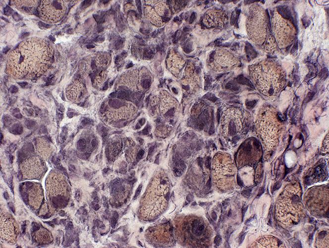
RIIM: Larger Vessels
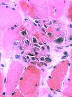
Wall is pale, thickened & cellular
Surrounded by histiocytes Perimysial connective tissue Contains histiocytic cells
Wall is pale, thickened & cellular Endothelial cells: Large
Regional Ischemic Immune Myopathy
Varied size Internal architectureL Irregular or Vacuoles Aggregates: Caveolin & LC3 aggregates
Cytoplasmic Aggregates: Caveolin & LC3
Regional Ischemic Immune Myopathy
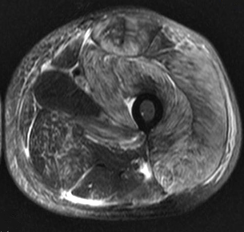
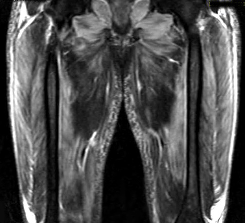
Regional Ischemic Immune Myopathy (RIIM): MRI
Muscle atrophy: R Vastus
Return to Neuromuscular Home Page Return to Inflammation Return to Inflammatory myopathies 10/6/2025 | |||||||||||||||||||||||||||||||||||||||