Cystinosis
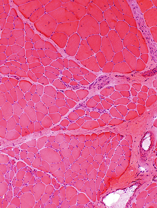 H&E stain |
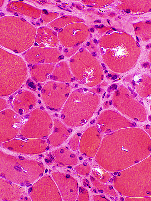 H&E stain |
|
Muscle fibers: Varied sizes
|
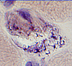
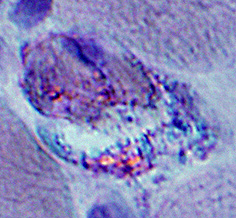
Muscle fibers: Vacuoles, irregular shaped; Large nuclei
|
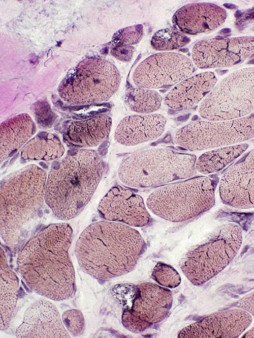 VvG stain |
Vacuoles in muscle fibers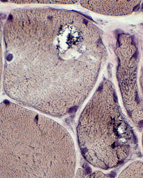 VvG stain |
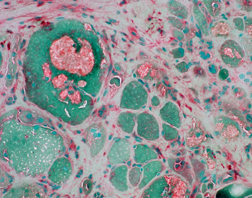
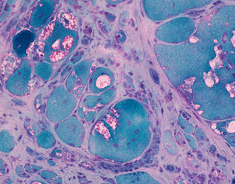 Gomori trichrome stain; From C Cai |
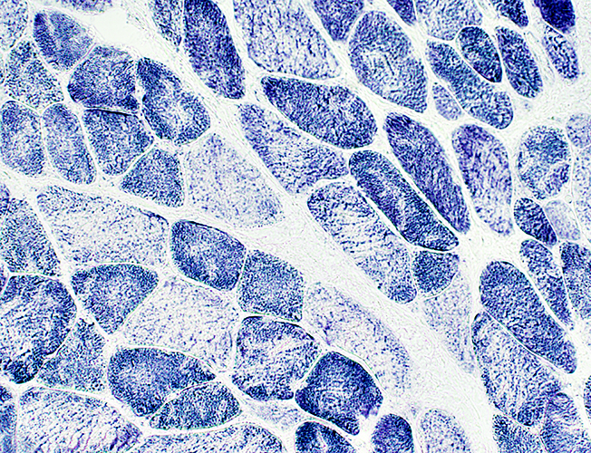 NADH stain Internal architecture Irregular Linearization in pale stained fibers |
|
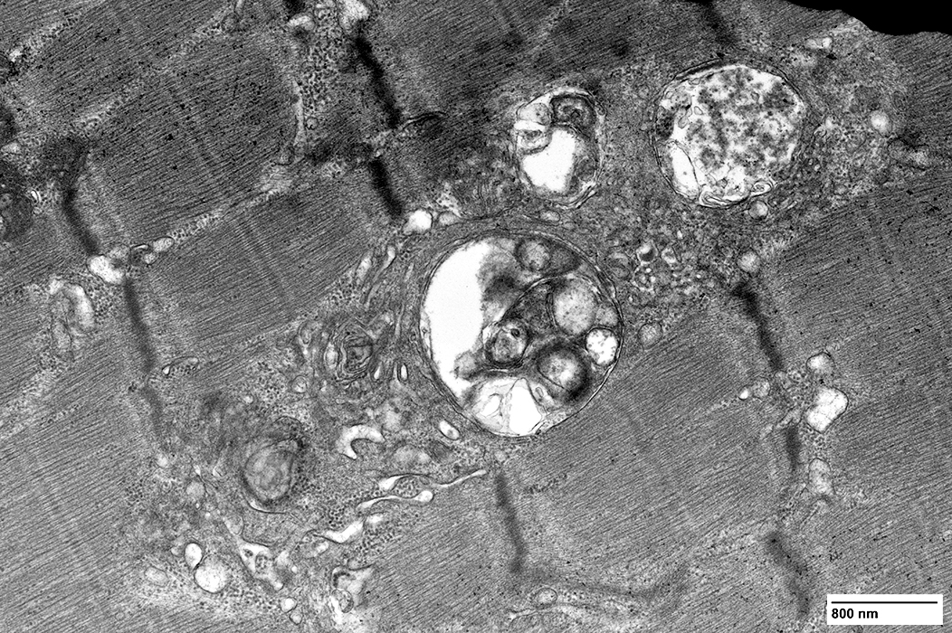
Vacuoles: Contain Basophilic granular debris Red-green birefringent material 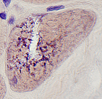 Congo red stain |
 Congo red stain |
  Congo red stain |
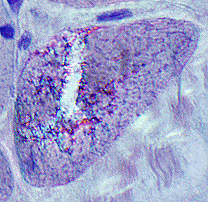
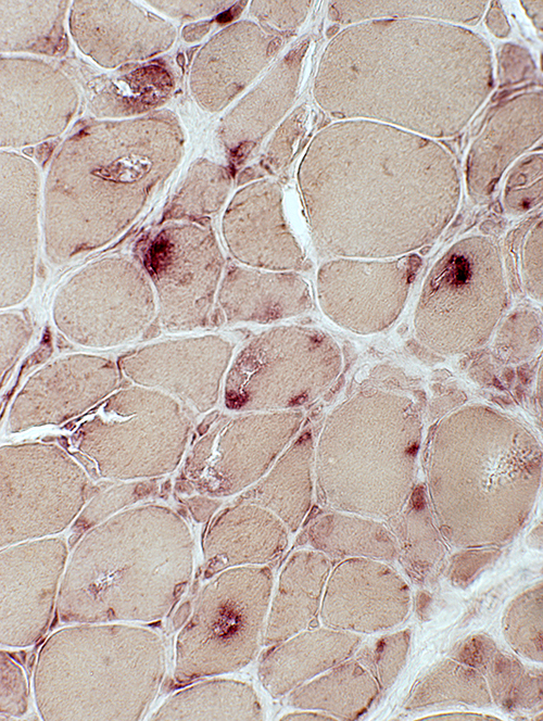
AMPDA positive agggregates: Some associated with vacuoles
 AMPDA stain |
 AMPDA stain;/ From C Cai |
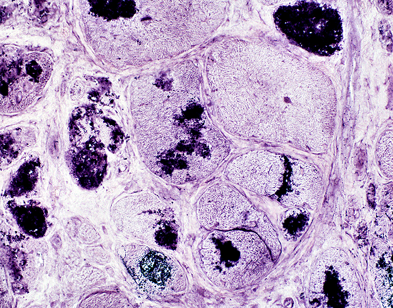
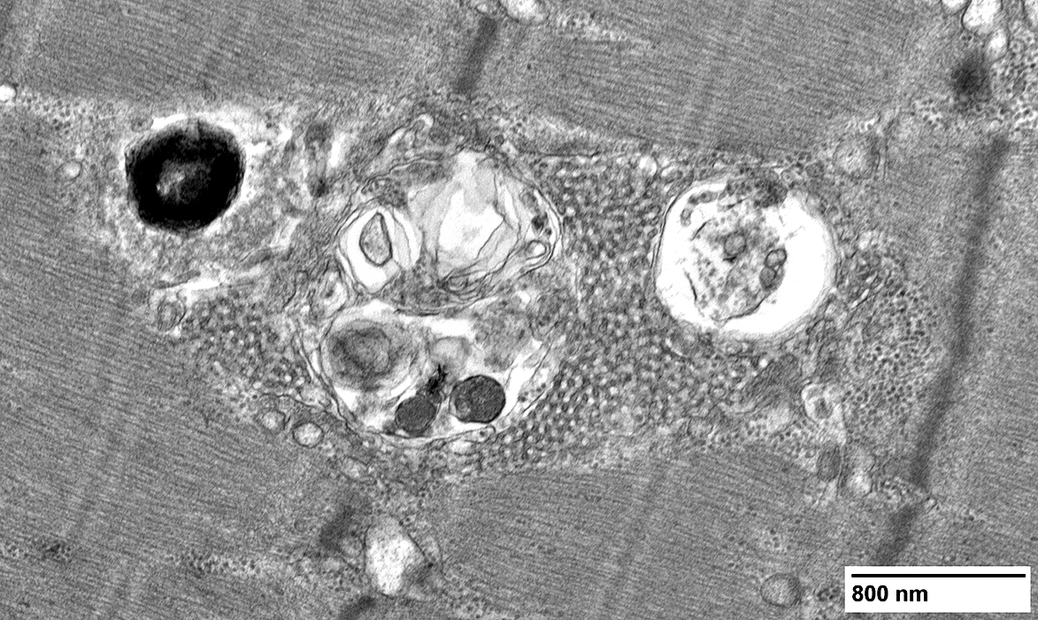
Acid phosphatase positive material: Some associated with, or within, vacuoles
 Acid phosphatase stain |
 Acid phosphatase stain; From C Cai |
Immature muscle fibers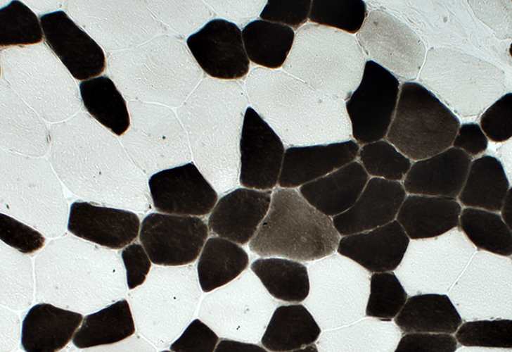 ATPase pH 4.3 stain |
 From C Cai |
 From C Cai |
 From C Cai |
 From C Cai |
Return to Cystinosis
Return to Muscle biopsies
10/8/2019