Intravascular Lymphoma (Angiotropic B-cell)
|
Brain Muscle |
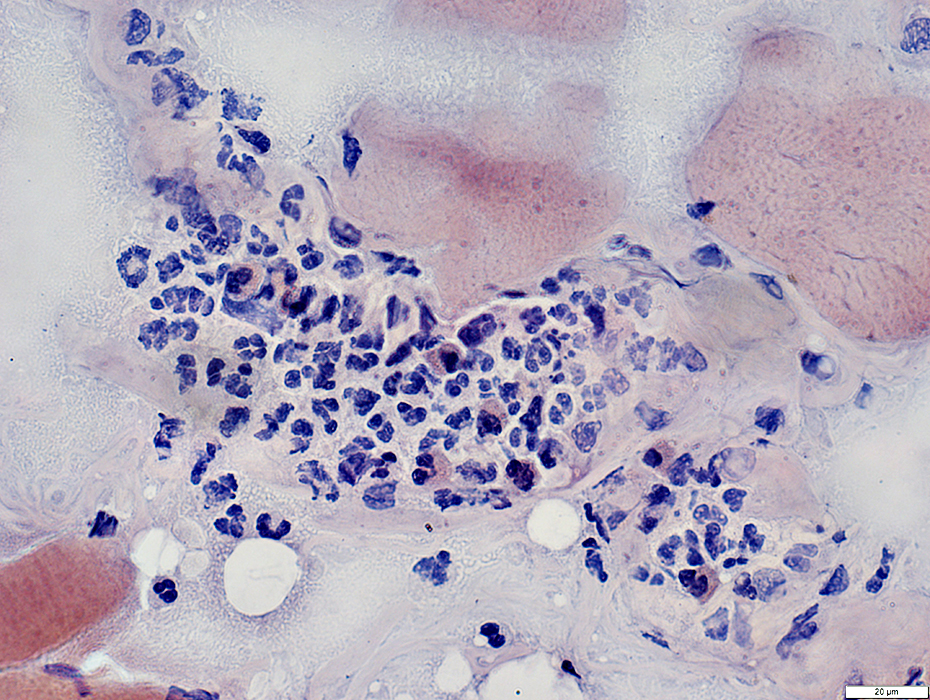
IV Lymphoma: Muscle
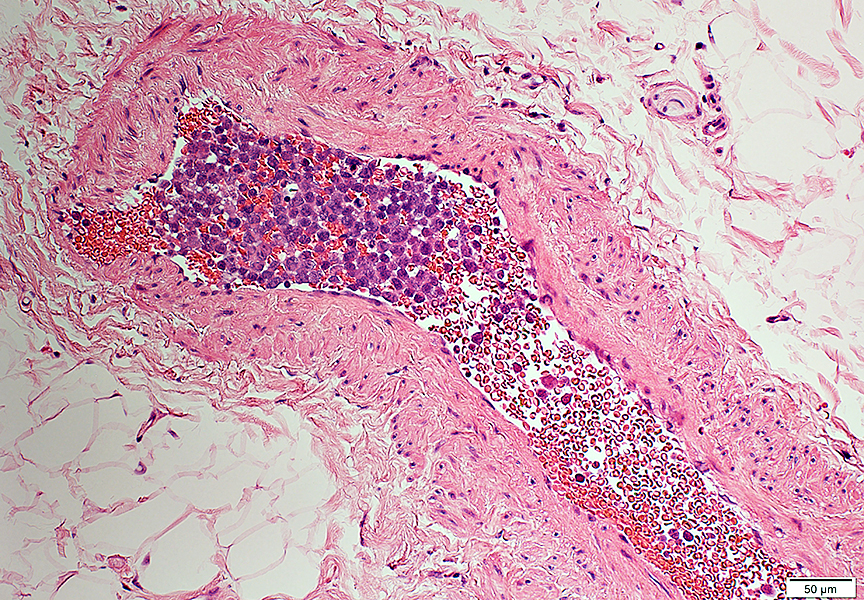 H&E stain |
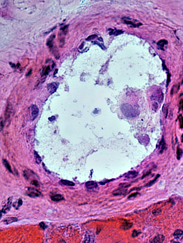
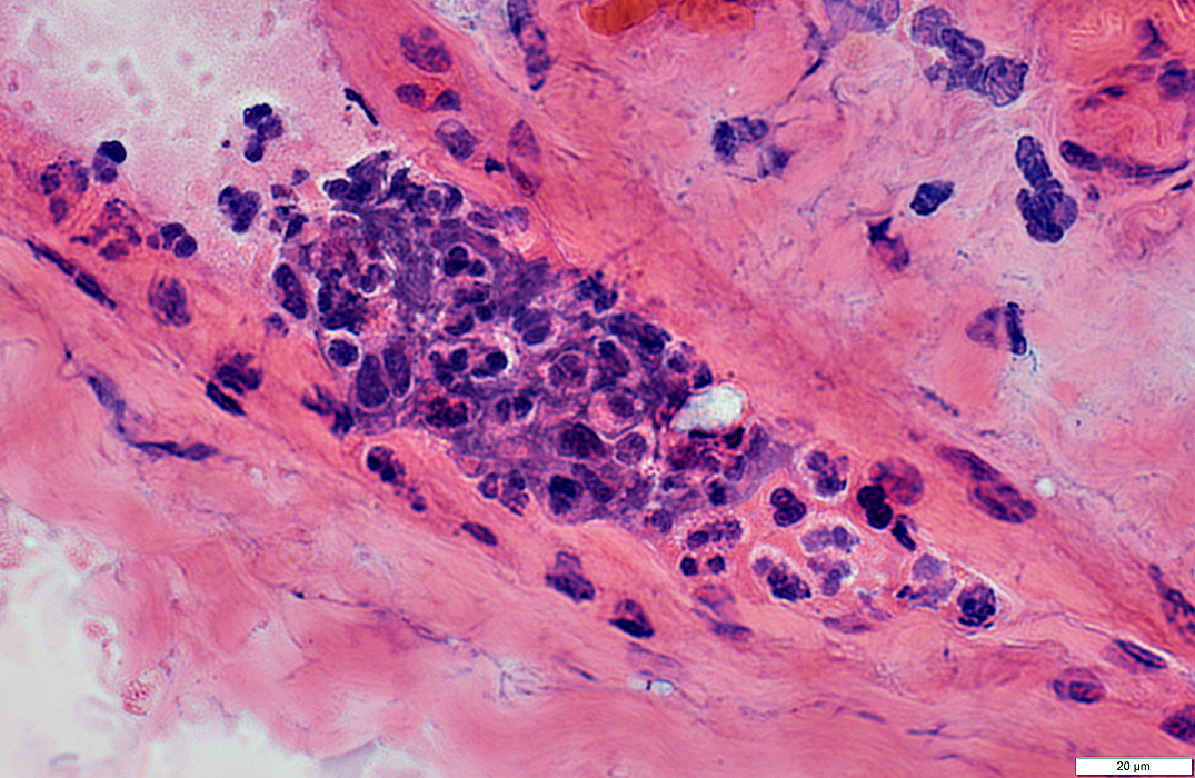
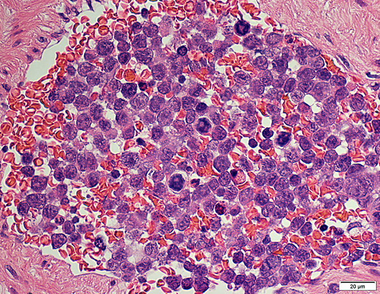
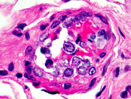
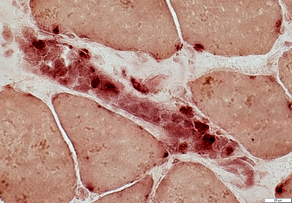
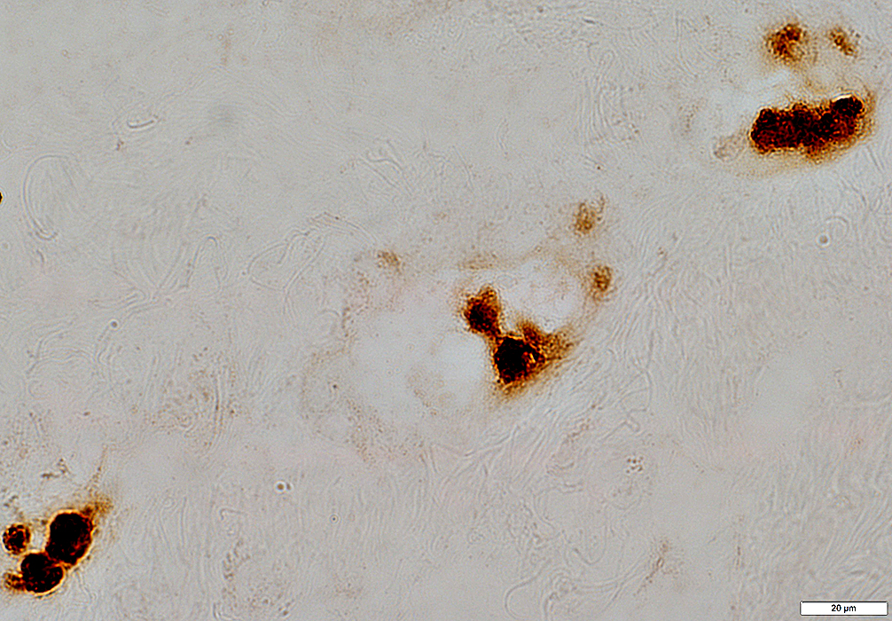
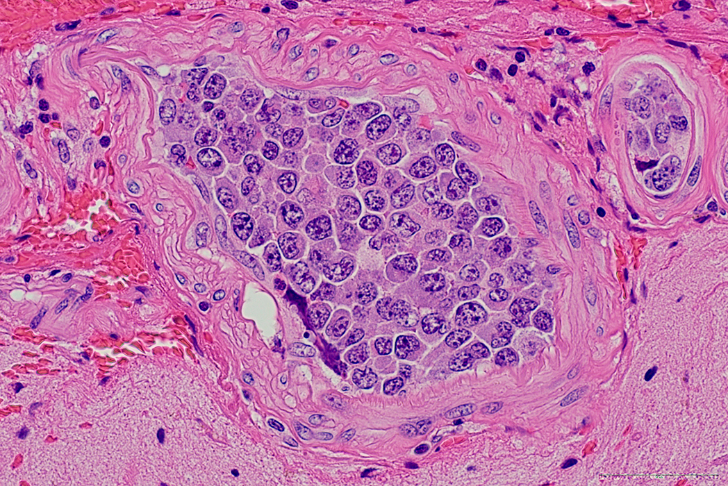
Cells aggregated in regions of vessels, often adhering to wall
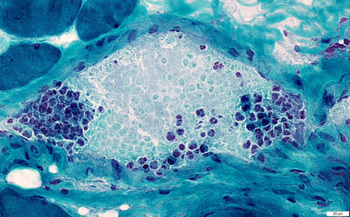 Gomori trichrome stain |
 H&E stain 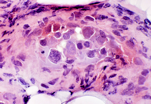 Lymphoma cells in vessels |
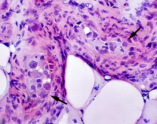
 H&E stain |
|
 H&E stain |
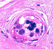 |
Large, pleomorphic neoplastic B-cells: In lumens of small & intermediate sized vessels 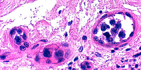
|
|
 H&E stain | ||
 H&E stain |
 Congo red stain |
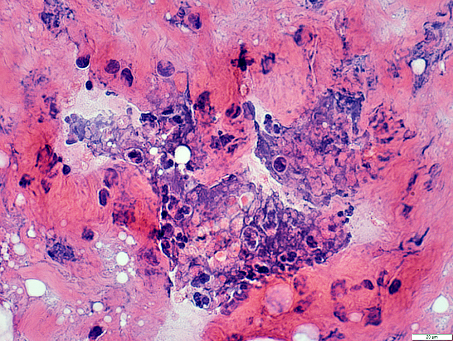
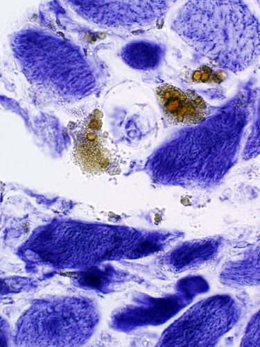 NADH stain Hemosiderin (Brown) in endomysium
|
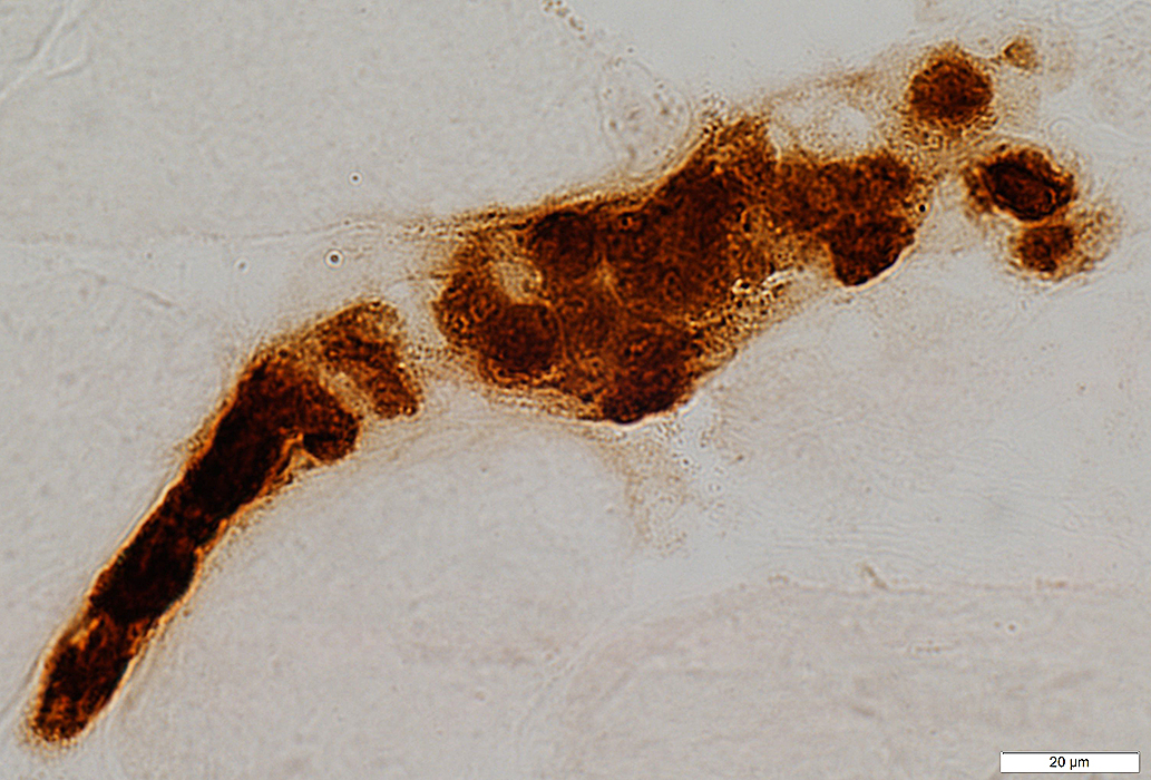
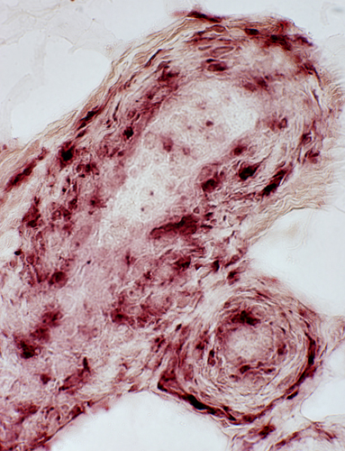 Acid phosphatase stain Histiocytic cells in vessel walls
|
 Intravascular cells in endoneurium stain for Leukocyte common antigen 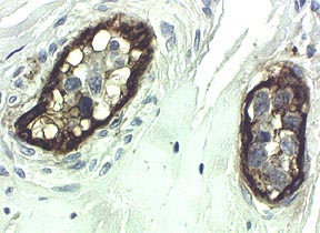 Vessel walls stain for Ulex lectin |
Intravascular lymphoma cells
Mild staining with acid phosphatase
 Acid phosphatase stain |
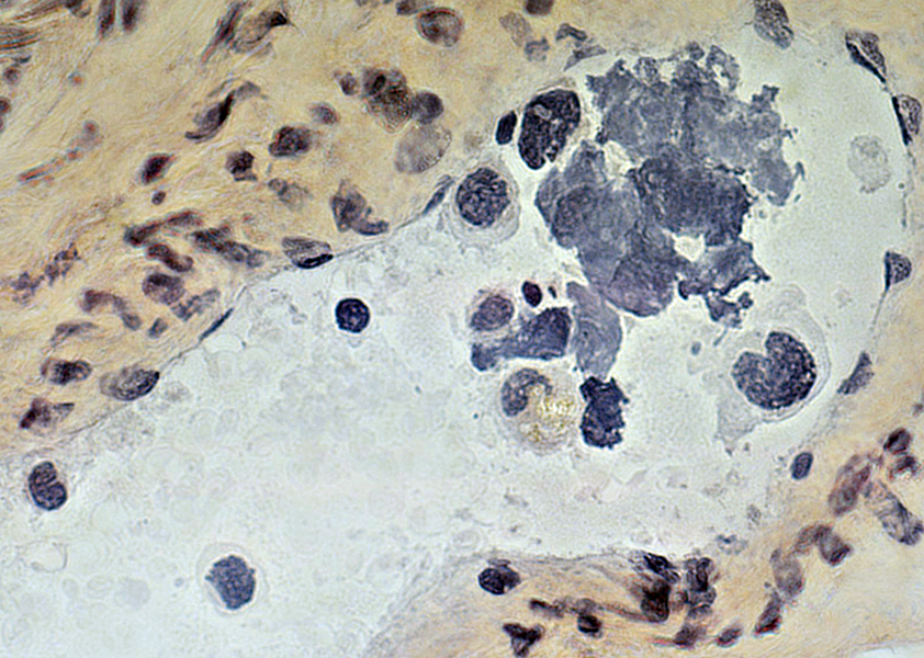
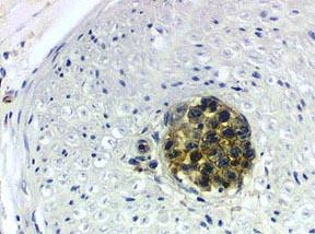
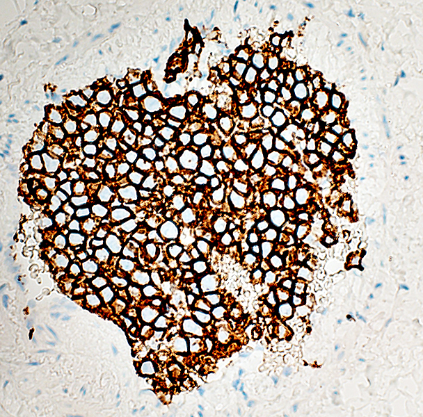
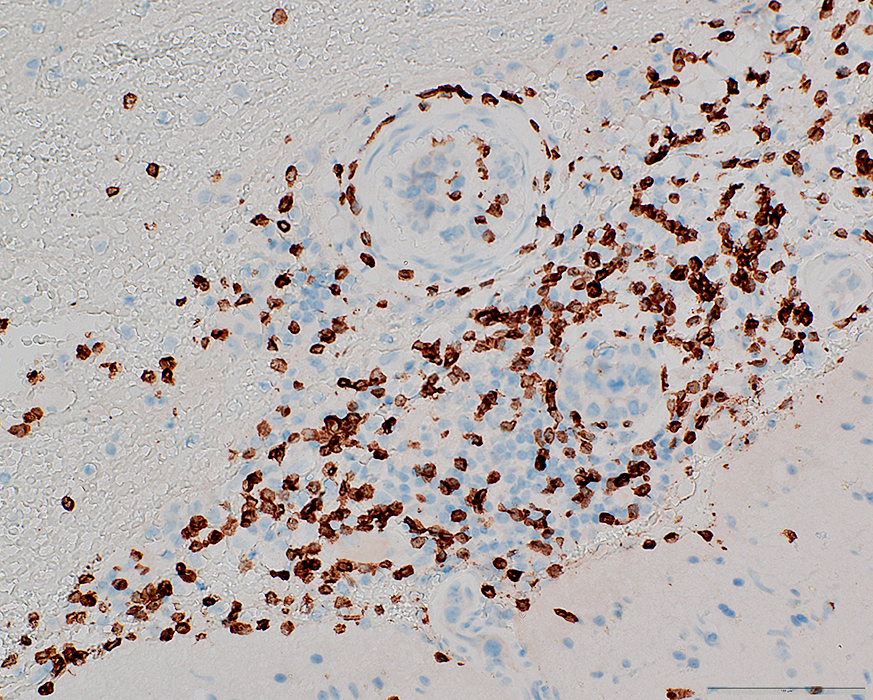
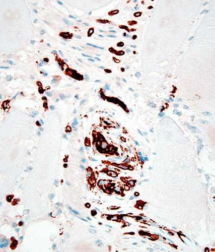 CD20 stain From: A Perry CD20+ cells in vessels & connective tissue
|
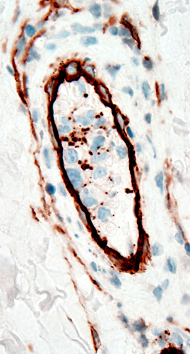 CD20 stain CD20+ cells in vessel walls
|
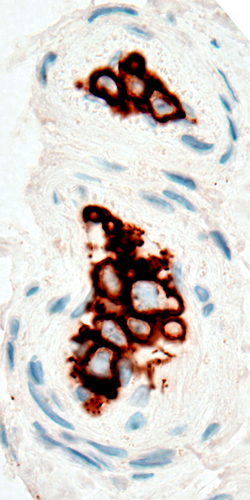 CD20 stain CD20+ cells in vessel lumens |
|
 CD20 stain |
 CD20 stain |
Intravascular B-cell lymphoma cells stain for: MUM-1 & BCL6
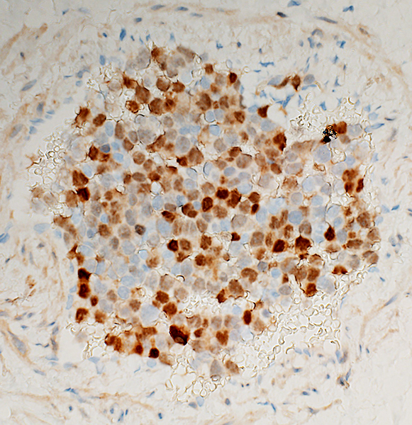 MUM-1 stain |
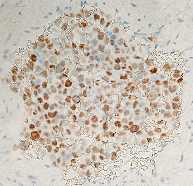 BCL6 stain |
Intravascular Lymphoma, B-cell: Brain
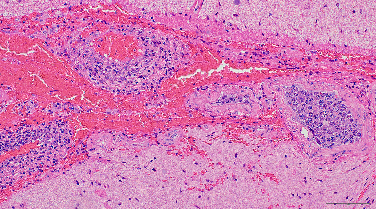 H&E stain From: R Bucelli |
Intravascular: Mostly B-cells (Right & Below)
Vessel wall: CD3 cells & Histiocytes
 H&E stain From: R Bucelli |
Intravascular: Mostly B-cells (Above)
Vessel wall: CD3 cells & Histiocytes (Below)
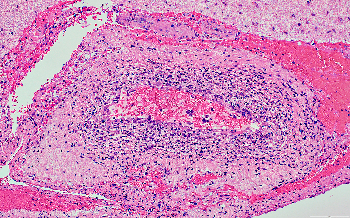 H&E stain From: R Bucelli |
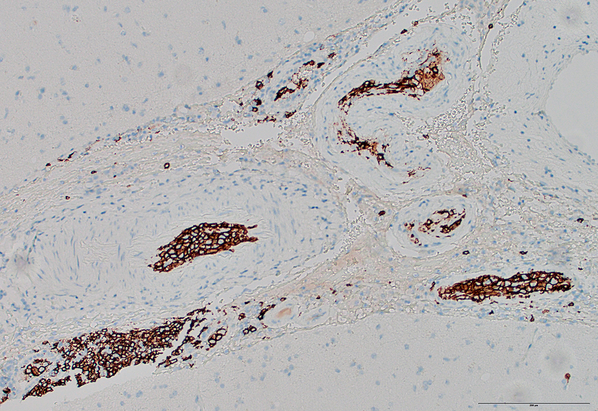 CD20 stain From: R Bucelli |
Intravascular: Mostly CD20+ B-cells (Above & Below)
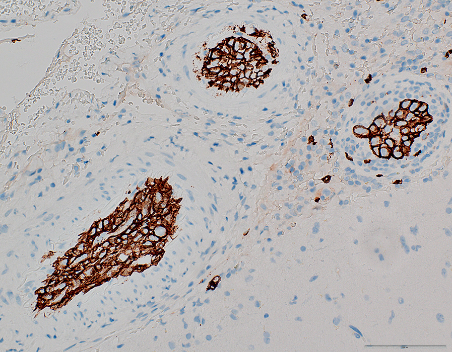 CD20 stain From: R Bucelli |
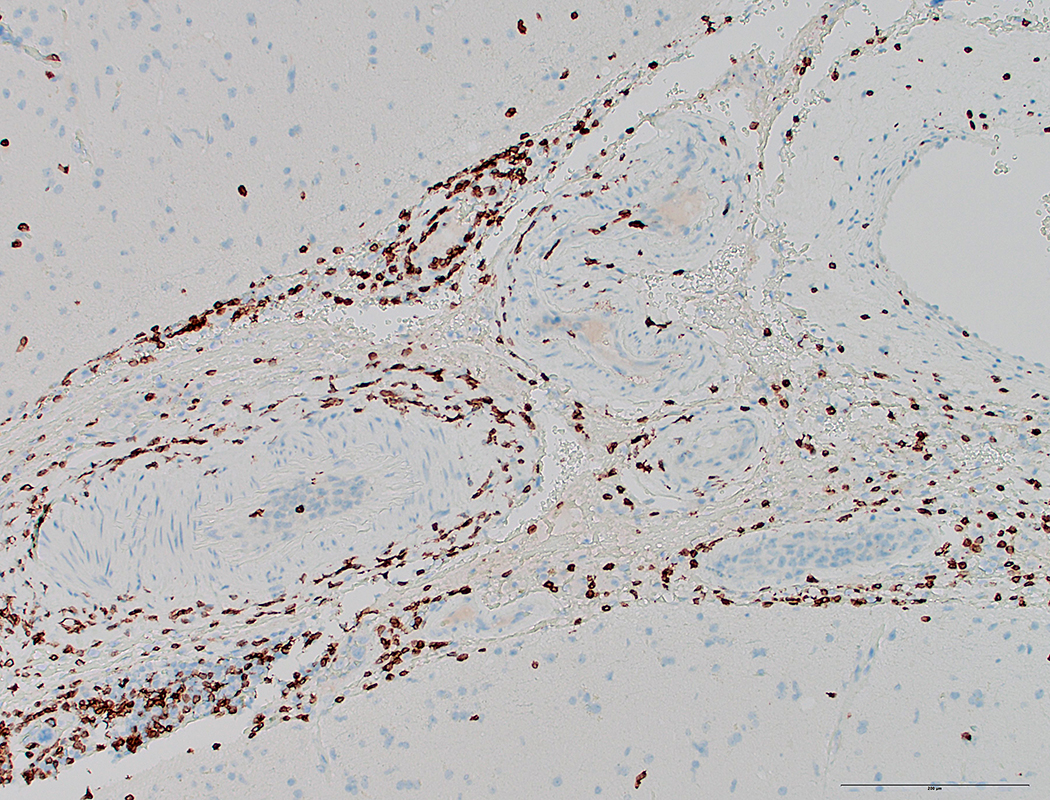 CD3 stain From: R Bucelli |
Vessel wall: Mostly CD3+ Lymphocytes (Above & Below)
 CD3 stain From: R Bucelli |
Return to: IV Lymphoma
Return to Neuromuscular Home Page
2/8/2021