SEPN1 (CMYO3)
Muscle Pathology
- Muscle fibers
- Fiber sizes: Varied
- Internal nuclei
- Internal architecture: "Minicores"; Aggregates (LC3)
- Fiber types: Scattered immature 2C fibers; 1 predominance, mild
- Endomysial connective tissue: Normal to mildly increased
- Young child: No prominent pathology
25 year old female
 H&E stain |
Internal nuclei: some muscle fibers
Endomysial connective tissue: Midly increased
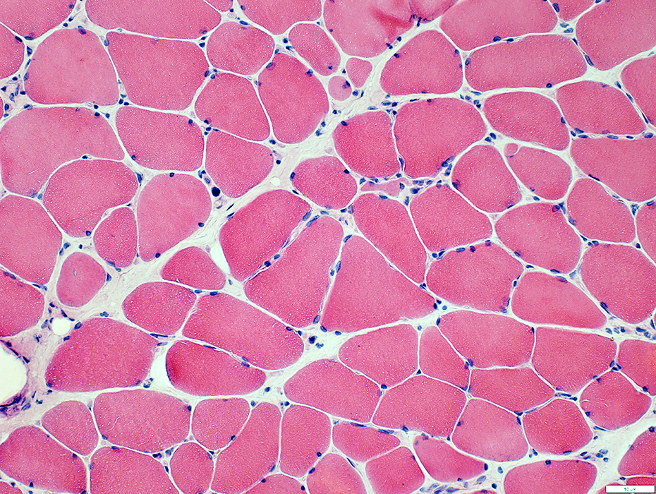 H&E stain |
 Gomori trichrome stain |
Endomysial connective tissue: Midly increased
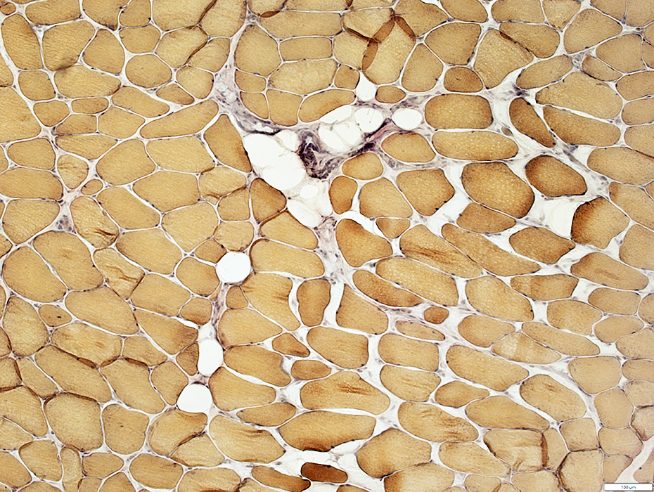 VvG stain |
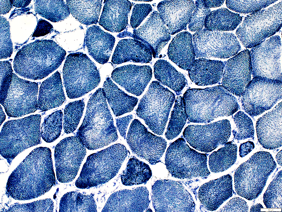 NADH stain |
Internal clear/pale regions with irregular borders
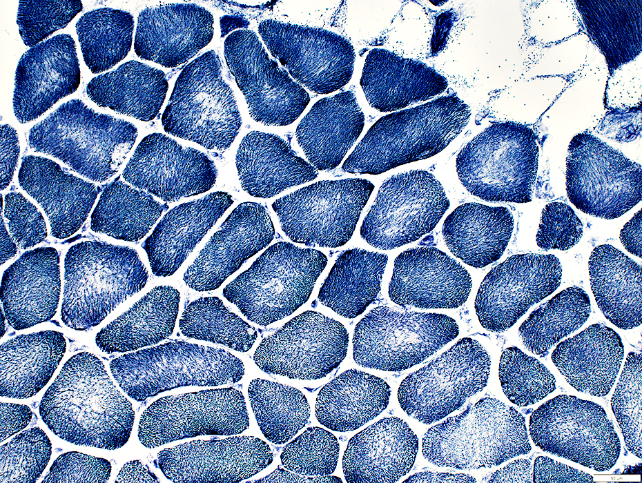 NADH stain |
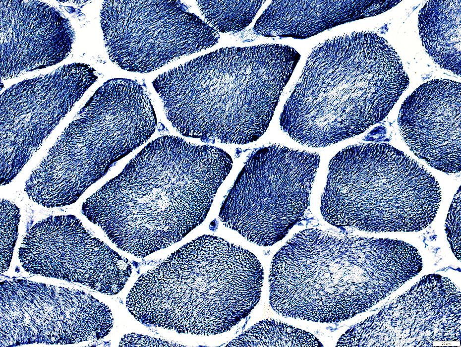 NADH stain |
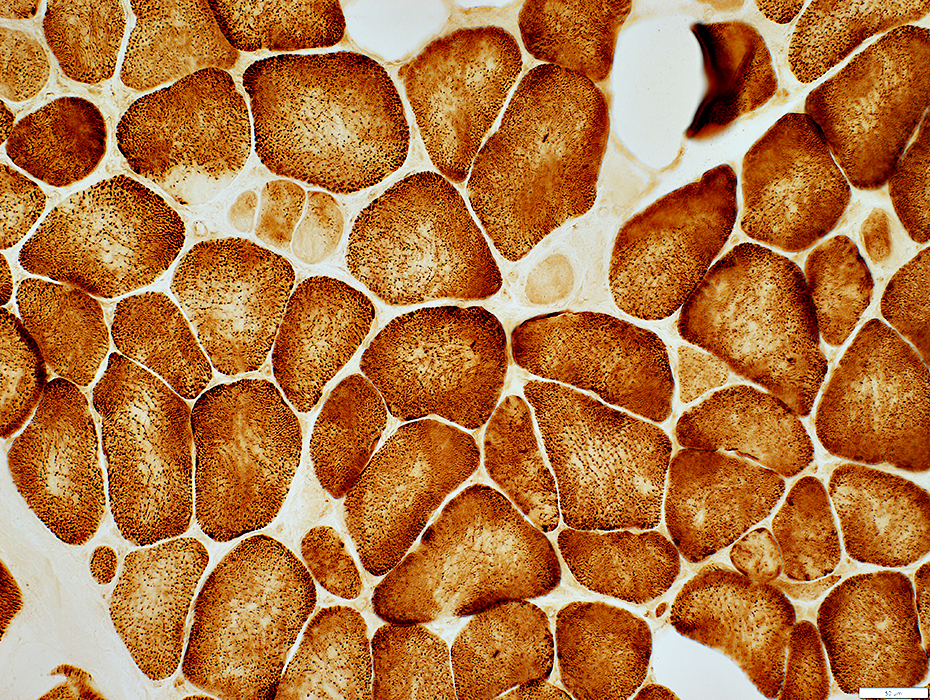 Cytochrome oxidase stain |
Internal clear/pale regions with irregular borders
 Cytochrome oxidase stain |
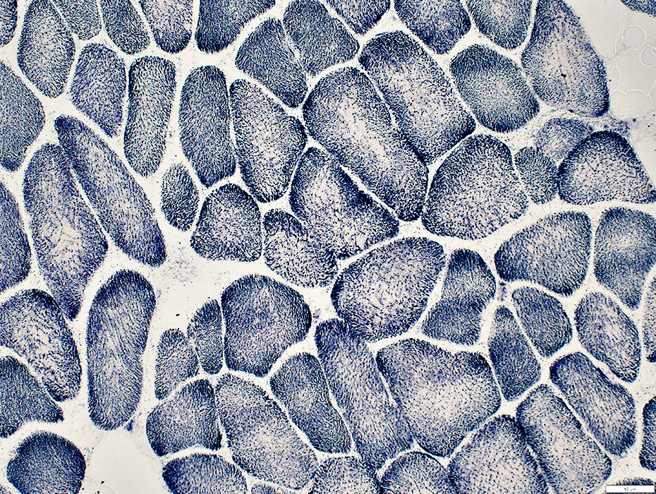 SDH stain |
 ATPase pH 4.3 stain |
 ATPase pH 4.3 stain |
 ATPase pH 9.4 stain |
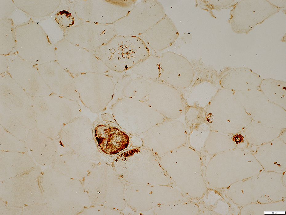 LC3 stain |
 LC3 stain |
 UEA I stain |
2 year old female
 H&E stain |
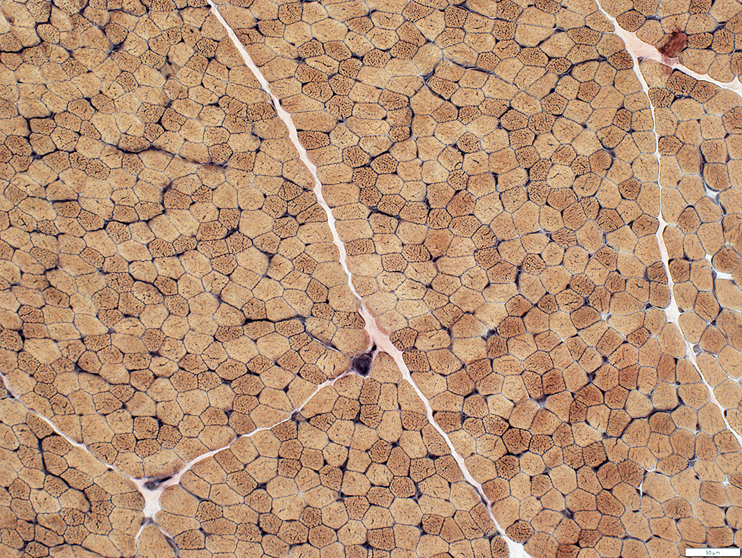 VvG stain |
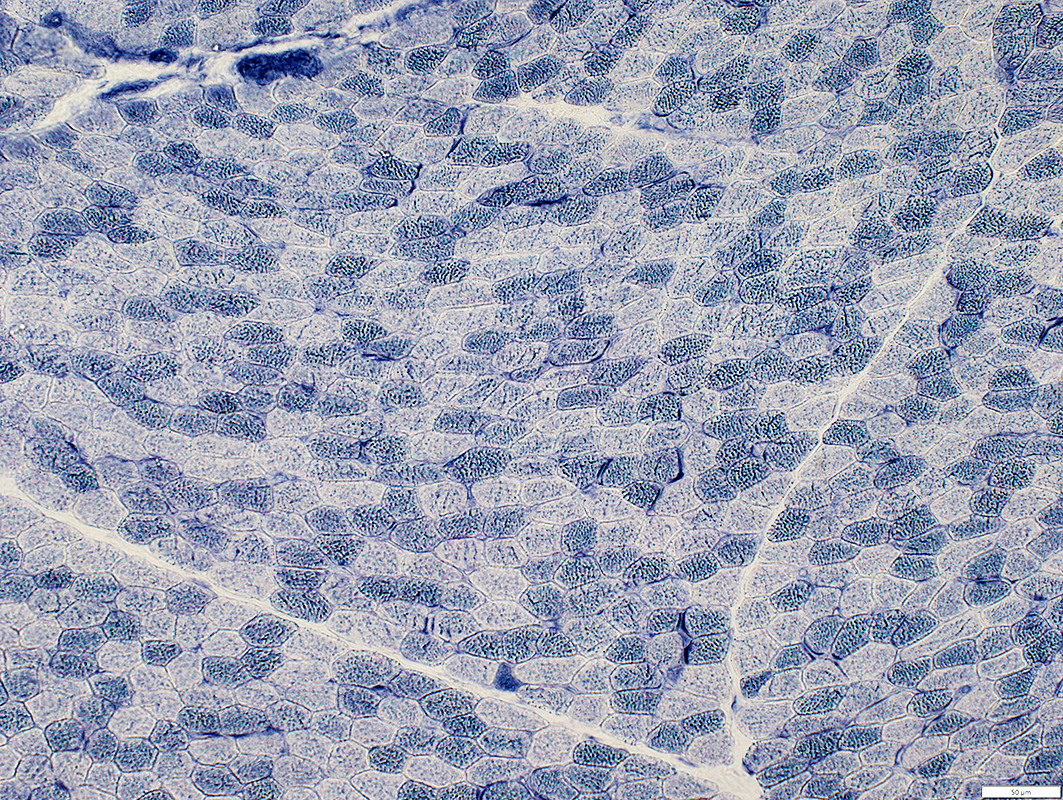 NADH stain |
 H&E stain |
 VvG stain |
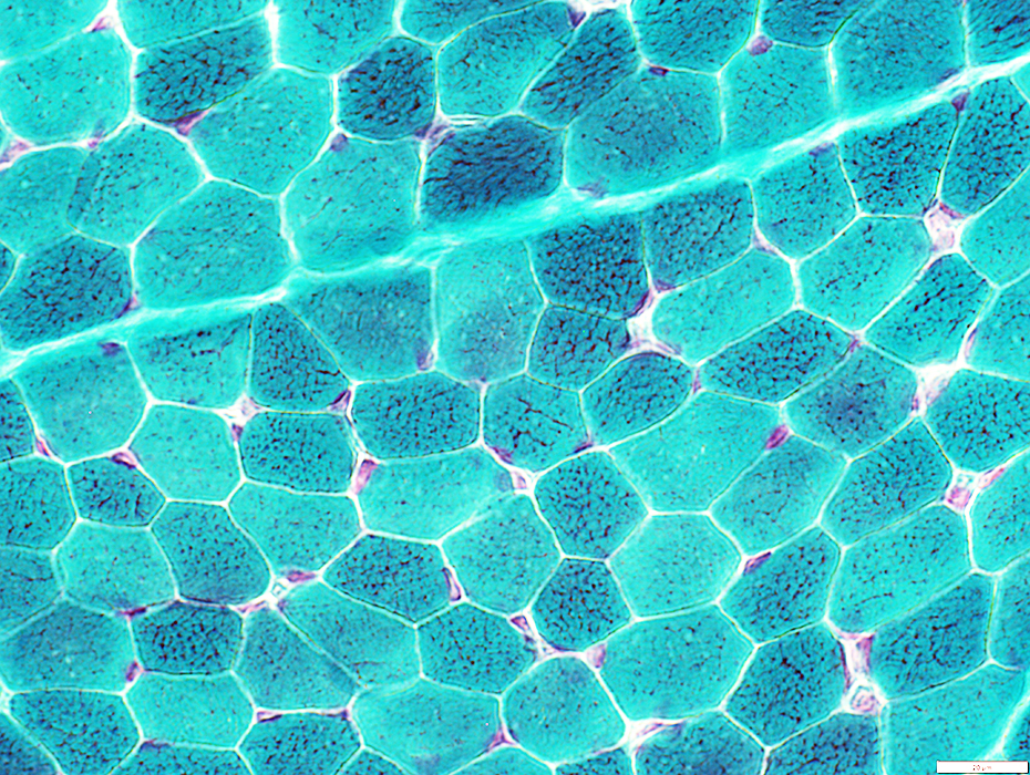 Gomori trichrome stain |
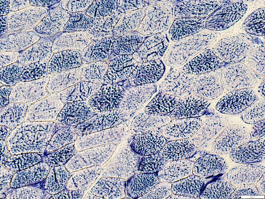 NADH stain |
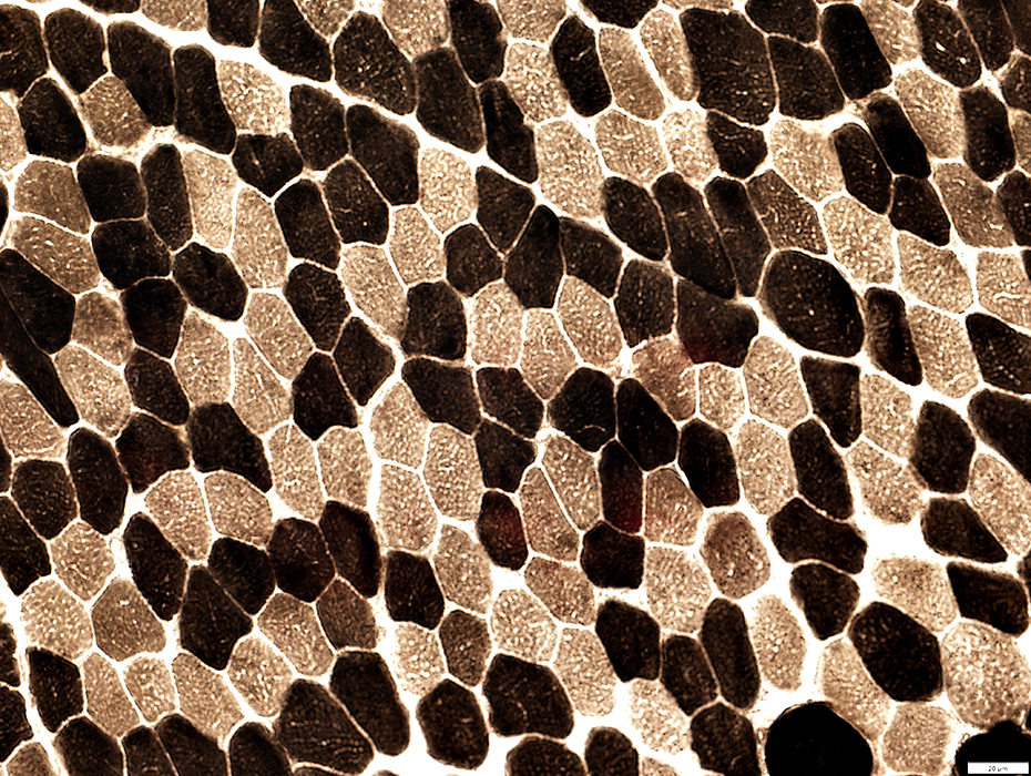 ATPase pH 9.4 stain |
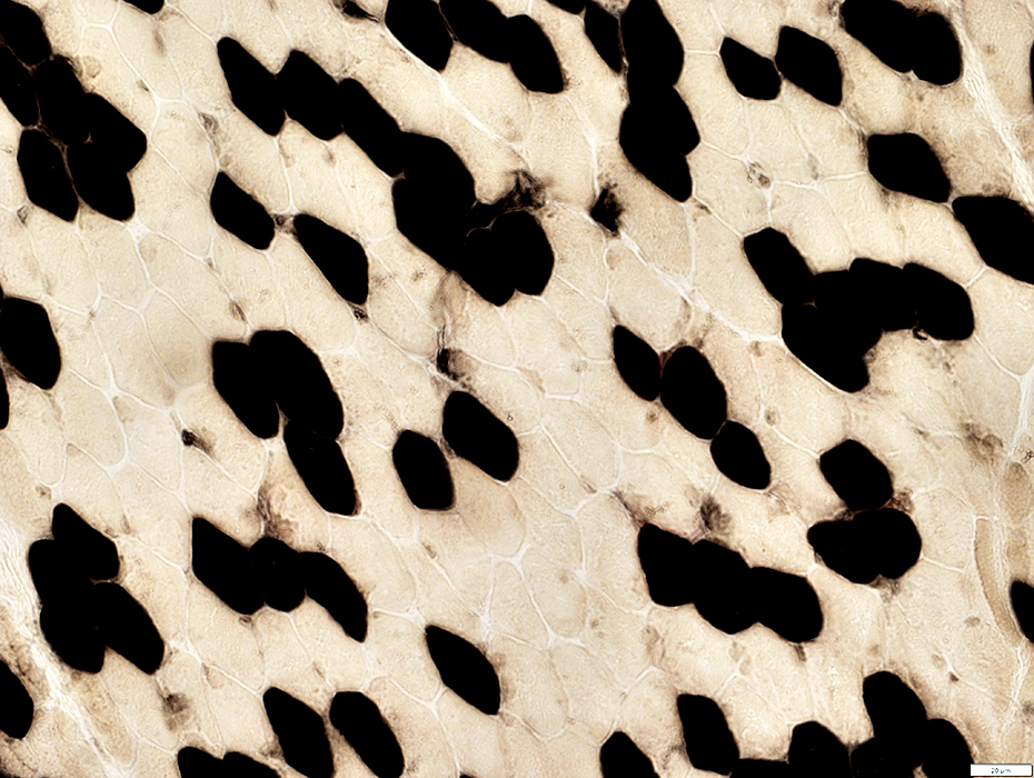 ATPase pH 4.3 stain |
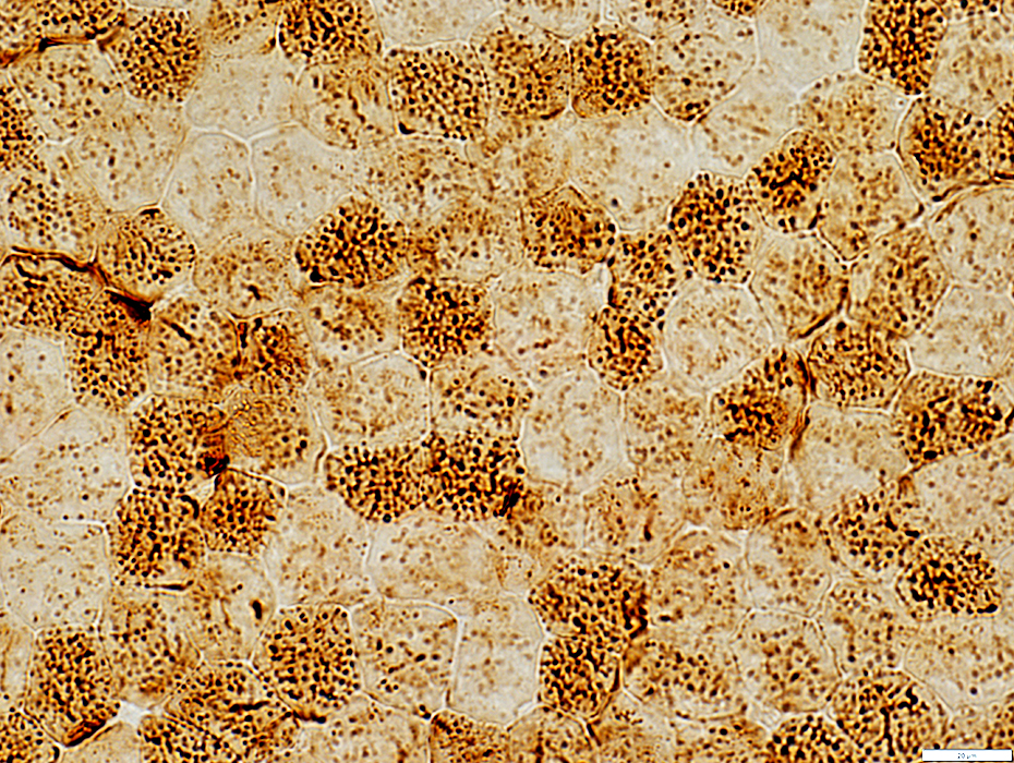 Cytochrome oxidase stain |
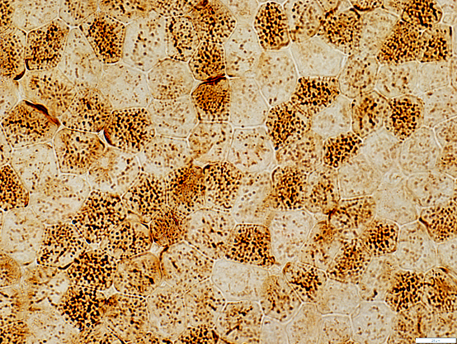 Cytochrome oxidase stain |
 SDH stain |
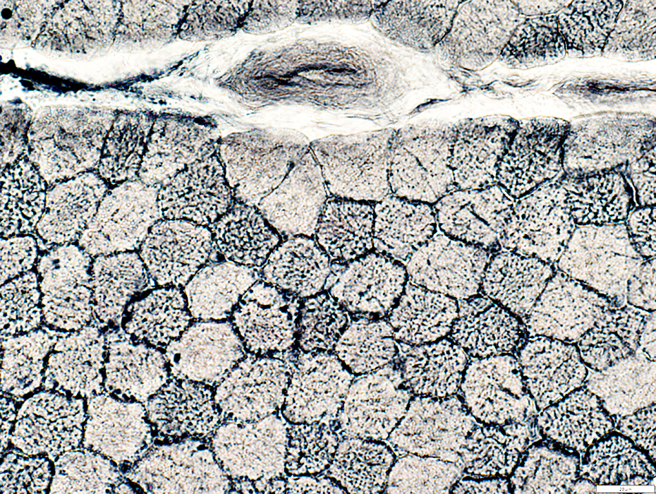 Sudan Black stain |
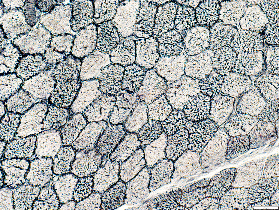 Sudan Black stain |
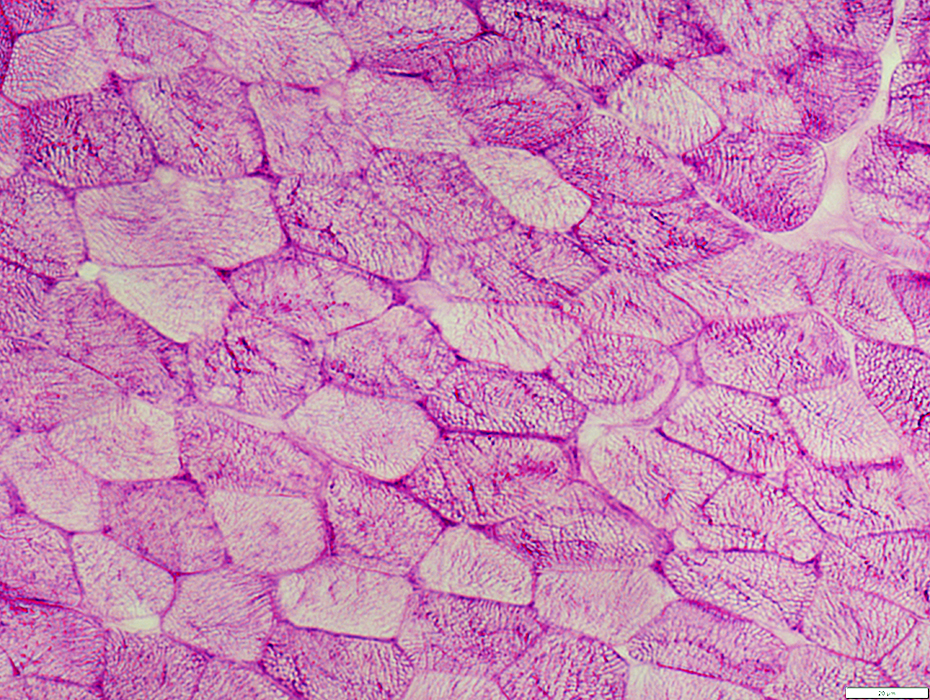 PAS stain |
 AMPDA stain |
Return to: SEPN1
12/21/2024