Ryr1 Recessive Myopathy
Muscle Pathology: General
- Fiber sizes: Bimodal distribution
- Fiber types: Type I small
- Morphology: No cores
Patient
3 week old male with: Ophthalmoplegia & Diffuse weakness + hypotonia
Ryr1 mutations: Bi-allelic; Missense; Leu3554Pro, Gly4782Arg
See: Same patient at 2 years of age
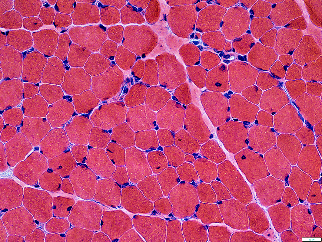
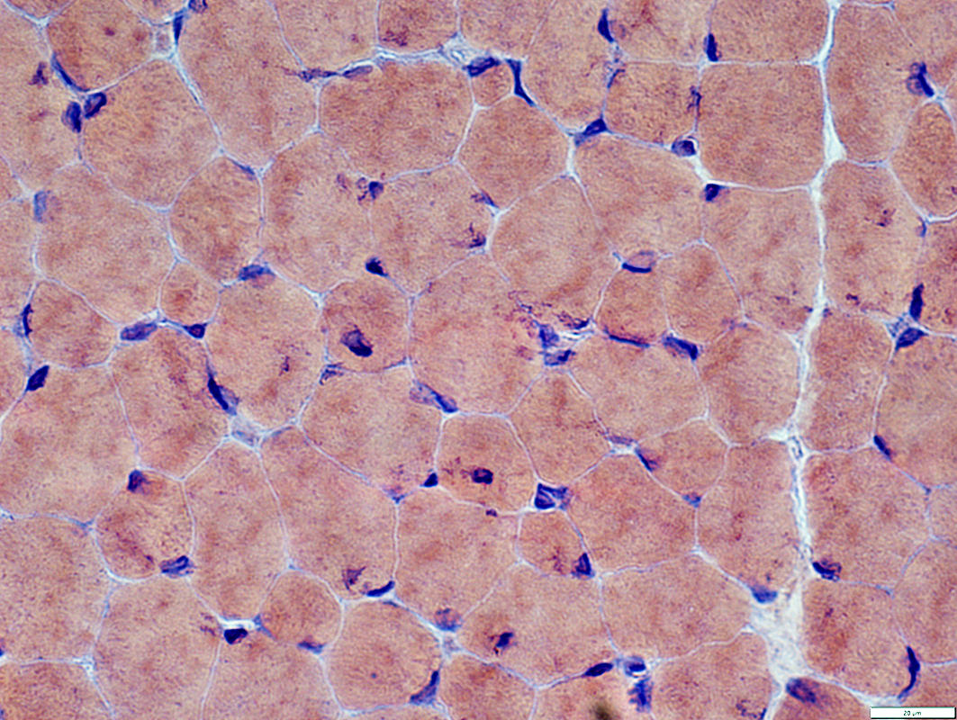
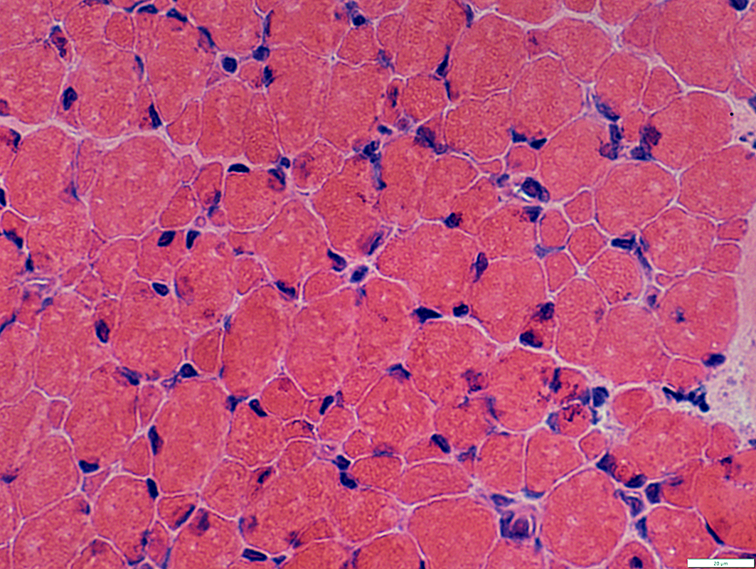 H & E stain |
Bimodal distribution
Nuclei
A few small fibers have central nuclei
Morphology: Some haver irregular shapes
 H & E stain |
 Congo red stain |
Bimodal distribution
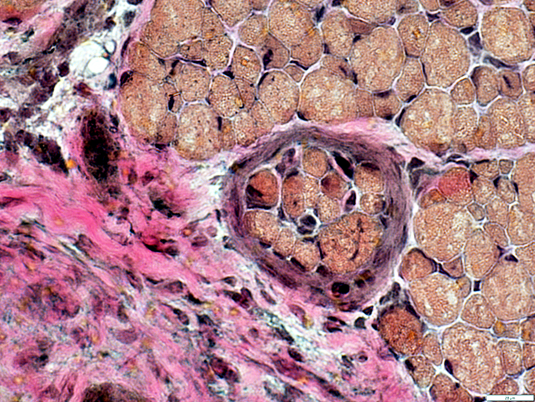
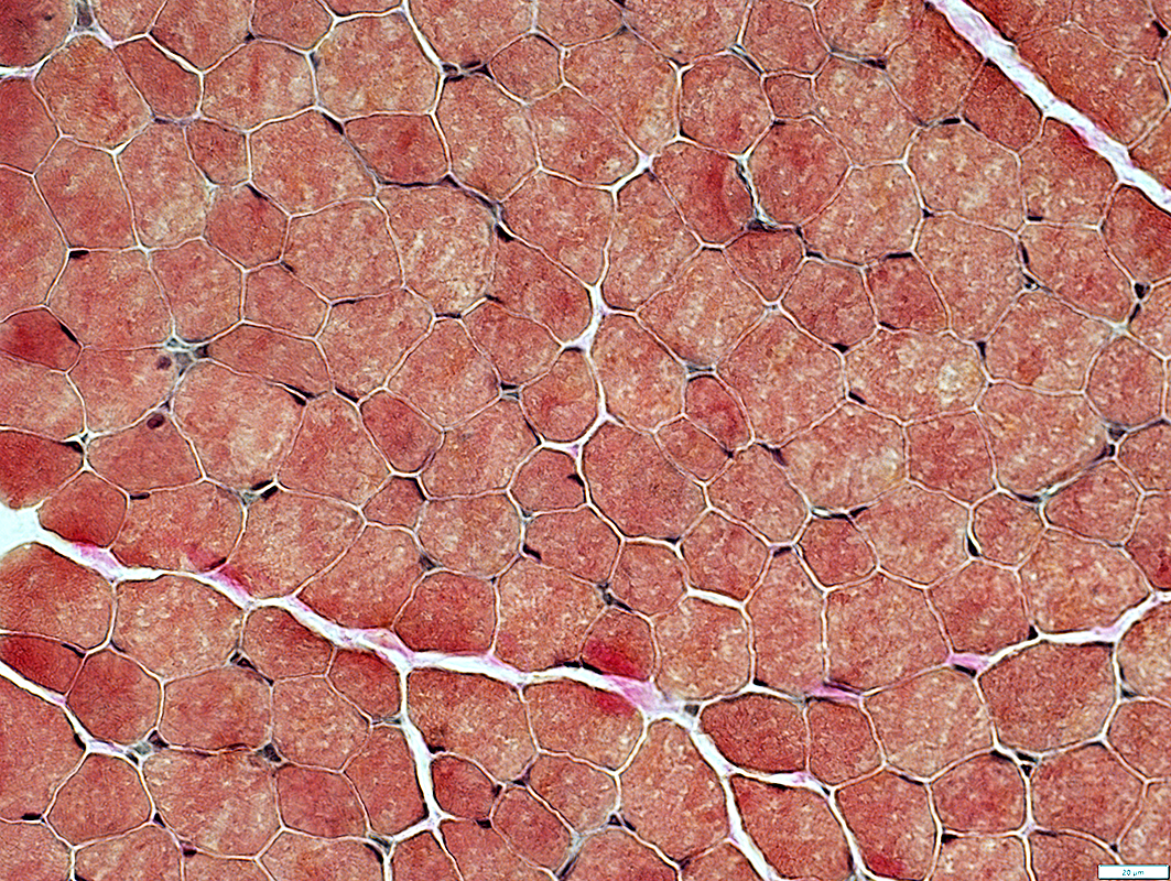
 Gomori trichrome stain |
 VvG stain |
Bimodal distribution
 VvG stain |
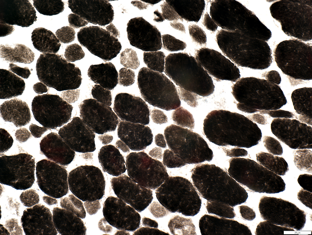
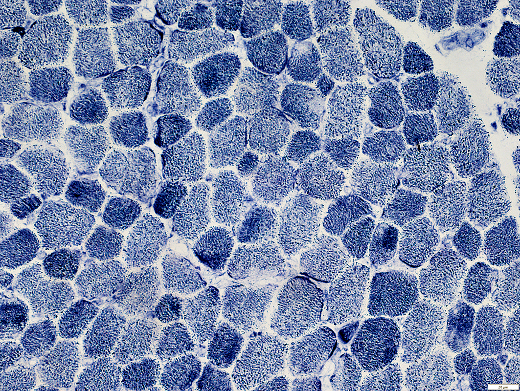
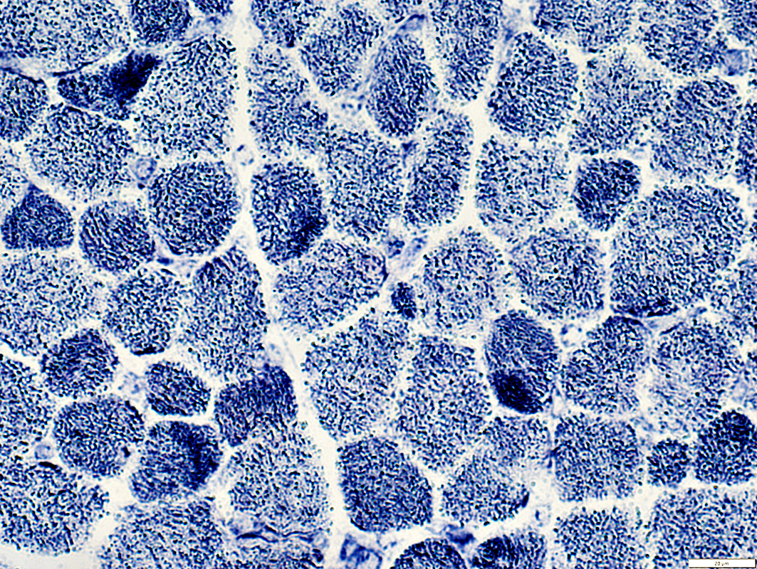
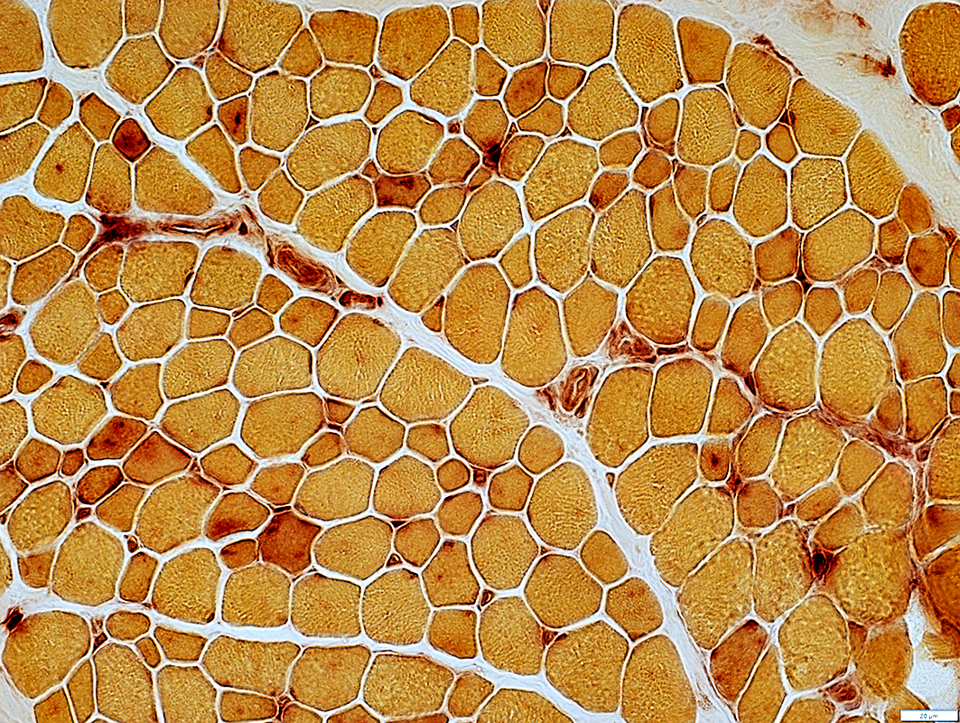
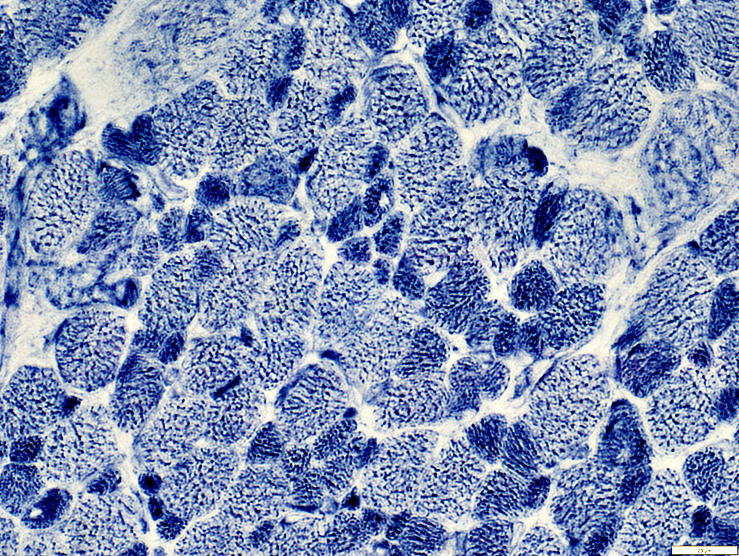 NADH stain |
Stain dark on NADH & COX
 COX stain |
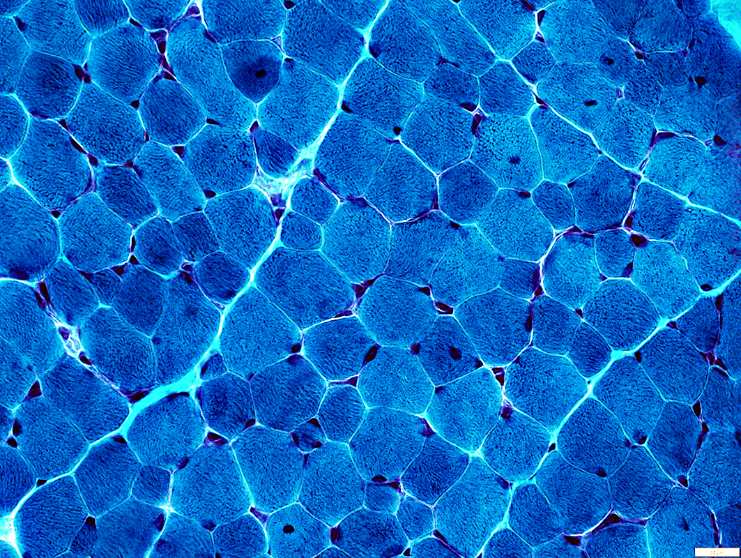
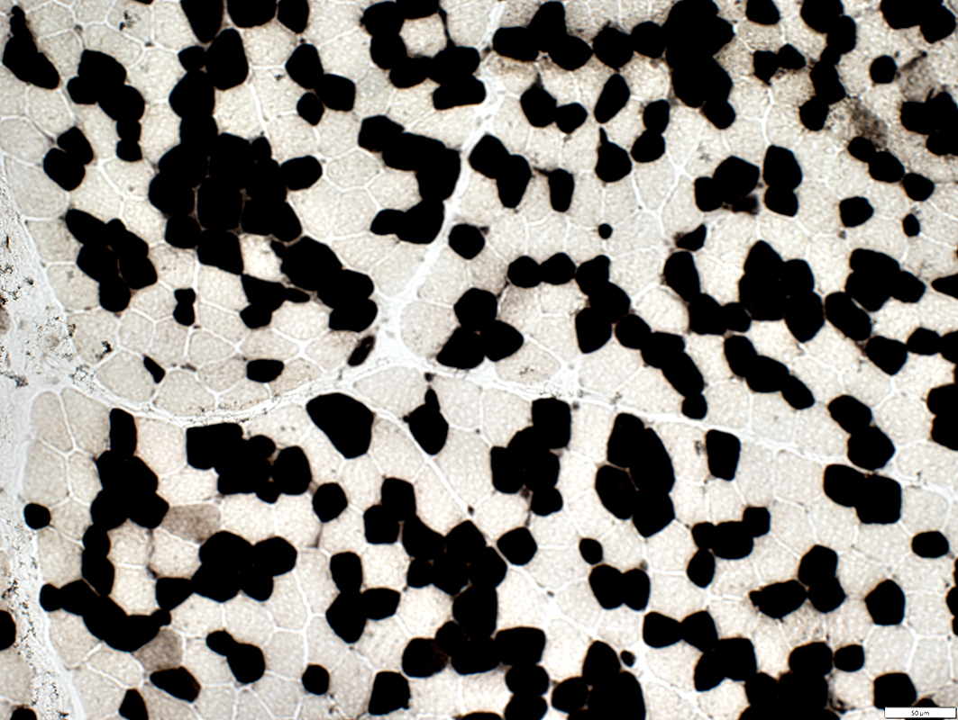
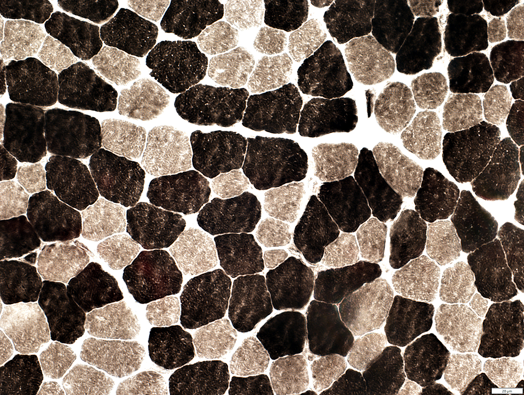
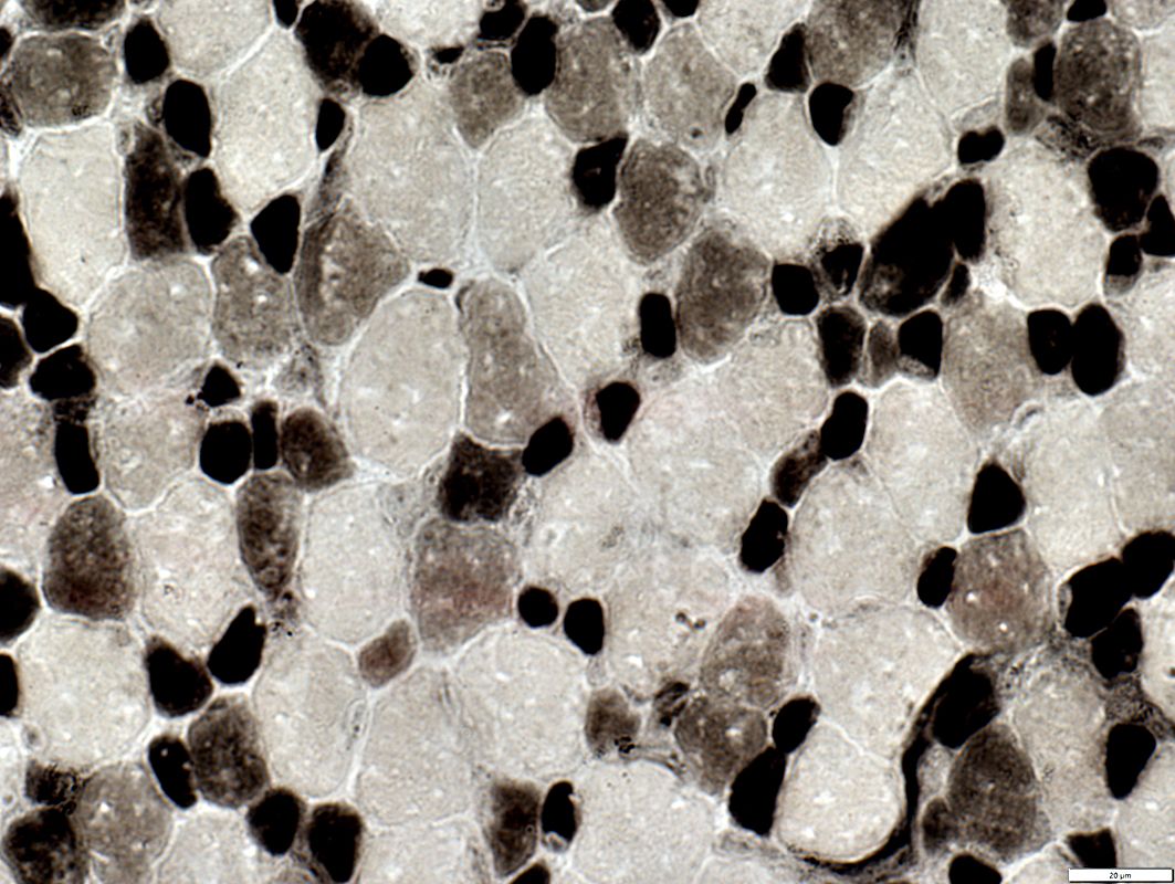 ATPase pH 4.3 stain |
Type 1: Stain dark on ATPase pH 4.3 & Intermediate on ATPase pH 9.4
Type 2C immature fibers
Many
Intermediate stained on ATPase pH 4.3
 ATPase pH 9.4 stain |
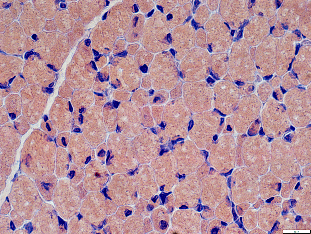
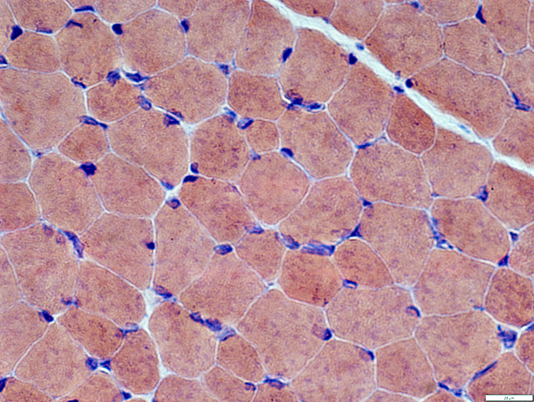
Small Muscle Fibers
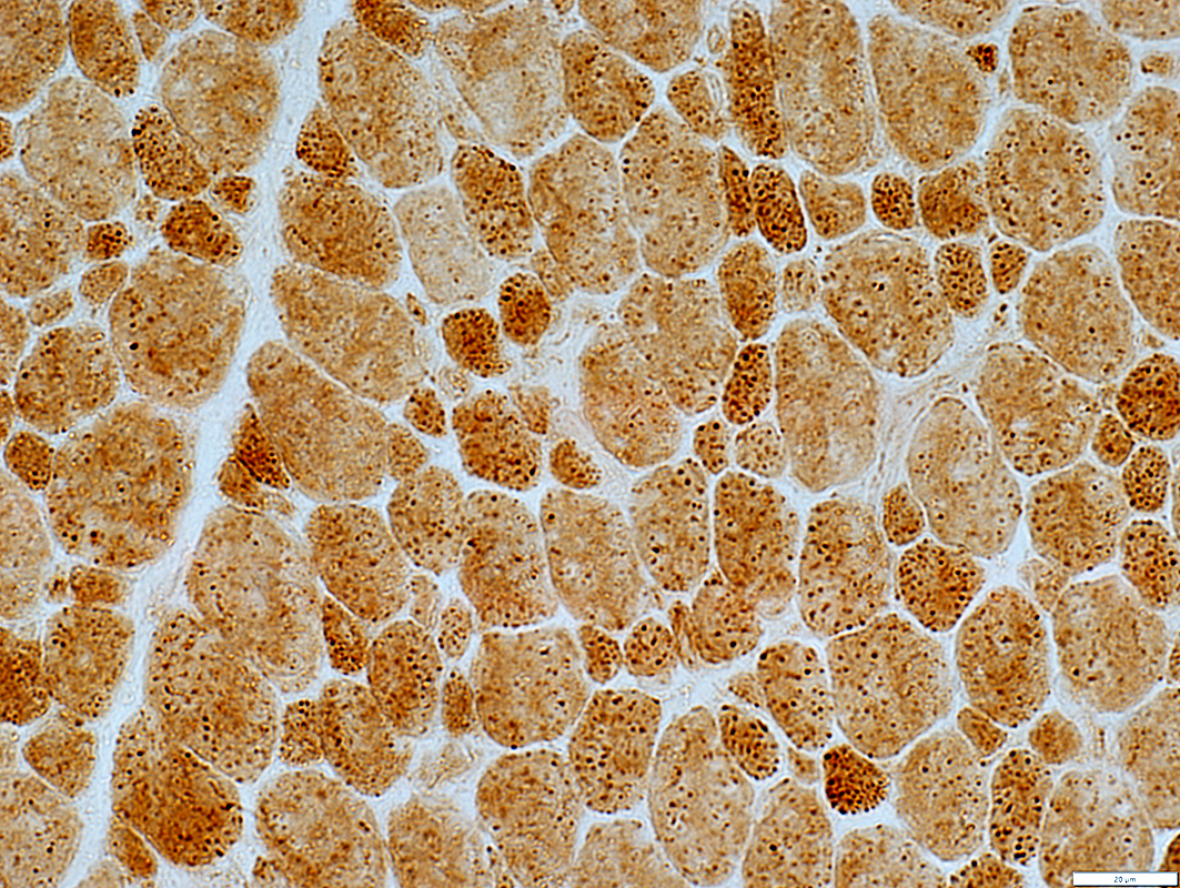
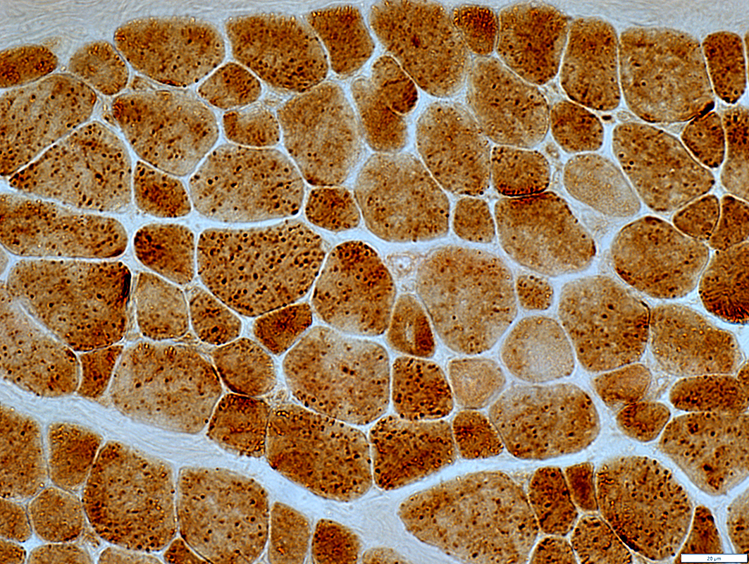
Acid phosphatase moderately stains cytoplasm
 Acid phosphatase |
Neuromuscular junctions
Dark esterase staining
 Esterase stain |
Ryr1 mutations
Same patient as above at 2 years of age
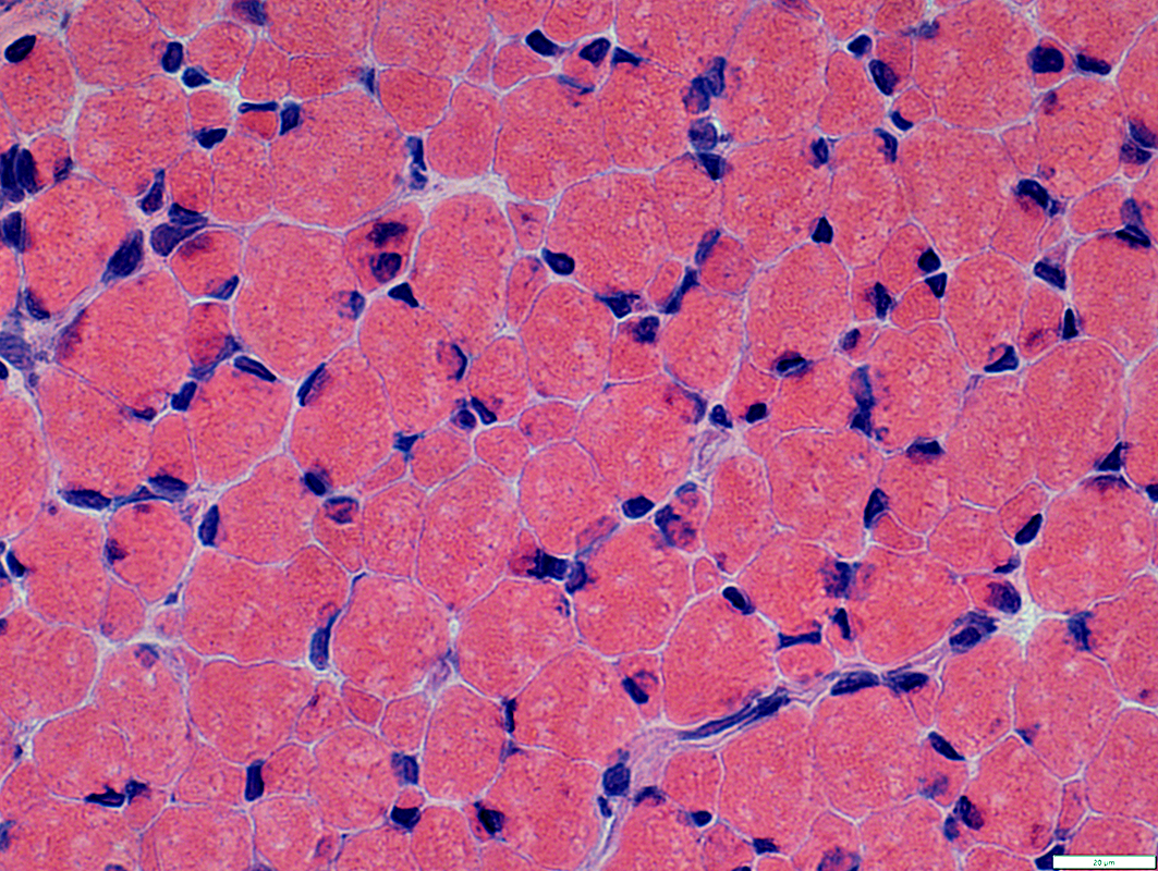
 H & E stain |
Bimodal distribution
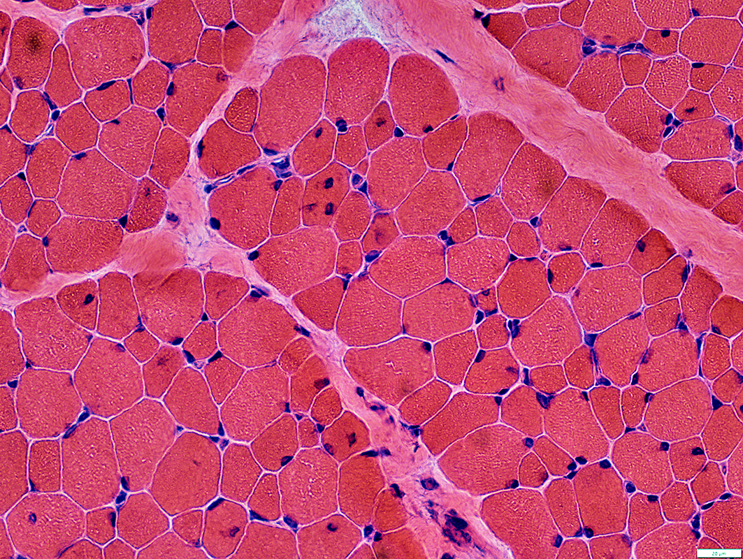 H & E stain |
 Congo red stain |
Bimodal distribution
Nuclear morphology
Irregular shapes
 Congo red stain |
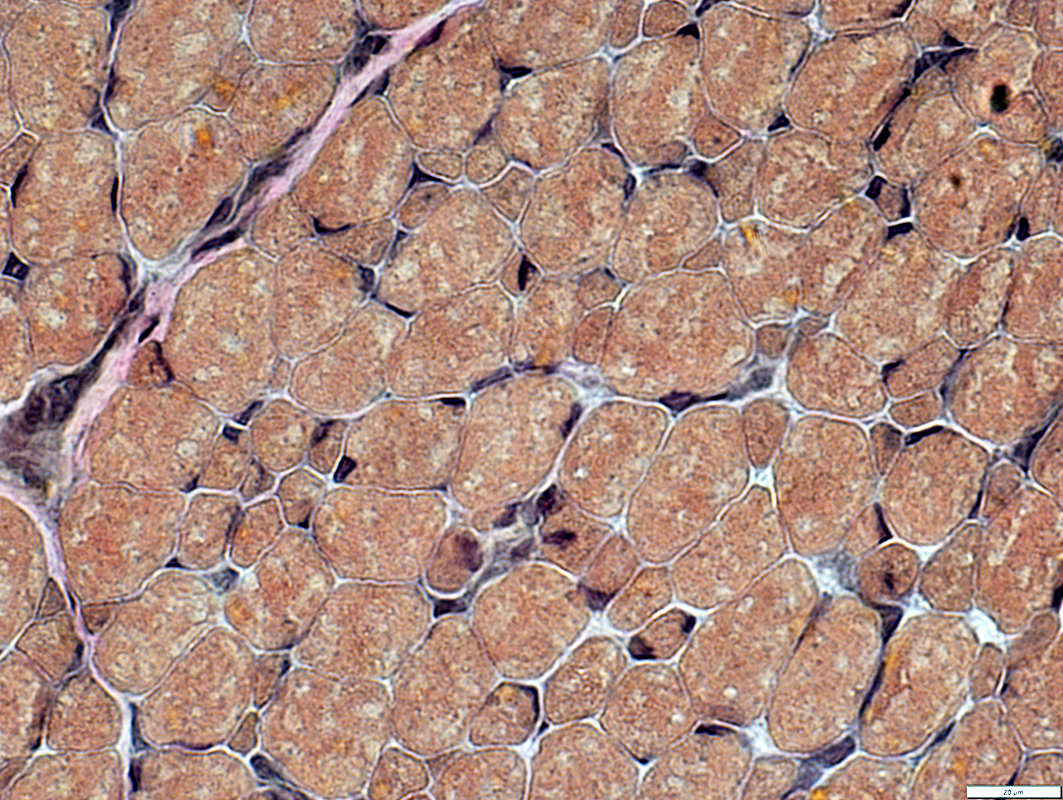
 Gomori trichrome stain |
Bimodal distribution
 VvG stain |
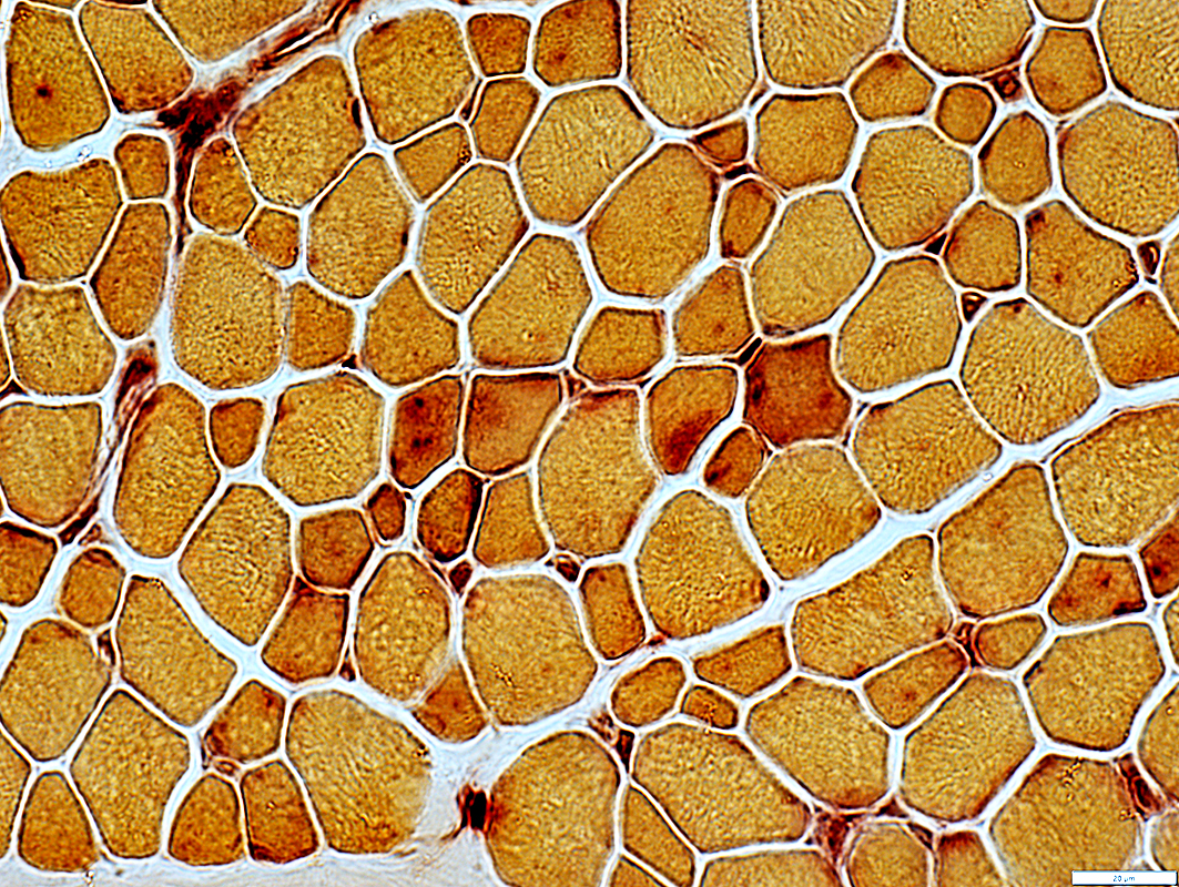
 NADH stain |
Moderately dark staining
Some have membrane aggregates
 NADH stain |
Small Muscle Fibers
Some have clear regions
 COX stain |
 ATPase pH 4.3 stain |
Immature type 2C fibers
Few
Scattered
Capillaries
Have moderate ATPase staining
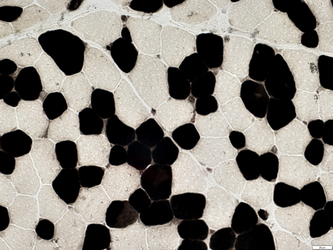 ATPase pH 4.3 stain |
 ATPase pH 4.3 stain |
 ATPase pH 9.4 stain |
 Acid phosphatase stain |
Acid phosphatase moderately stains cytoplasm
 Acid phosphatase stain |
Return to Ryr1
6/22/2026