RFC1: CANVAS
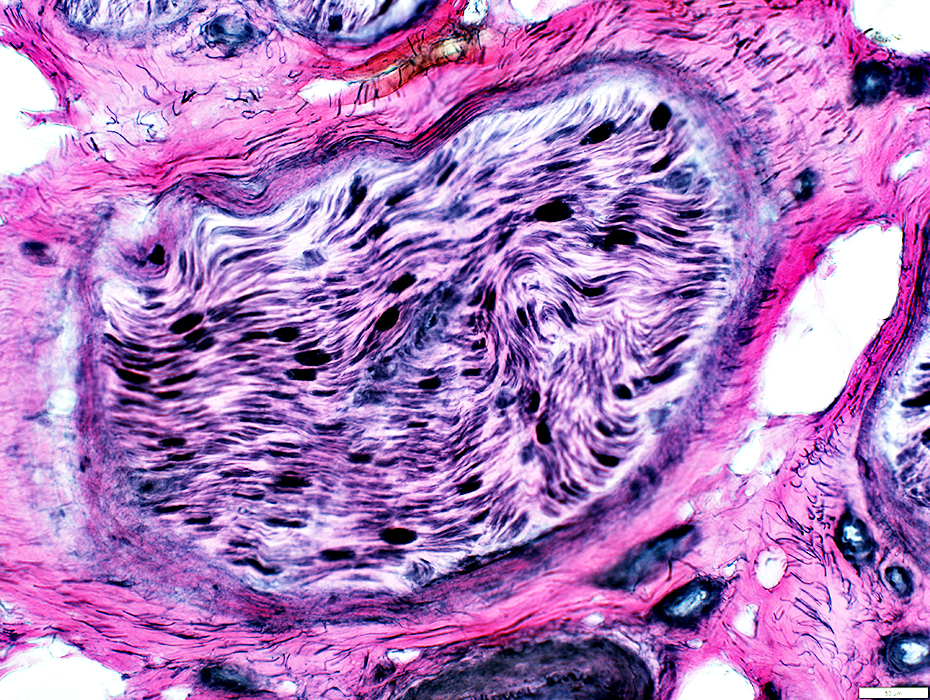
 H&E stain |
Normal structure of connective tissue & vessels
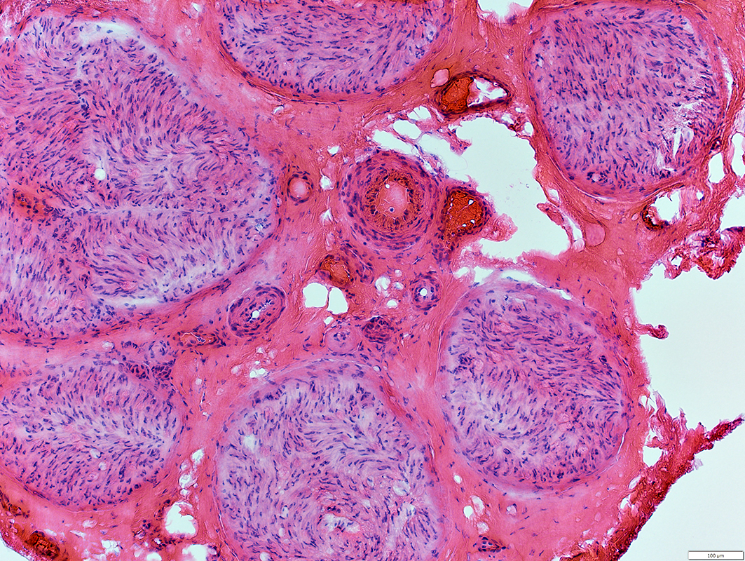
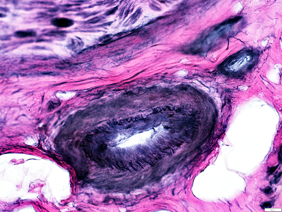
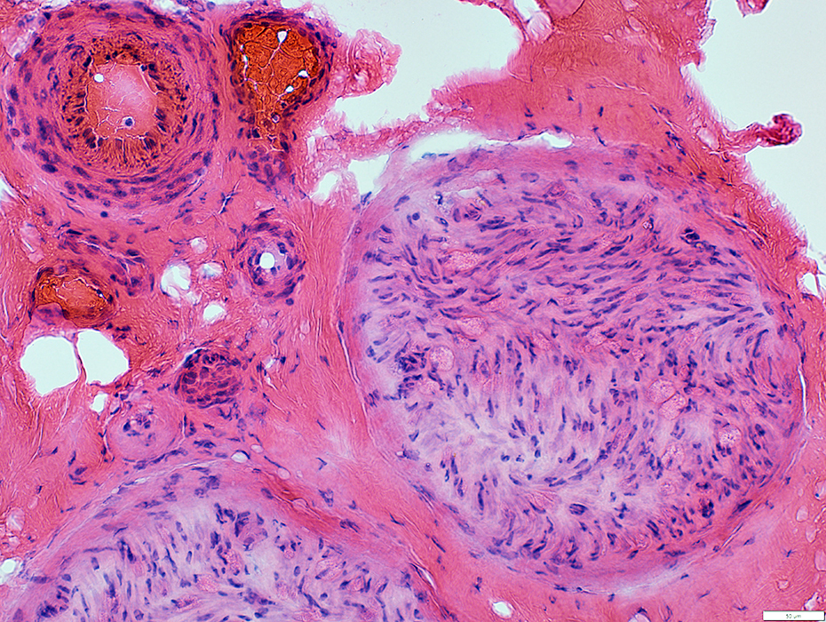 H&E stain |
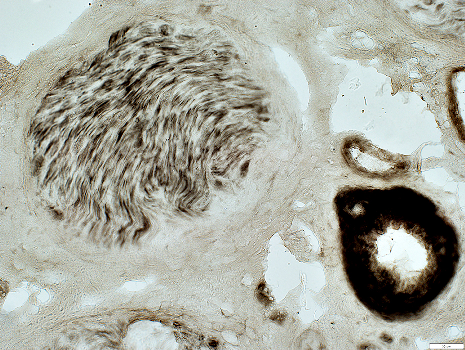
 VvG stain |
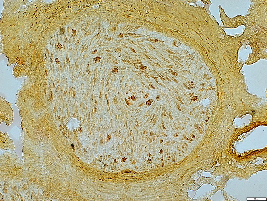
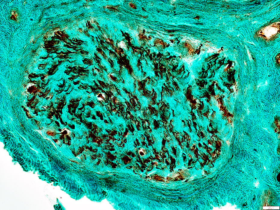
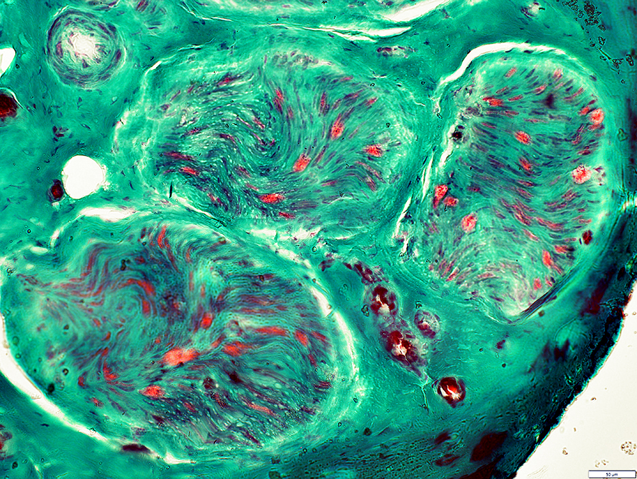 Gomori trichrome stain |
Reduced numbers
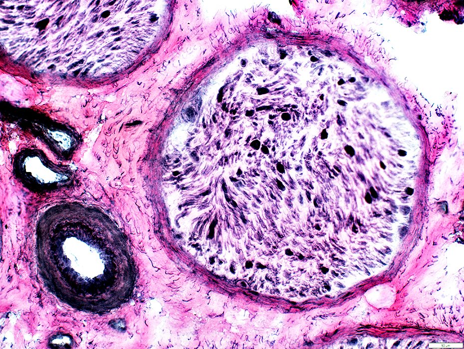
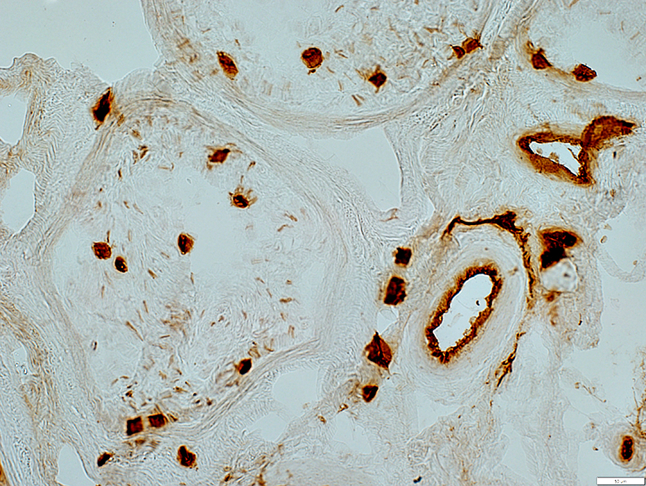
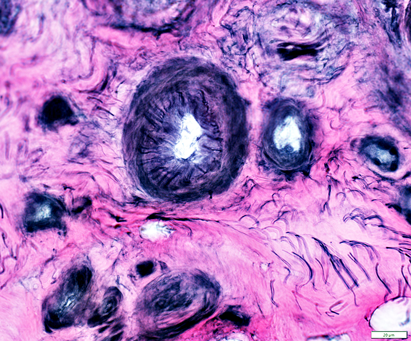
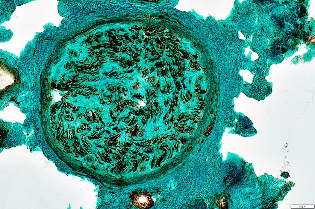
 VvG stain |
Endomysium
Normal ATPase staining
 ATPase pH 4.3 stain |
 Alkaline phosphatase stain |
Numbers: Normal to Increased
Alkaline phosphatase staining: Reduced
 UEAI stain |
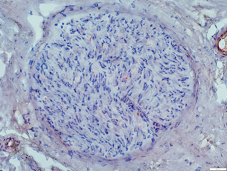
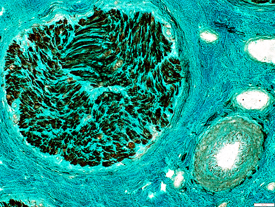
 VvG stain |
Irregular inner fibril layer
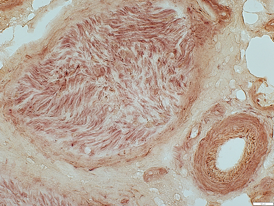
 VvG stain |
 Congo red stain |
No endoneurial histiocytes
 Acid phosphatase stain |
 Neurofilament stain |
Large: Severe loss
Small, Unmyelinated: Relatively preserved
 Neurofilament stain |
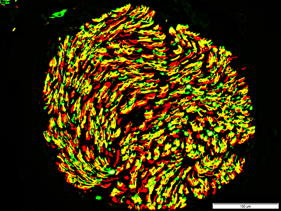
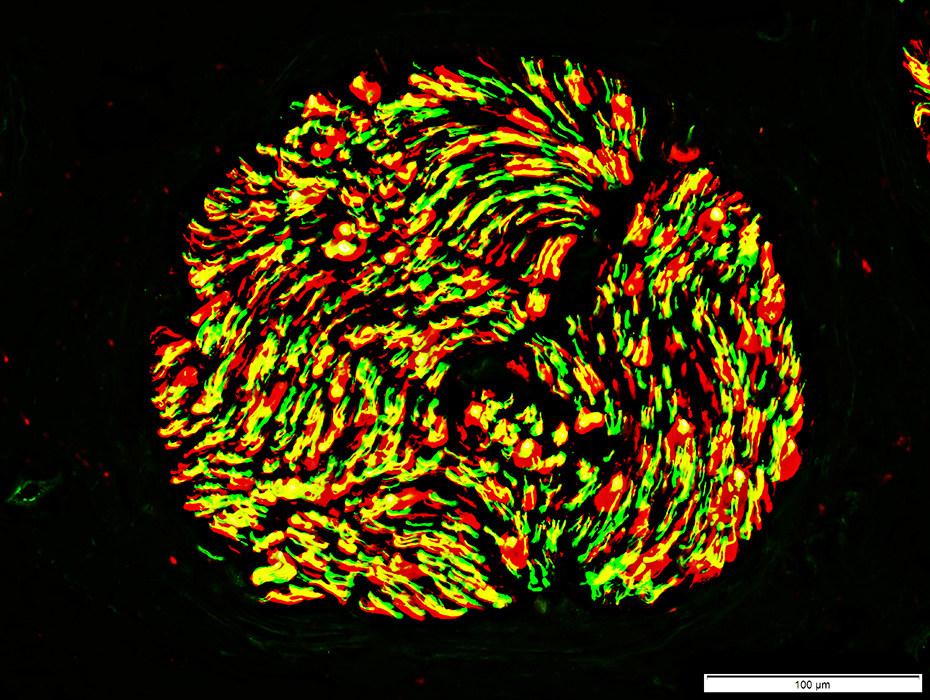 Neurofilament (Green)-P0 (Red) stain |
Large: Severe loss
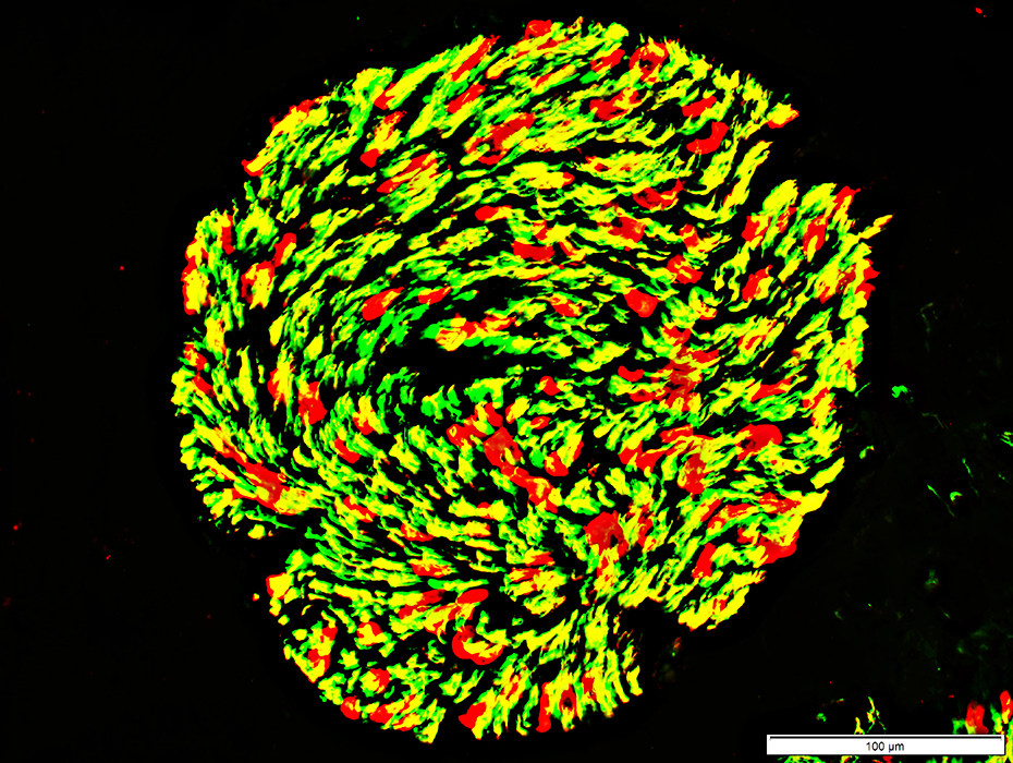
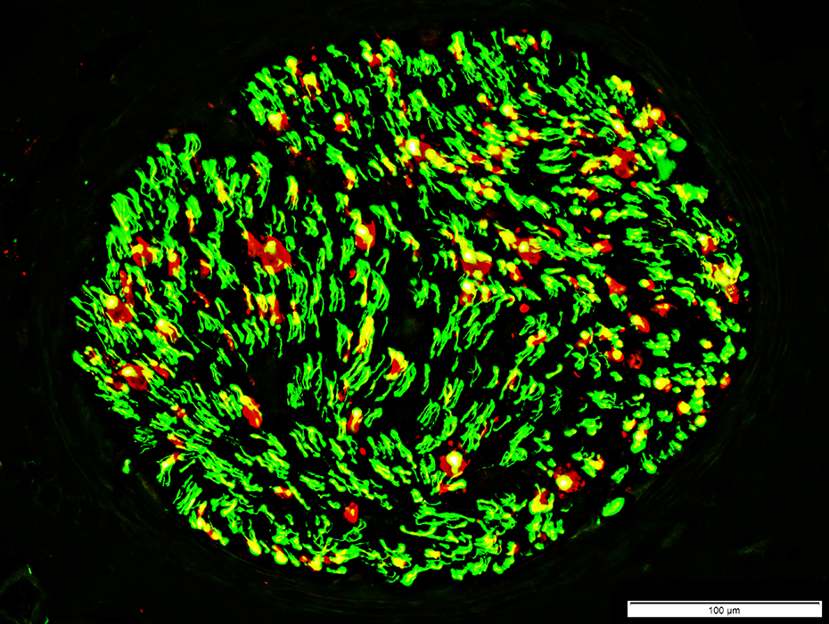 Neurofilament (Green)-MBP (Red) stain |
 Neurofilament (Green)-NCAM (Red) stain |
Small: Mild loss; Some non-myelinating Schwann cells have no axons (Red)
 NCAM stain |
 NCAM (Green) - P0 (Red) stain |
Büngner band cells (Yellow) present
Schwann cell pathology: Chronic
Büngner band cells (Red) present
Myelinated axons (Yellow): Reduced numbers
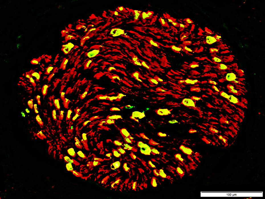 Periaxin (Green) - P0 (Red) stain |
RFC1: Axons
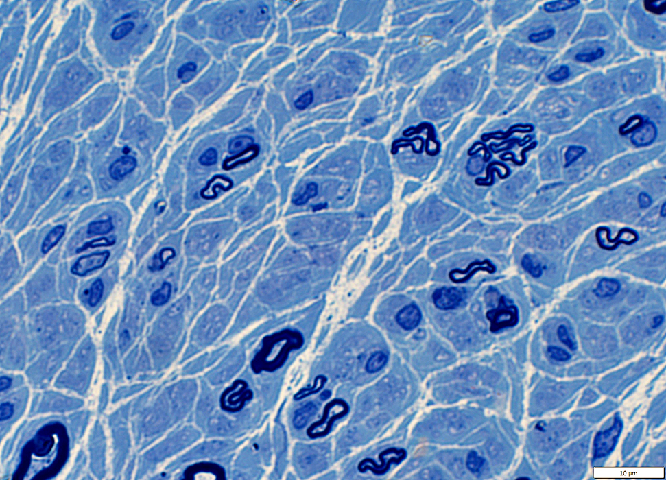
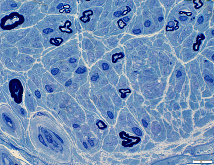
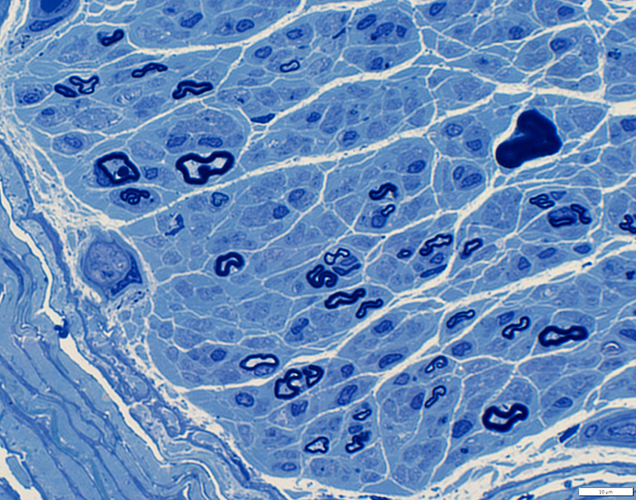 Toluidine blue-stained Plastic sections |
Large & Small myelinated axons
Axon regeneration
Clusters of small, thinly myelinated axons
Axon shapes
Many irregularly shaped small myelinated axons
 Toluidine blue-stained Plastic sections |
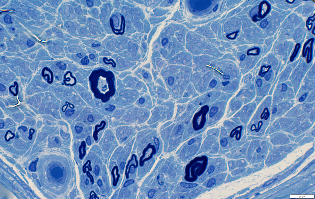
 Toluidine blue-stained Plastic sections |
Large & Small myelinated axons
Axon regeneration
Clusters of small, thinly myelinated axons
Axon shapes
Many irregularly shaped small myelinated axons
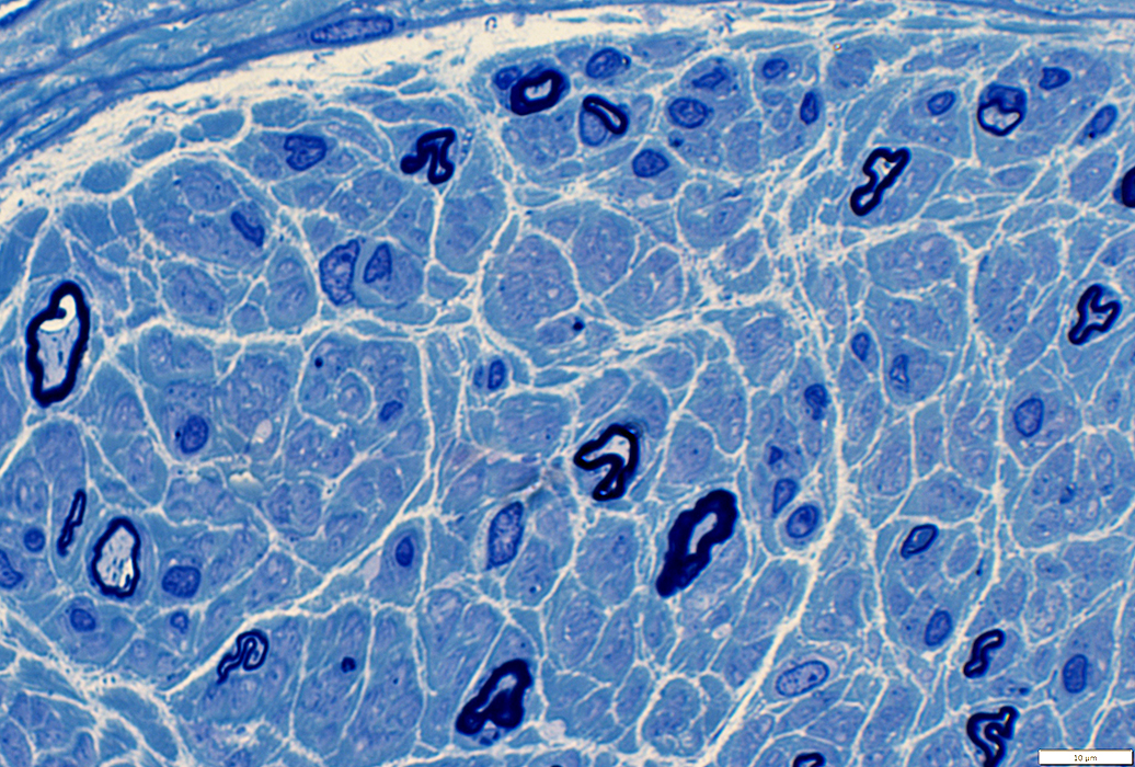
 Toluidine blue-stained Plastic sections |
 Toluidine blue-stained Plastic sections |
Return to: RFC1
Return to: Neuromuscular Home Page
3/22/2022