Renaut Bodies 1
|
Patterns Confluent Internal Typical Renaut illustration |
 Reference 2 |
Renaut bodies: Features
- Frequency
- 2% of nerve biopsies
- Increased age: Possibly more frequent
- Structure
- Shape: Round, Elliptical (Flattened) or Elongated (Along length of nerve)
- Internal: Whorled
- Contents
- Cell: Fibroblasts, often with long processes; "Spider-like" (cellules godronées)
- Other: Mucopolysaccharides; Collagen; Elastin
- Edge: May be bounded by fibroblast process
- Size: 20 to 130 μM
- Location
- Often multiple: In several fascicles in nerve
- Usual: All in similar region of different fascicles
- Atypical: Confluent
- May be 1, or several, in individual fascicles
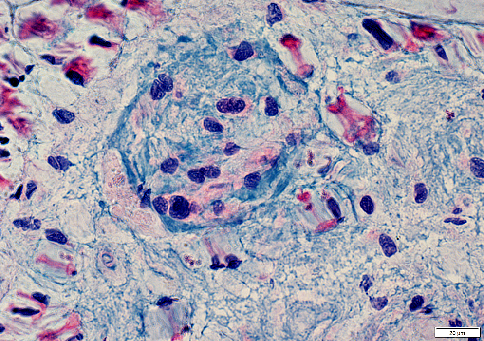
- Stains: Alcian blue; Gomori trichrome; ± EMA
- Associated vessels: May have thick wall
- Possible cause: Chronic nerve compression
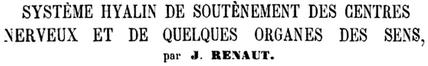
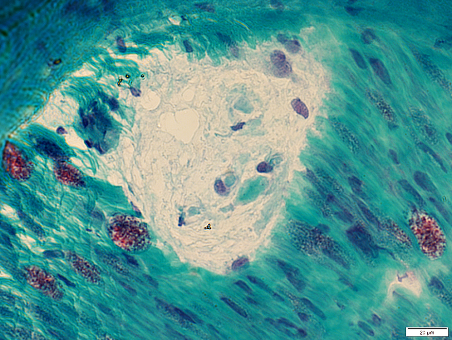
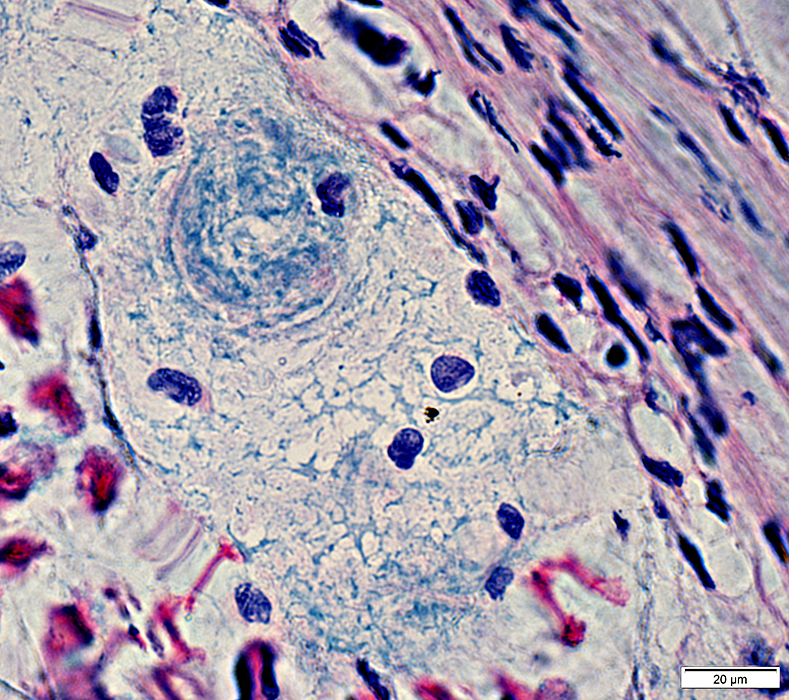
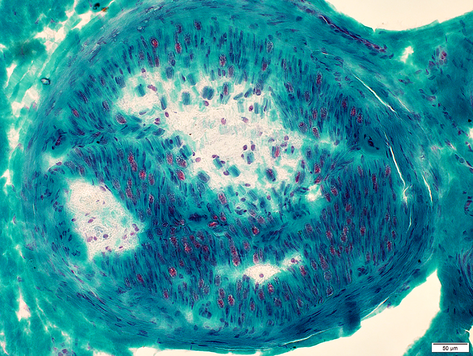
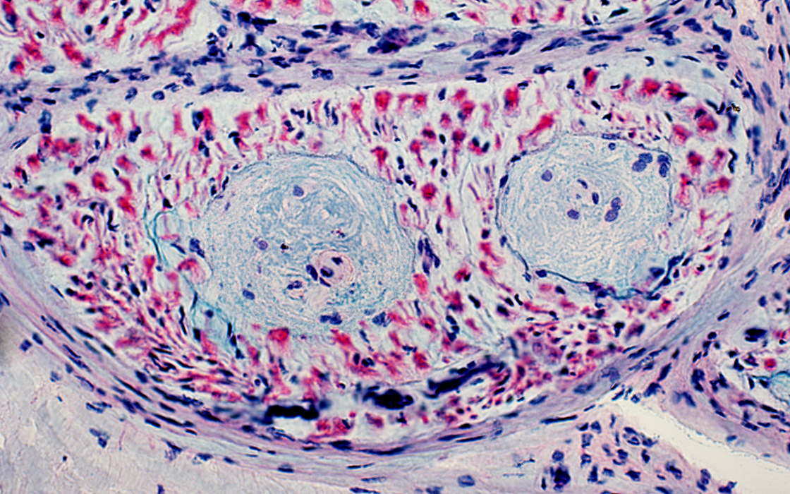
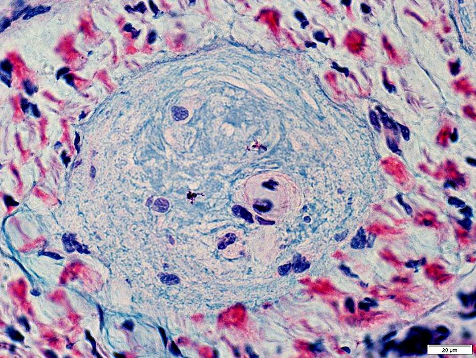
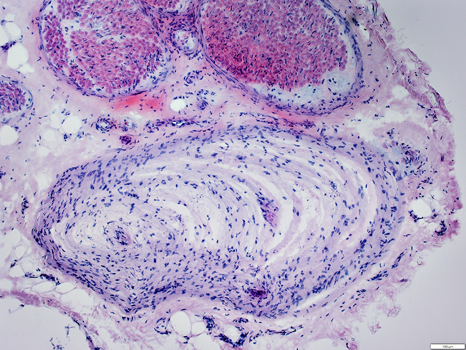
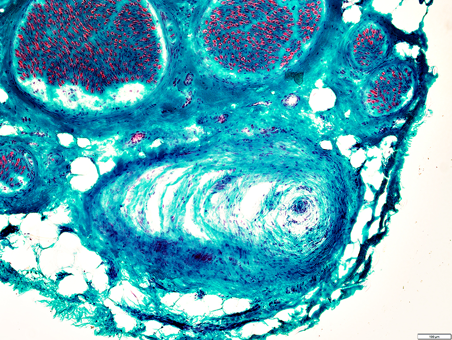
Renaut Bodies: Typical
 Gomori trichrome stain |
Rounded
Subperineurial
Present in several fascicles
All located in similar endoneurial regions
 Toluidine blue stain |
 VvG stain |
 Gomori trichrome stain |
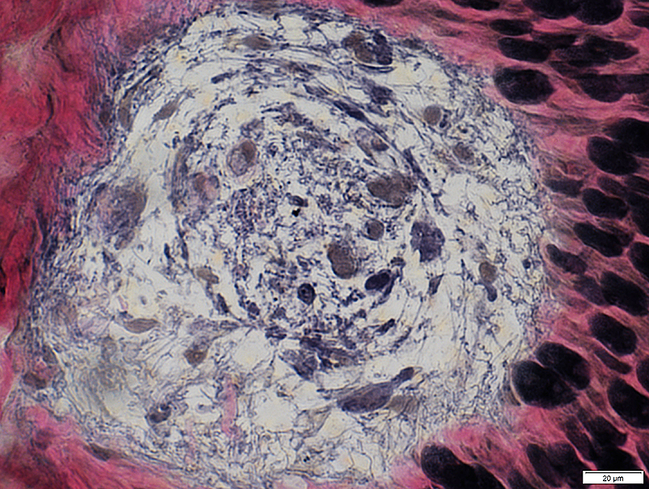
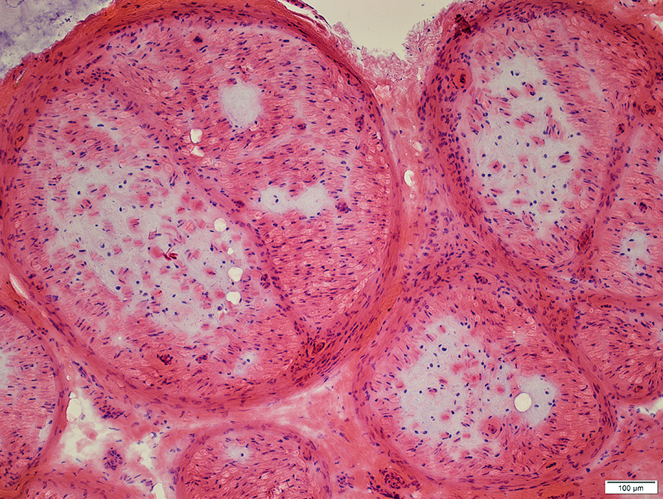
 H&E stain |
 H&E stain |
 Gomori trichrome stain |
 Gomori trichrome stain |
 VvG stain |
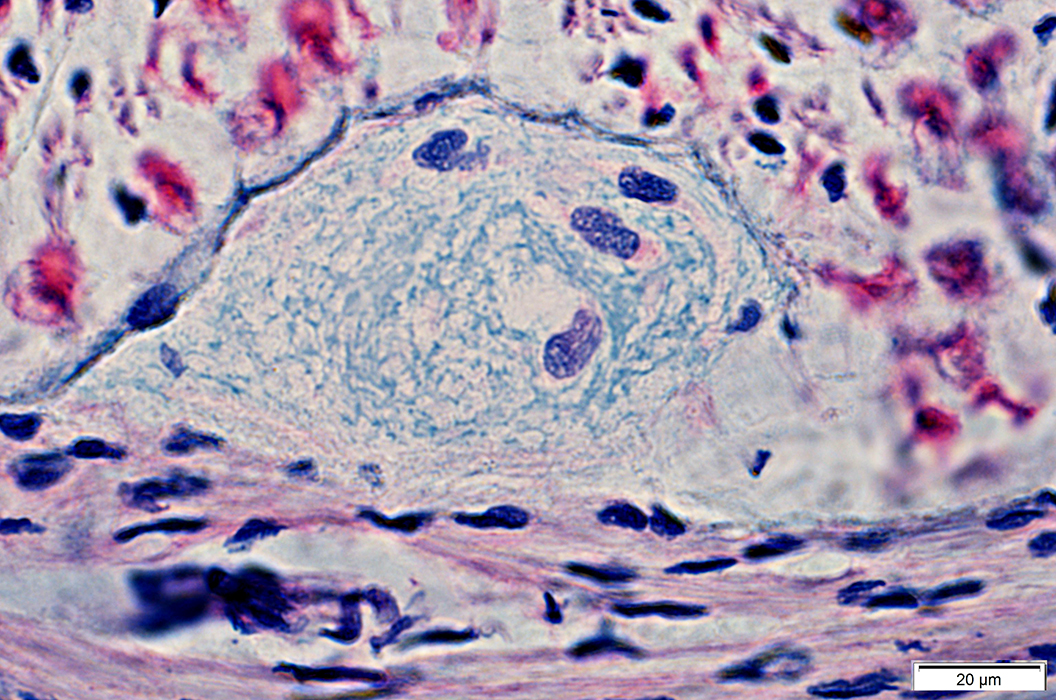
Rounded
Filamentous layers
Rounded sub-structures
Cells, Scattered
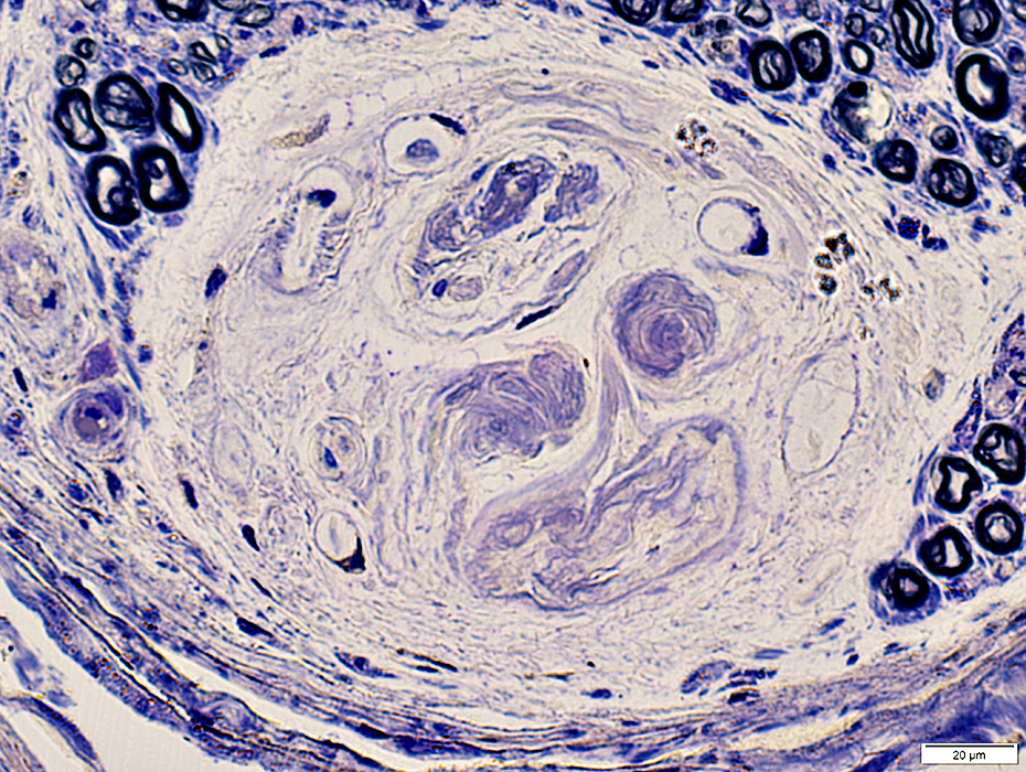 Toluidine blue stain |
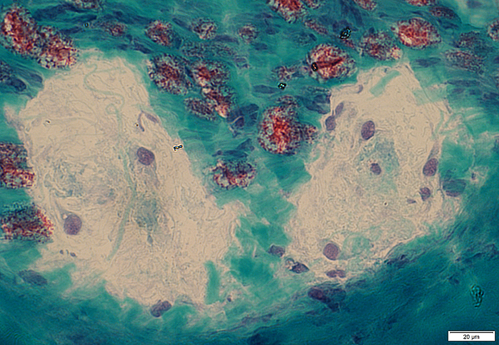
 Alcian blue stain |
 Alcian blue stain |
 Alcian blue stain |
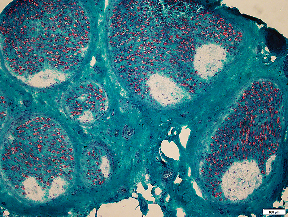
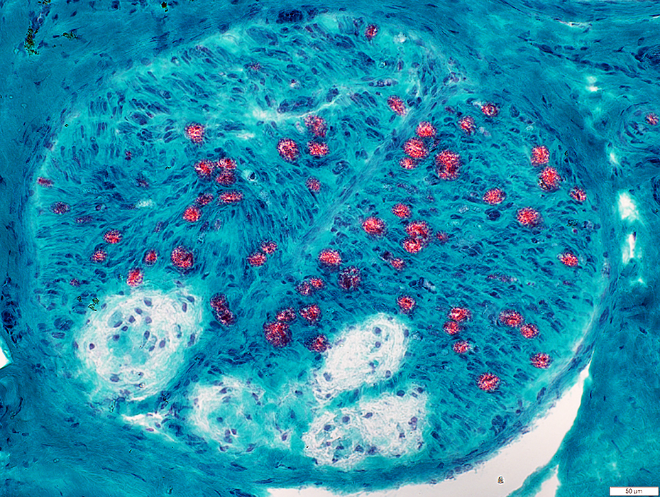
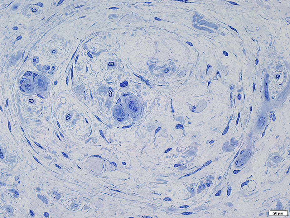
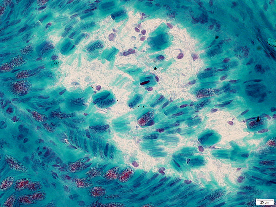
Renaut Bodies: Confluent
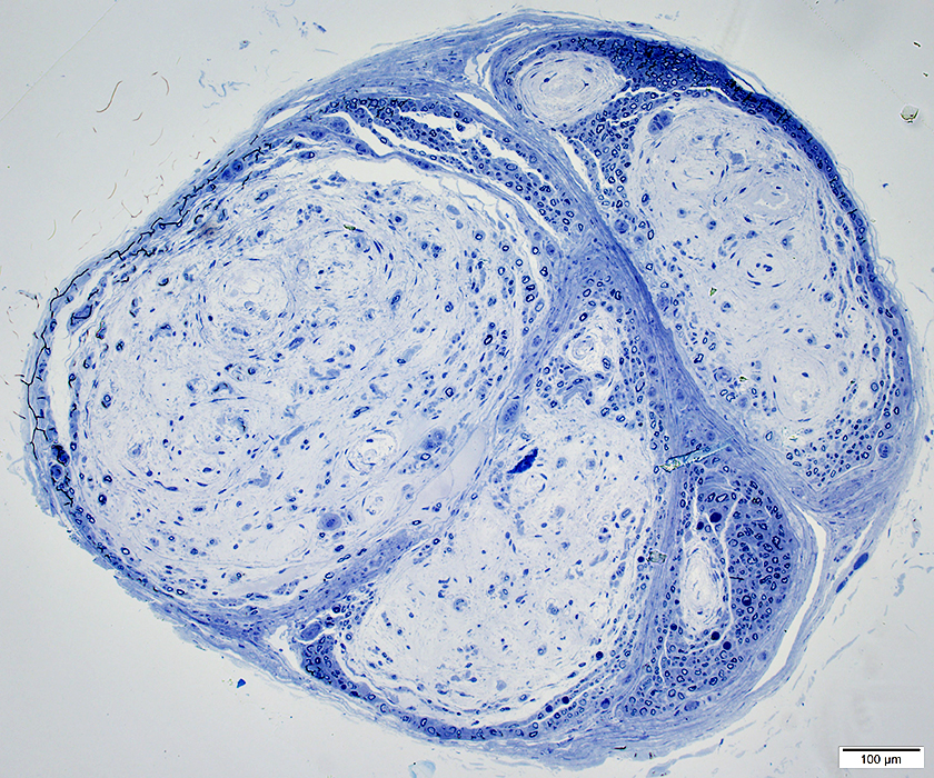 Toluidine blue stain |
Multiple pale rounded regions
Probable area of chronic nerve damage
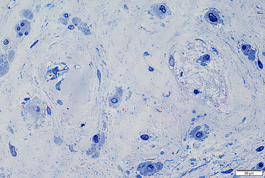
 Toluidine blue stain |
 Toluidine blue stain |
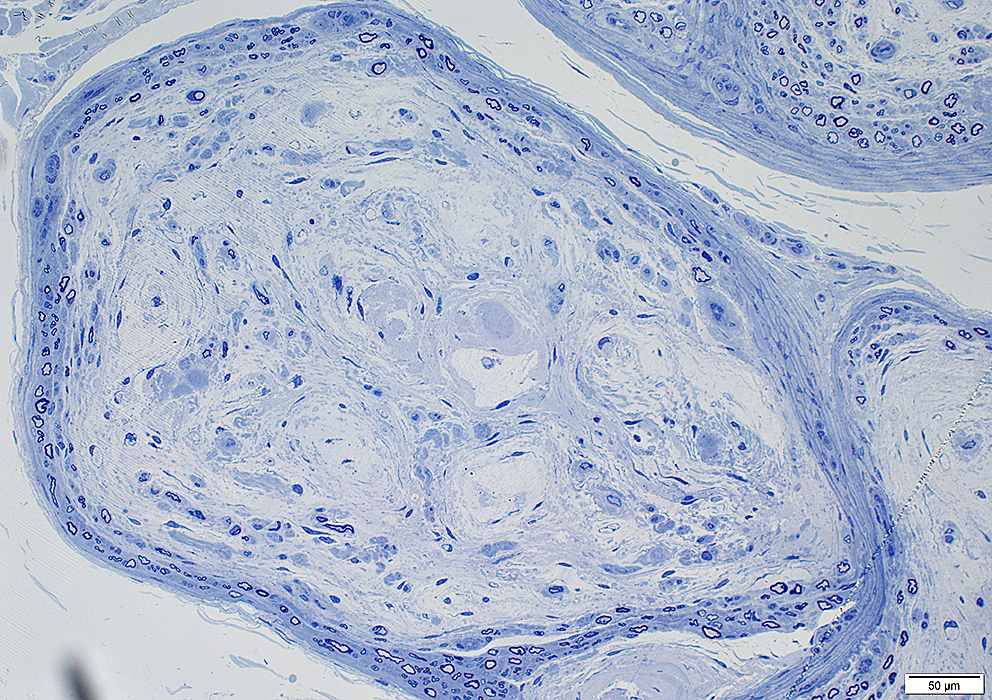
Rounded
Filamentous layers: Variable structure
Rounded sub-structures
Cells, Scattered
 Toluidine blue stain |
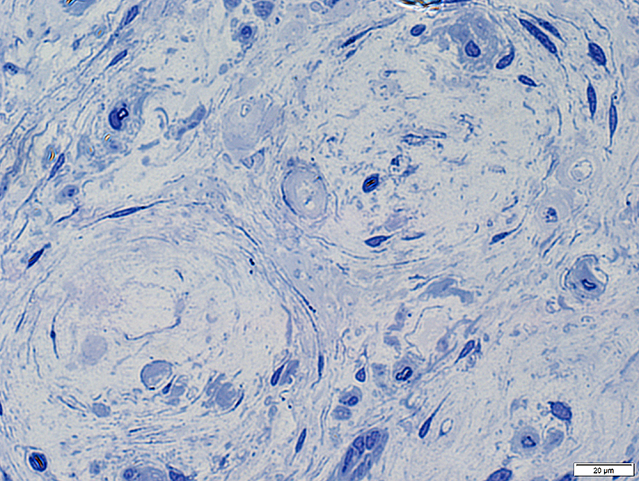 Toluidine blue stain |
 Alcian blue stain |
 Alcian blue stain |
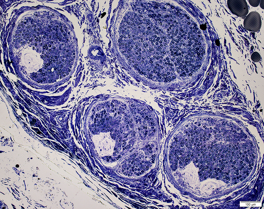
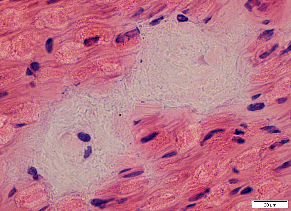
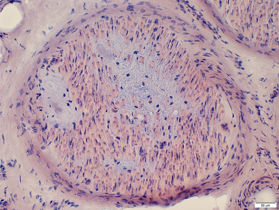
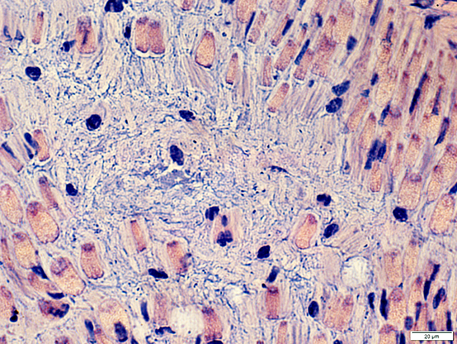
Renaut Bodies: Internal
 H&E stain |
Within endoneurium
Shape: Irregular
Contain occasional axon
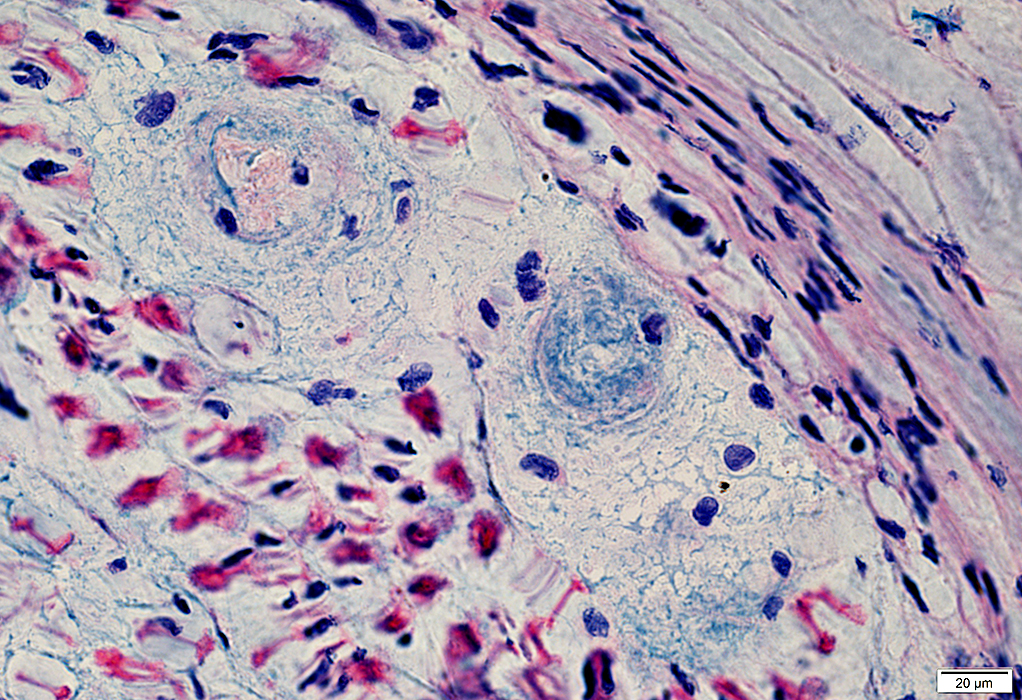
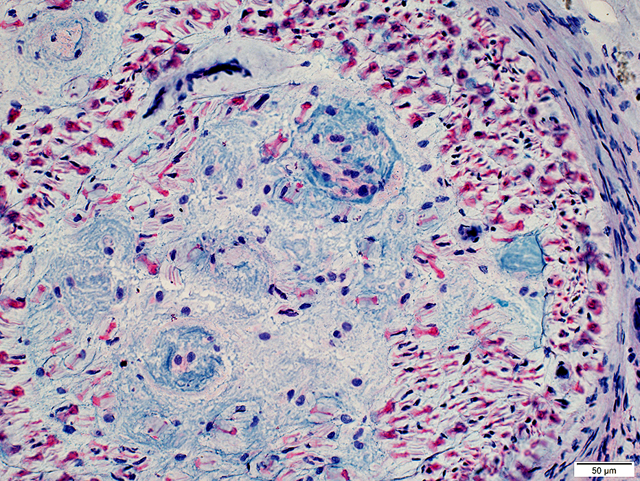
 Gomori trichrome stain |
 Congo red stain |
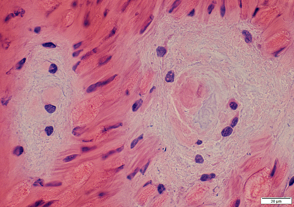
 Gomori trichrome stain |
Within endoneurium
Shape: Irregular
Contain: Irregular extracellular material; Cells; Occasional axon
 Congo red stain |
Renaut Bodies: Contain mucopolysaccharides (Alcian blue positive)
 Alcian blue stain |
 Alcian blue stain |
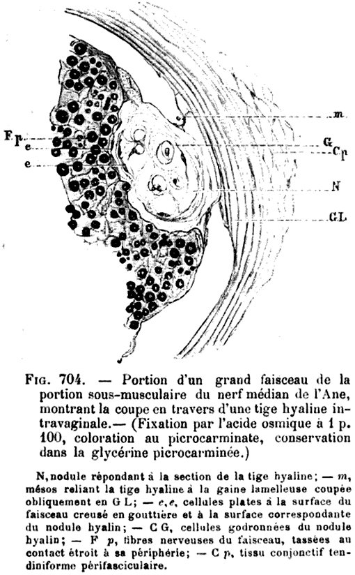
Renaut body: Renaut illustration
|
|
Pacinian corpuscle: Epineurial
 H&E stain |
 gomori Trichrome stain |
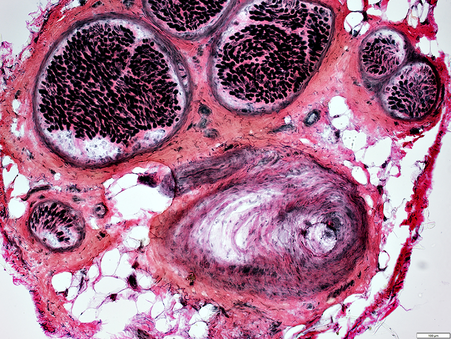 VvG stain |
Return to Neuromuscular Home Page
References
1. Can J Neurol Sci 2017;44:184-189, J Neuropathol Exp Neurol 1973;32:334-343
2. Renaut J. Systčme hyalin de soutčnement des centres nerveux et de quelques organes des sens. Arch Physiol Normale et Pathologique (Paris). 1881;8:846-59.
3/25/2017