Fukuyama Congenital Muscular Dystrophy
|
Adult Child |
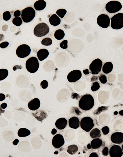
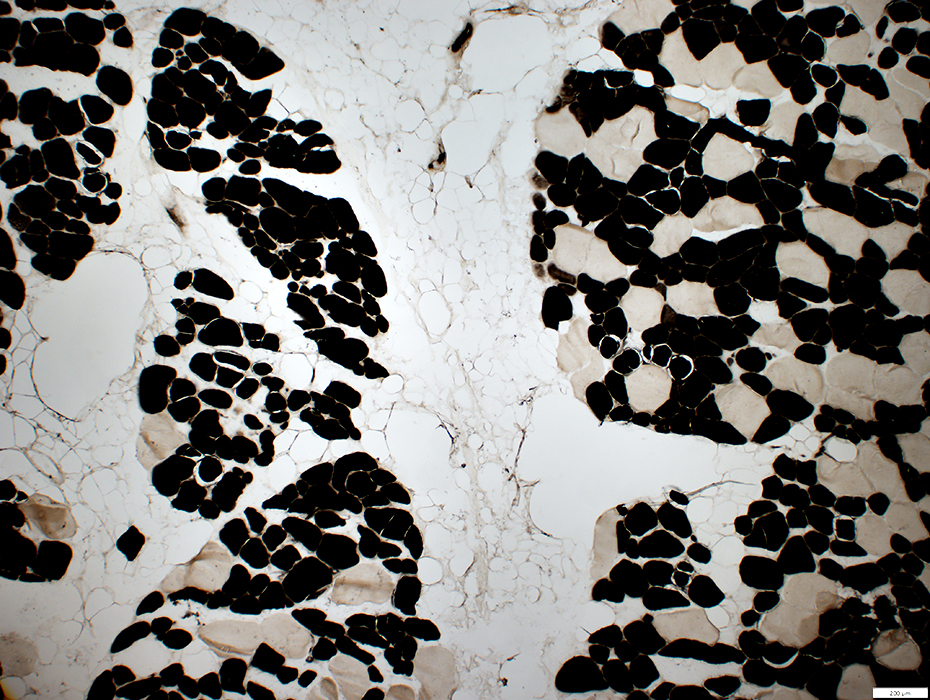
Childhood Pathology
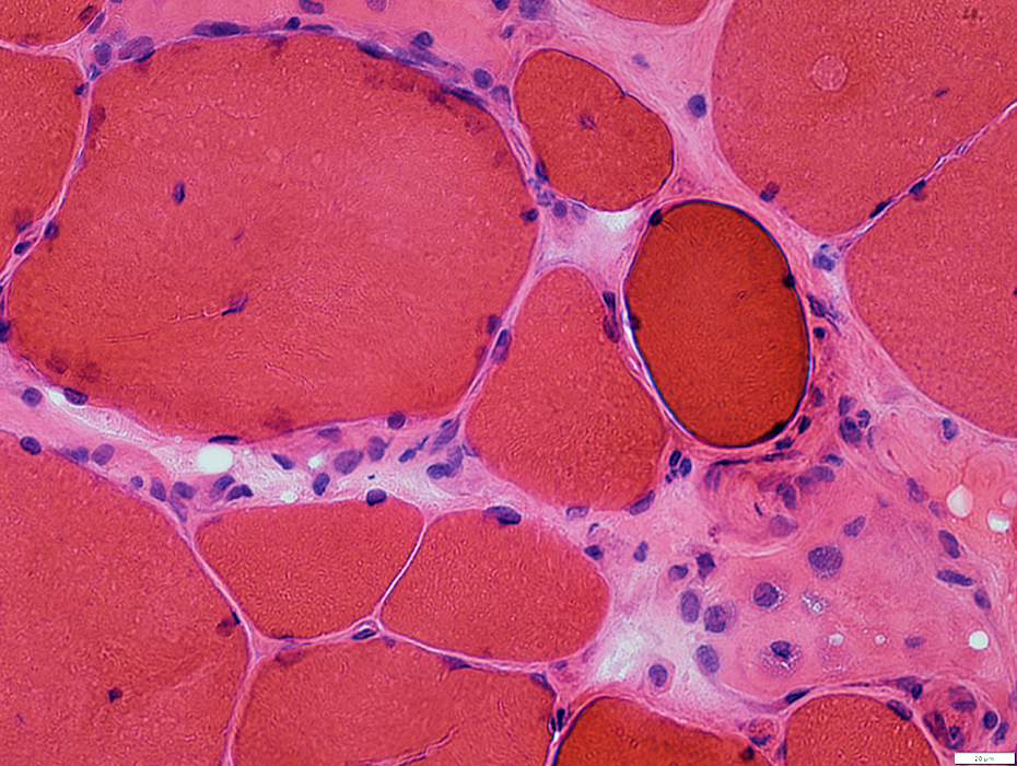
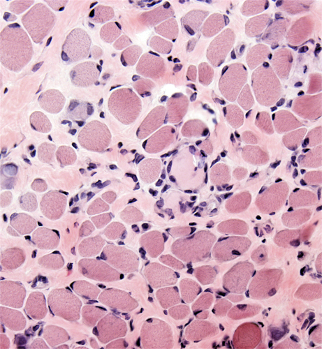 H&E stain From: I Nishino |
 H&E stain From: I Nishino |
Sizes: Varied
Small fibers: Scattered
Basophilic fibers: Scattered
Internal nuclei: Common
Endomysial connective tissue: Increased
Perimysium: May be replaced by fat
 Gomori trichrome stain Muscle fiber size: Varied Endomysial connective tissue: Increased |
 Acid phosphatase stain Positive staining cells associated with necrotic fiber |
 NADH stain Scattered small immature muscle fibers with coarse internal architecture |
 ATPase pH 11 stain |
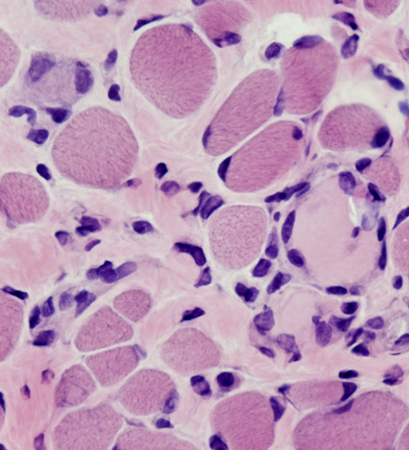
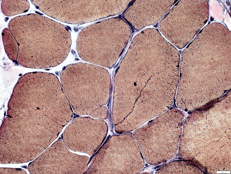
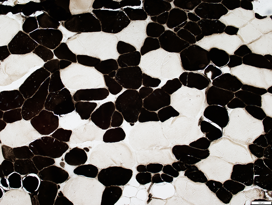
Adult Pathology
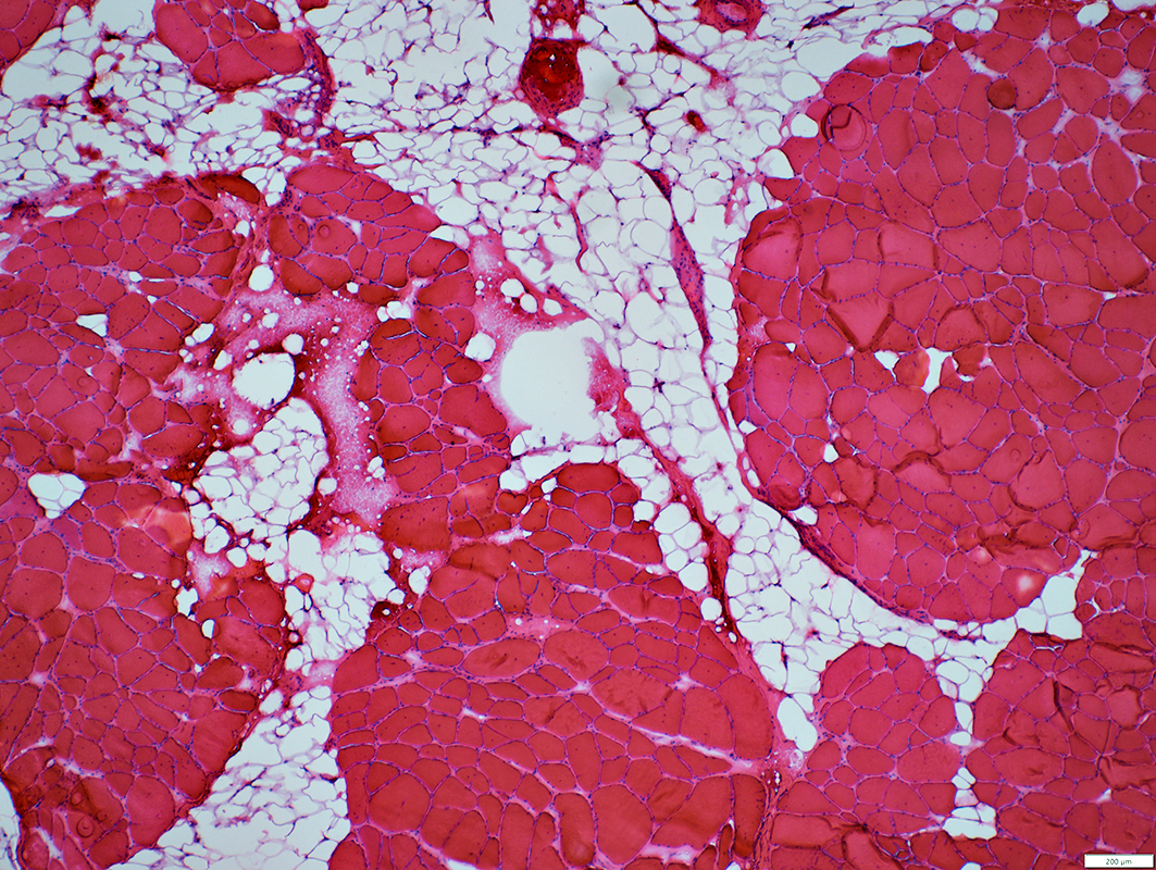 H&E stain |
Sizes: Varied
Small & Hypertrophic fibers: Scattered
Basophilic fibers: Scattered
Internal nuclei: Common
Necrotic fibers: Few; Scattered
Partially fused (Split) fibers
Endomysial connective tissue: Increased
Perimysium: May be replaced by fat
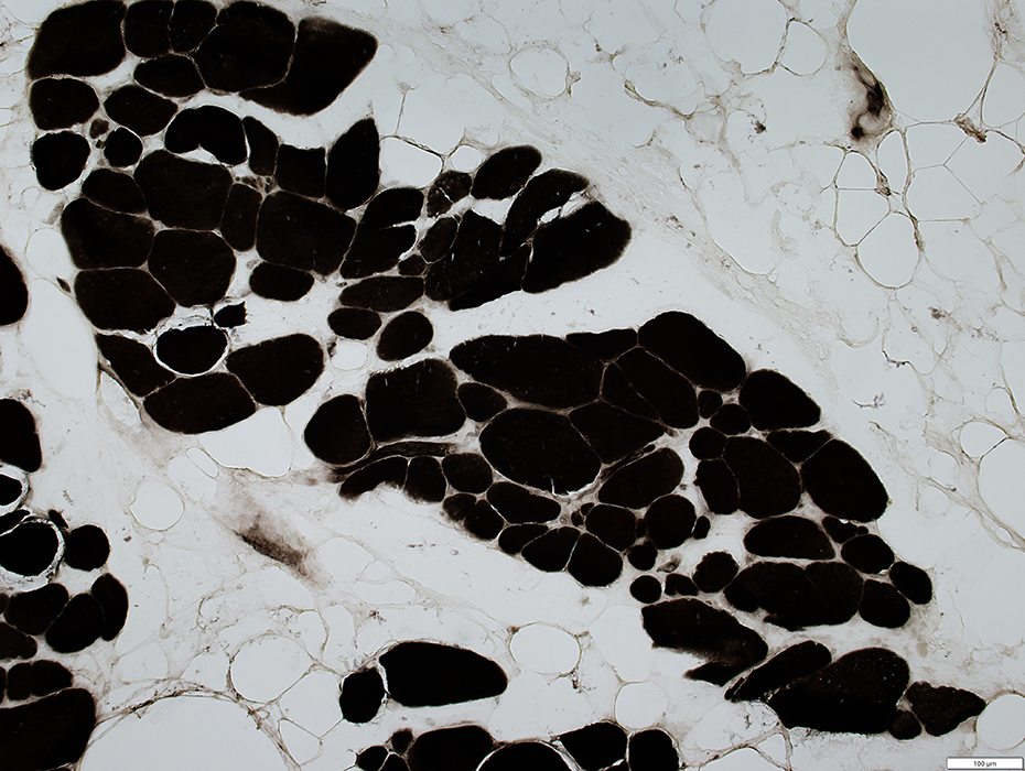
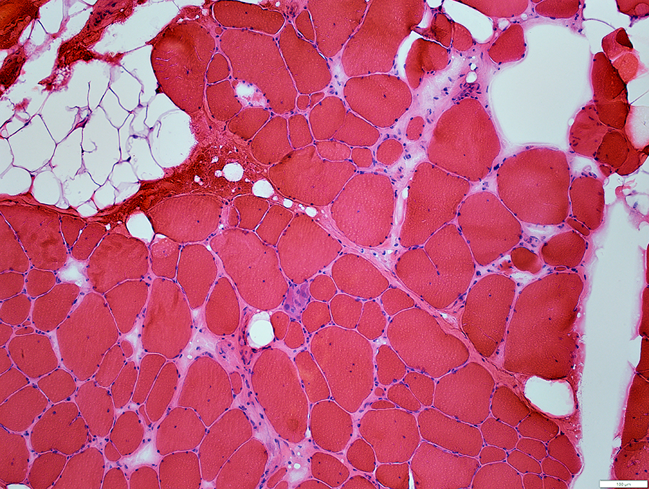 H&E stain |
 H&E stain |
Small & Hypertrophic fibers: Scattered
Internal nuclei: Common
Necrotic & Regenerating fibers: Few; Scattered
Partially fused (Split) fibers
Endomysial connective tissue: Increased
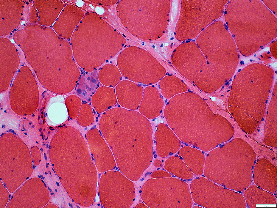
 H&E stain |
 VvG stain |
Partially fused (Split) fibers
Non-fused small fibers closely neighboring other fibers
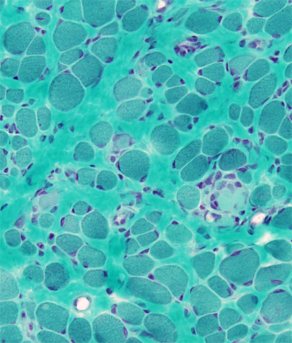
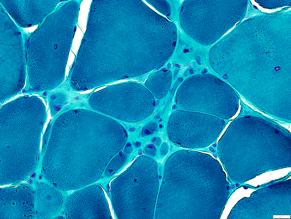 Gomori trichrome stain |
Partially fused (Split) fibers
Non-fused small fibers closely neighboring other fibers
Endomysial connective tissue: Increased
 NADH stain |
Pale-stained fibers: Linear pattern; Pale centers
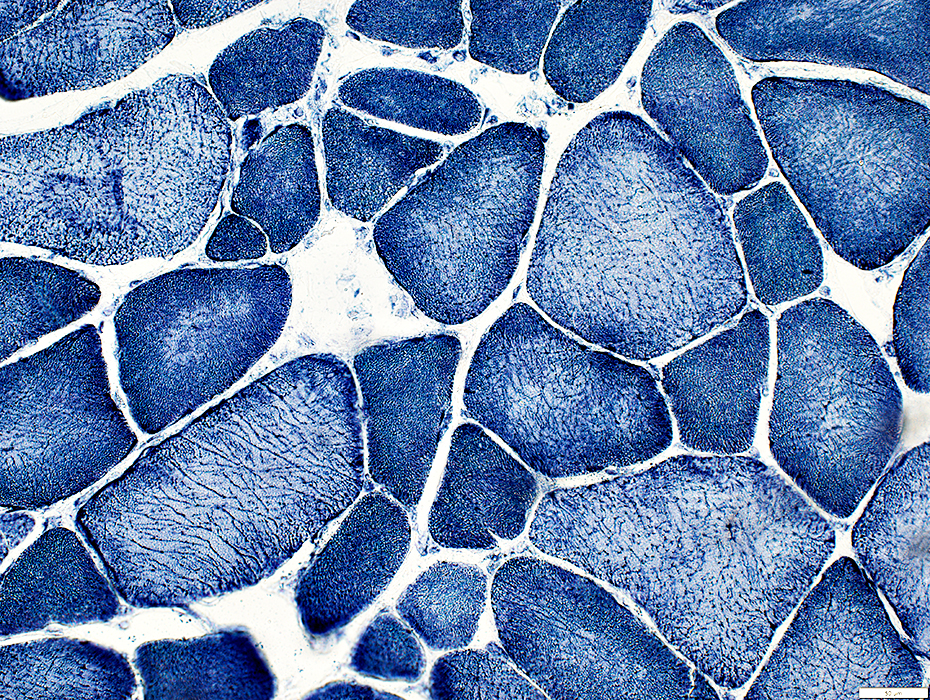
 ATPase pH 4.3 stain |
Type 1 fiber predominance
Type 1 fibers smaller than type 2
 ATPase pH 4.3 stain |
 ATPase pH 4.3 stain |
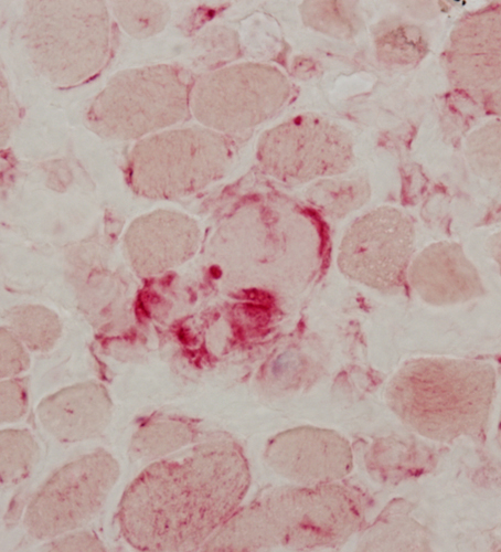
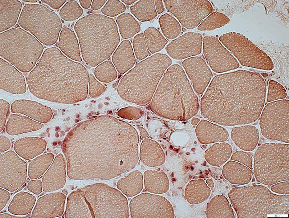 Acid phosphatase stain |
Histiocytic cells replace necrotic fibers
 Alkaline phosphatase stain |
May be mis-oriented: Longitudinal orientation
Mildly increased numbers stained
Return to Neuromuscular Home Page
Return to Fukuyama Congenital MD
3/21/2023