MULTICORE MYOPATHY: Pathology
Histology
- Multiple small areas with reduced oxidative enzyme staining
- Absent: Mitochondria
- Present in both type 1 & 2 fibers
- Length: Shorter than cores
- Myofibrils: Focal disruption of few sarcomeres
- Hereditary
- Denervation
- Tenotomy
- Rule out: Motheaten
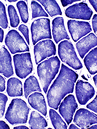 NADH stain |
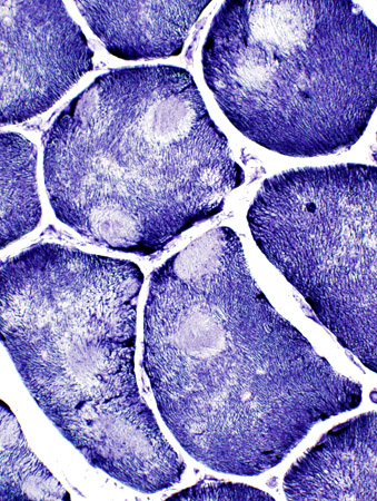 |
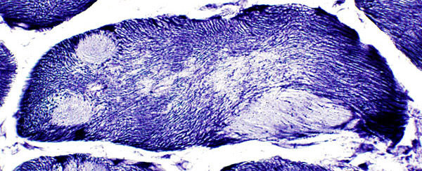 NADH stain |
|
Mutliple internal clear zones in muscle fibers
|
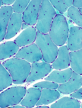
|
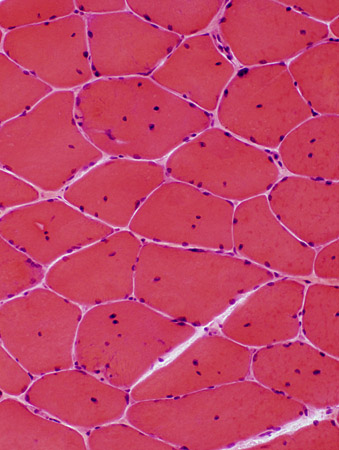
- Fiber size: Variability
- Internal nuclei: Many fibers
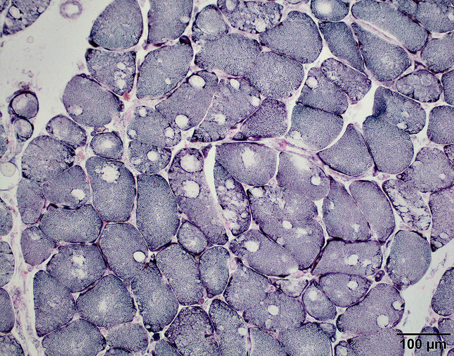 NADH stain |
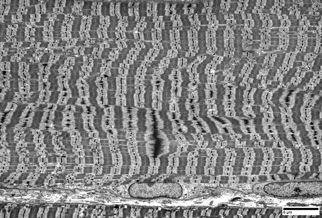
|
Multicore/Minicore: Titin mutations
Patient: 2 year old female with noncompaction cardiomyopathy, hypotonia & developmental delayGenetics: Titin mutations K2186C & D19486Y
From Chunyu (Hunter) Cai
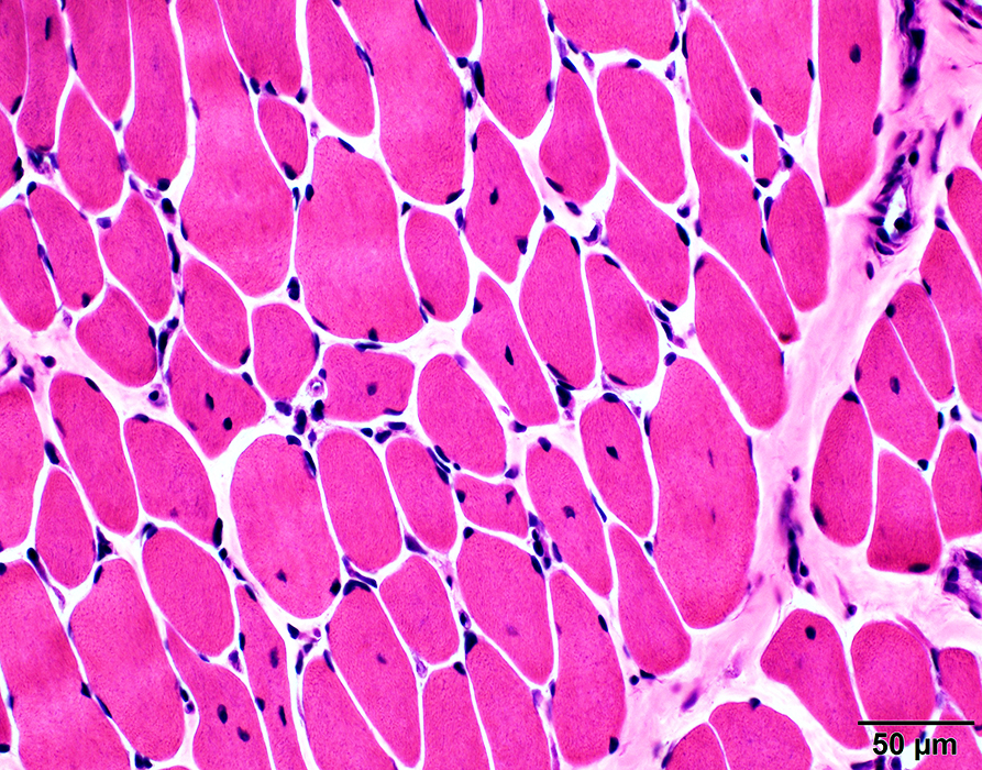 H&E stain |
Fiber sizes: Abnormal variation
Internal nuclei
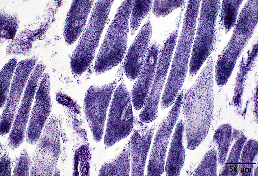 NADH stain |
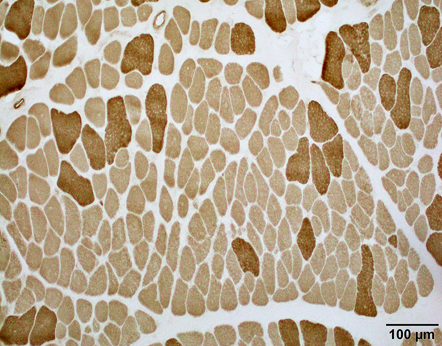 ATPase pH 9.4 stain |
Type 1 fibers: Predominance; Smallness
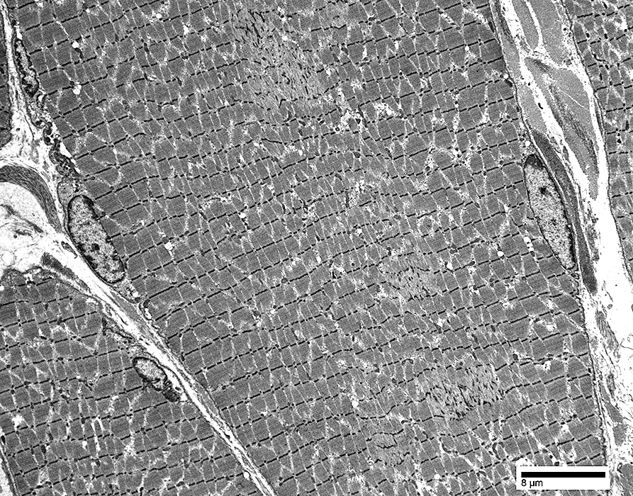
|
Horizontal Z band streaming: Multifocal regions
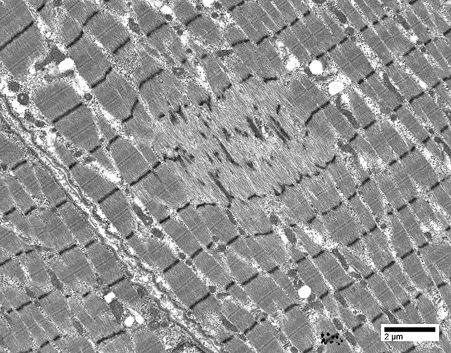
|
Return to Multicore myopathy
Return to Neuromuscular Home Page
10/23/2023