EMERY-DREIFUSS MUSCULAR DYSTROPHY 1 (EMD1)
|
Affected males Carrier female |
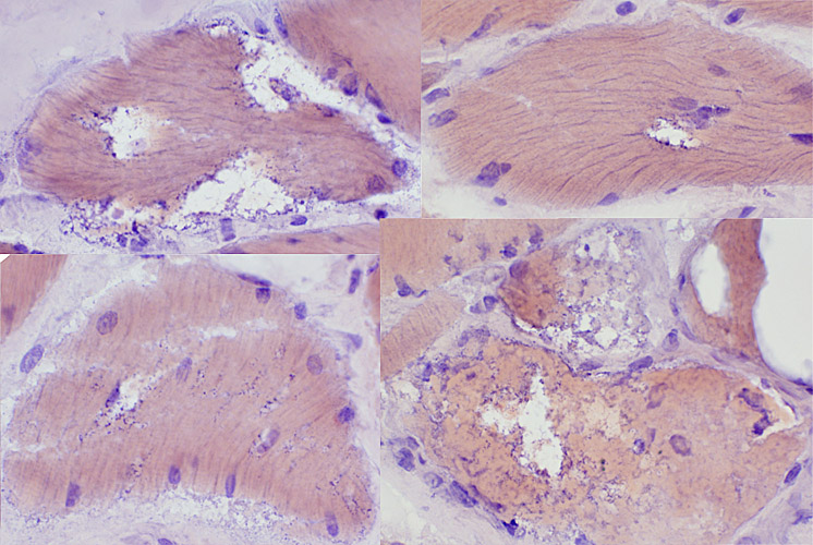
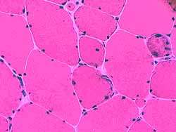
Affected Males
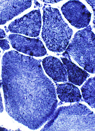
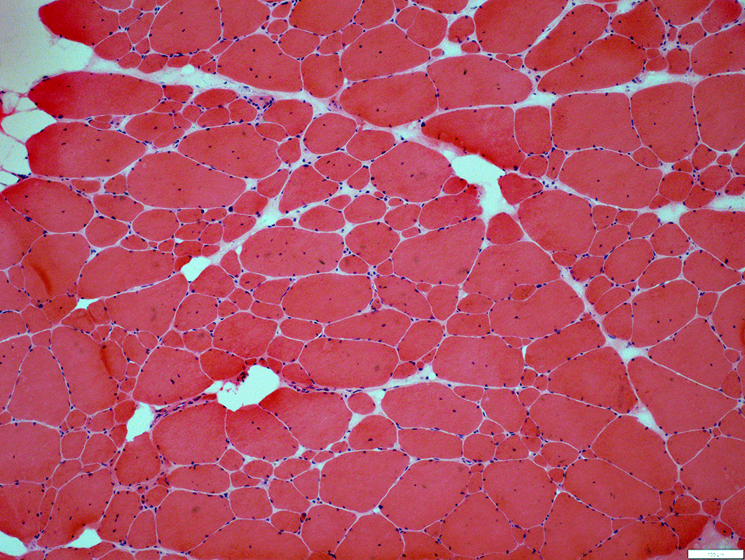 H & E stain |
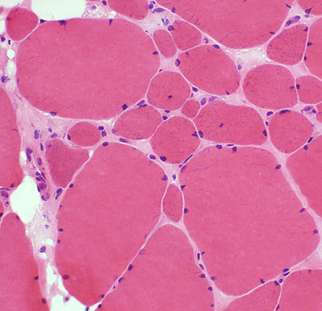
Moderately affected muscle (Above)
Fiber sizes: Varied
Internal nuclei
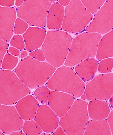
Severely affected muscle (Below)
Perimysial connective tissue: Replacement by fat
Endomysial connective tissue: Increased between muscle fibers
Fiber sizes: Varied
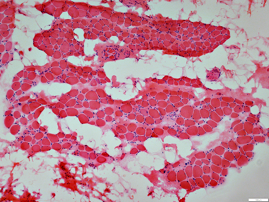 H & E stain |
 VvG stain |
 H & E stain |
 |
|
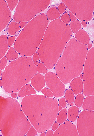
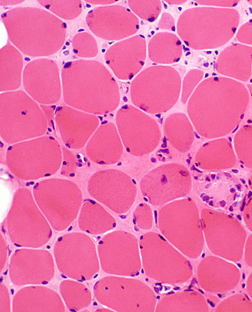
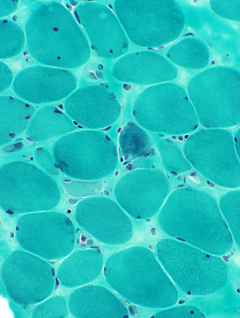
Myopathy Bimodal variation of fiber size Small fibers: Rounded Large fibers: Hypertrophied Increased endomysial connective tissue Internal nuclei: Some fibers |
|
 H & E stain |
 |
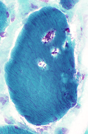
 Gomori trichrome stain |
 |
|
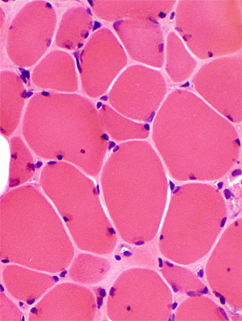
Chronic myopathy Varied fiber size Increased endomysial connective tissue Replacement of muscle by fat |
|
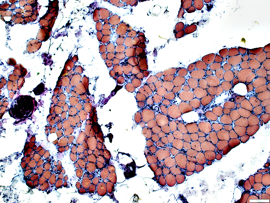
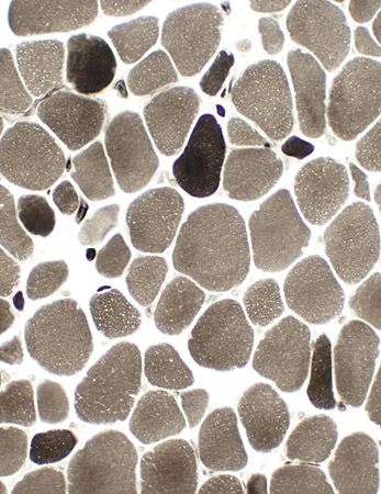
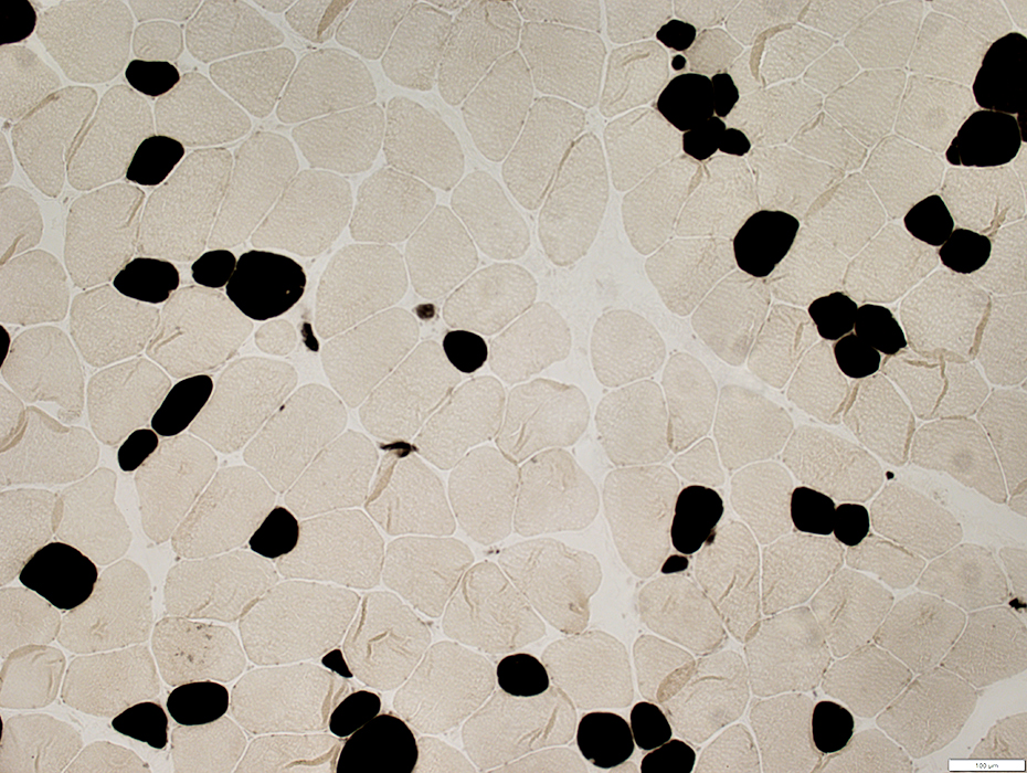
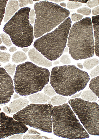 ATPase ph 9.4 stain |
 |
|
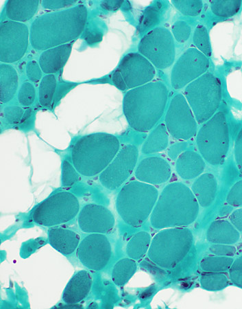
Fiber type abnormalities Small type 1 (Left) Type 1 predominance (Right) |
|
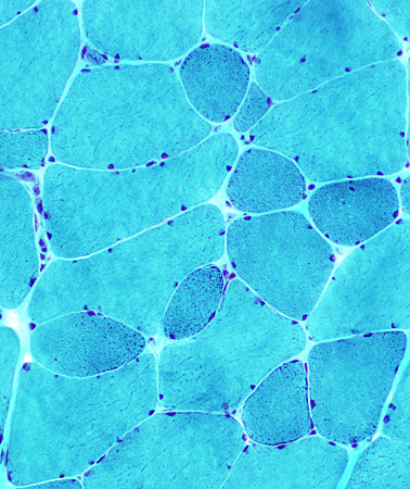
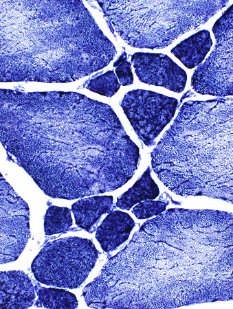 NADH stain |
 |
|
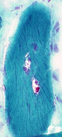
Abnormal internal architecture |
|
 Gomori trichrome stain |
 |
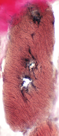 VvG stain |
 Congo red stain |
||
Emerin Stain
Normal muscle
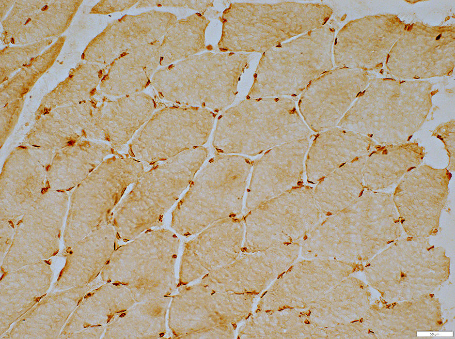
Emerin stain |
|
 Emerin stain |
Muscle Fiber Nuclei Subsarcolemmal Stain for emerin |
Emerin: Normal muscle
Emerin protein: Present in varied amounts in different nuclei
Myonuclei, Subsarcolemmal: Abundant emerin (Green; Yellow arrow)
Capillary nuclei: Little emerin (Blue; White arrow)
Muscle fiber sarcolemma: Dystrophin (Red)
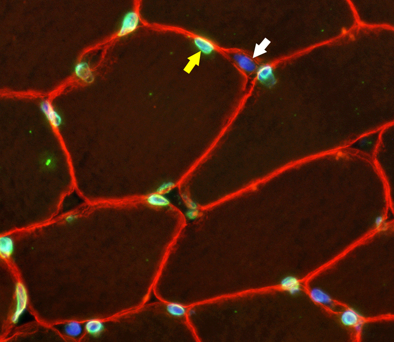 Emerin - Green; DAPI - Blue; Dystrophin - Red |
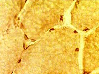
EDMD muscle: No emerin staining of nuclei
 Emerin stain |
|
 Emerin stain |
Muscle Fiber Nuclei No emerin staining |
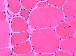
Manifesting Carrier (Female)
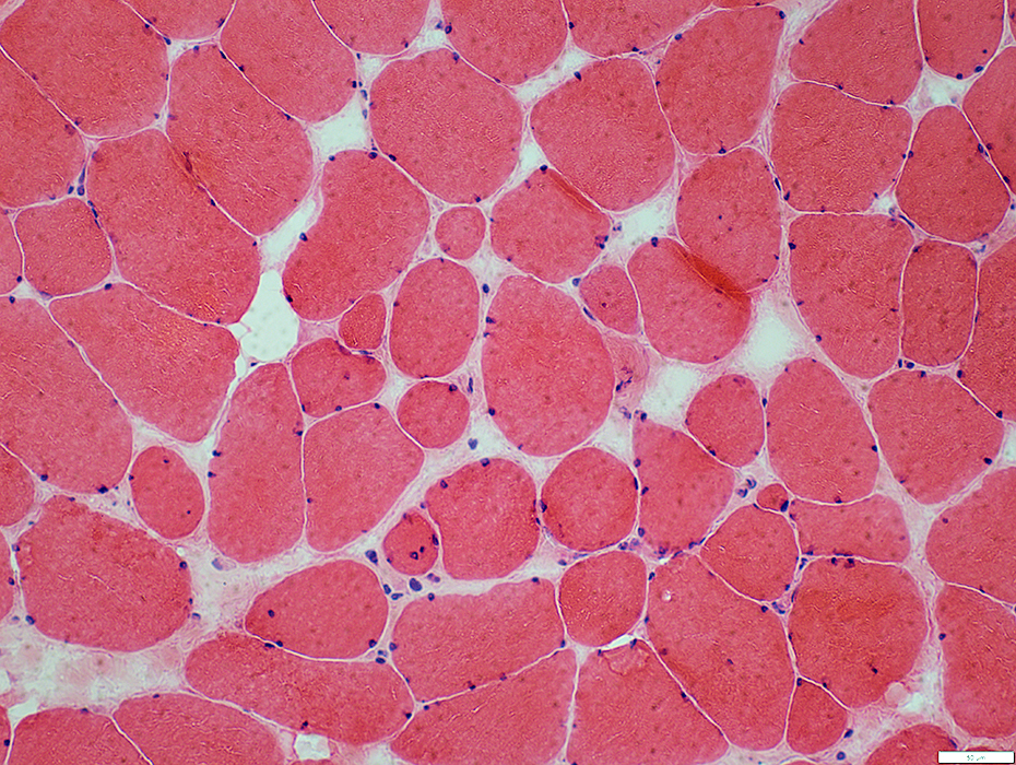 H & E stain |
  H & E stain |
|
Muscle fiber sizes: Varied Small fibers Rounded Some basophilic regenerating Large fibers: Hypertrophied. Internal nuclei: Some fibers Connective tissue: Mild increase 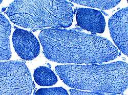 NADH |
 Gomori trichrome |
 H & E stain |
|
Abnormal internal architecture |
 ATPase, pH 4.3 |
|
Fiber type disorder Most small fibers are type I Larger fibers are type I & II 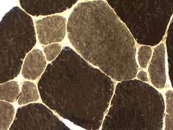 ATPase, pH 9.4 |
 |
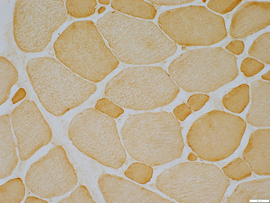
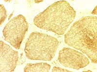
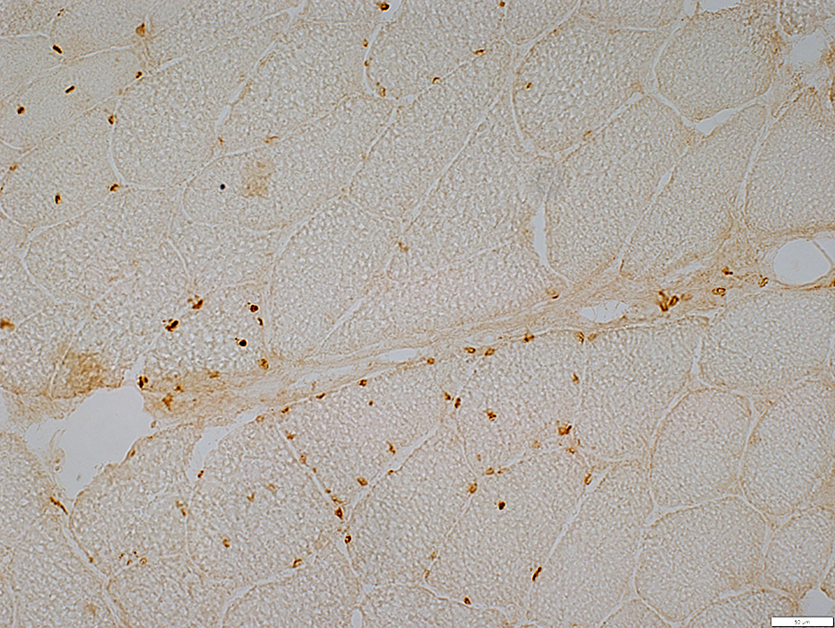
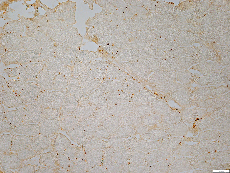 Emerin Immunohistochemistry
|
Only some myonuclei stain for emerin.
Emerin staining of nuclei is non-random and patchy.
Some muscle fibers have many nuclei with emerin.
Other muscle fibers have no nuclei with emerin.
|
Return to Emery-Dreifuss.
Return to Neuromuscular syndromes
Return to Neuromuscular home page
11/12/2025