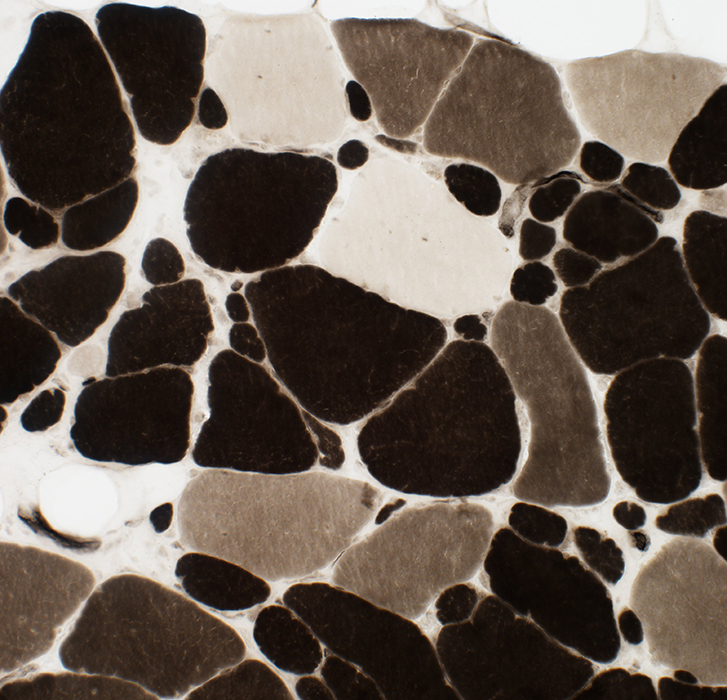
SQSTM1: Distal Myopathy
Digenic: SQSTM1 c.1165 G>A; TIA1 N357S
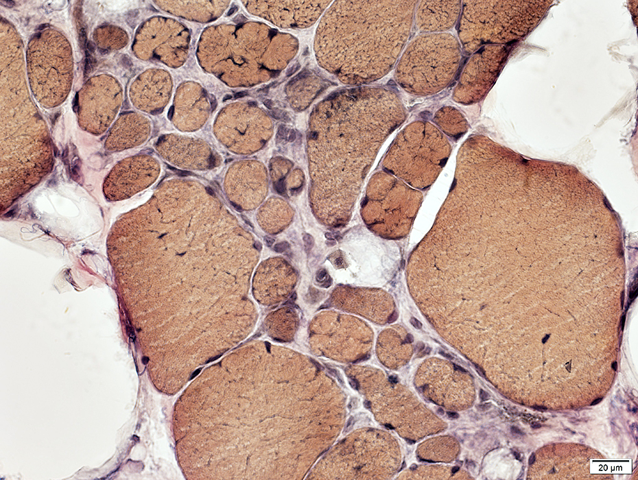
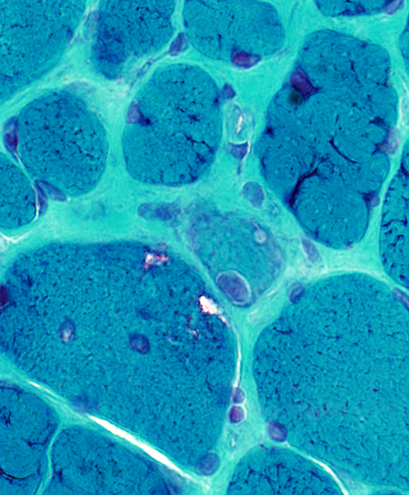
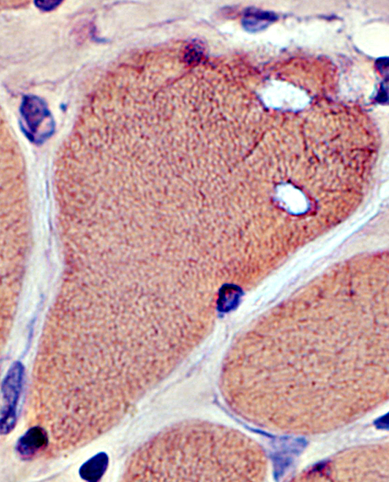
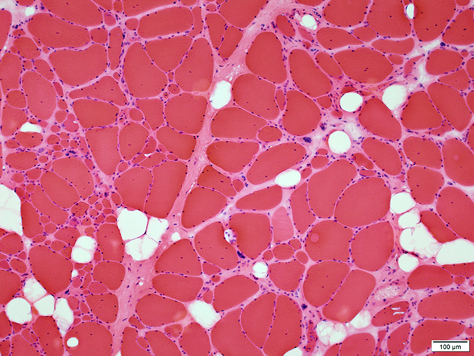 H&E stain |
Fiber sizes
Varied from Small to Large, Occasional pyknotic nuclear clumps
More small fibers in some areas than others
Perimysium & Endomysium: Replaced by fat
No necrosis or regeneration
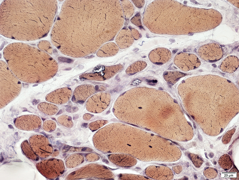
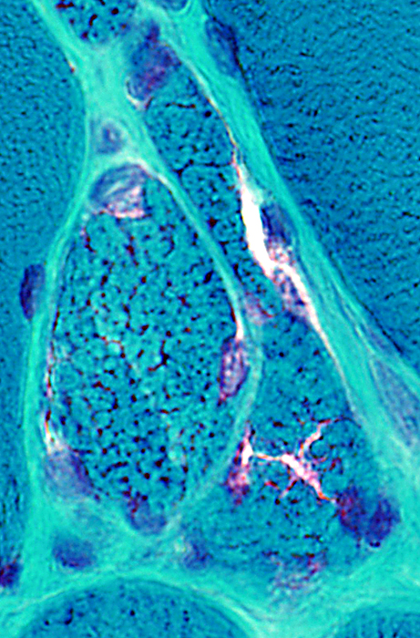
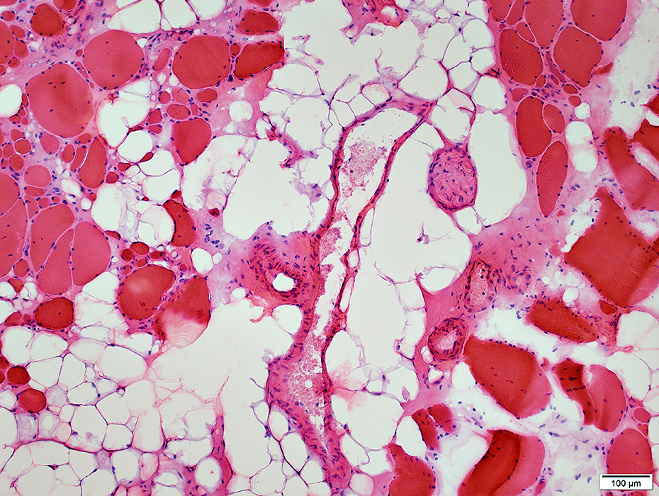 H&E stain |
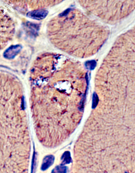
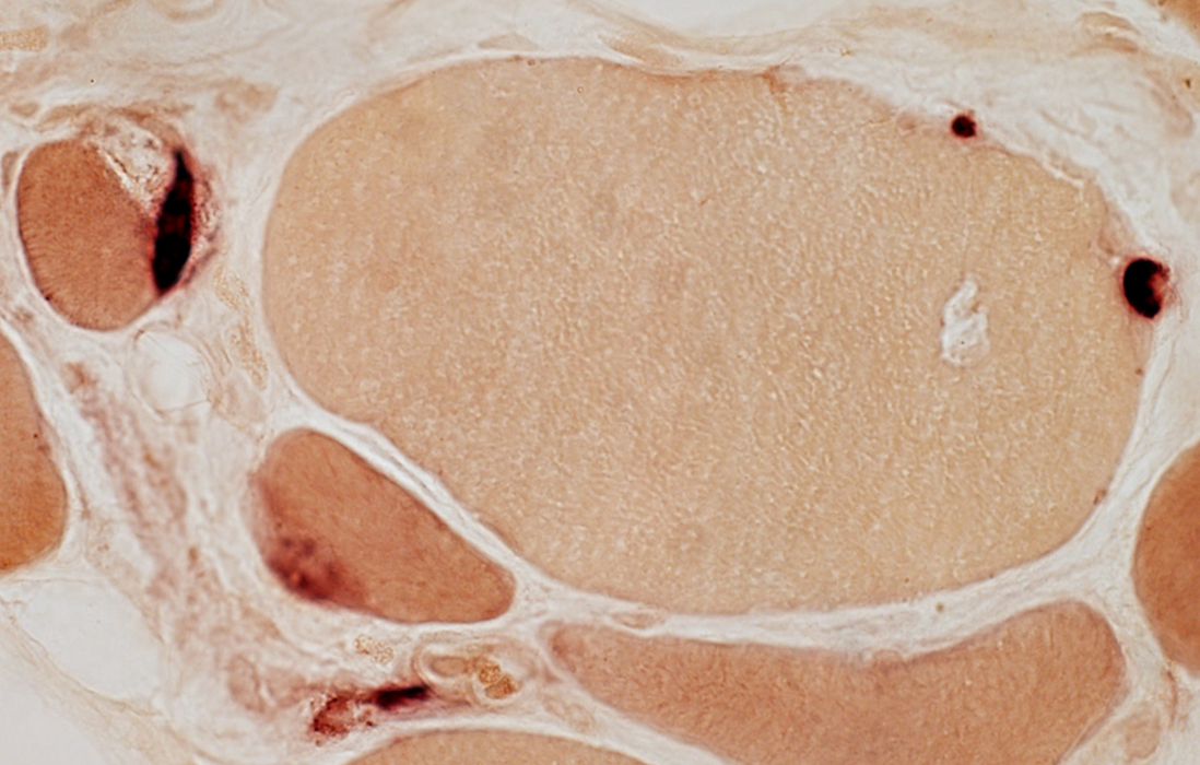
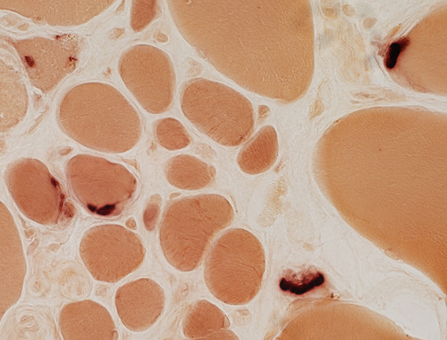
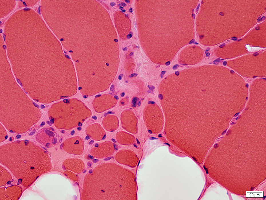 H&E stain |
May occur in clusters or groups
Some have basophilic cytoplasm & large nuclei
Irregular internal architecture or Dark staining
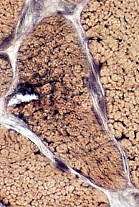
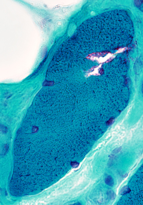
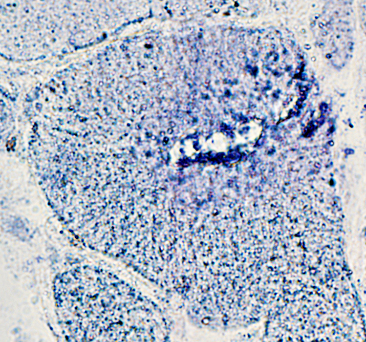
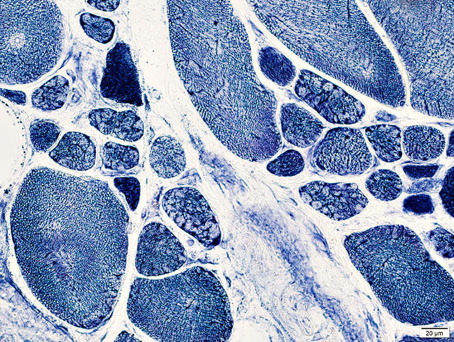 NADH stain |
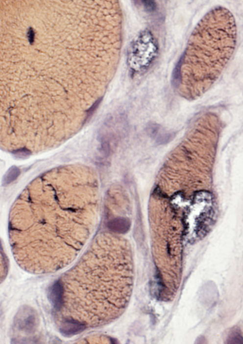
 VvG stain |
Vacuoles
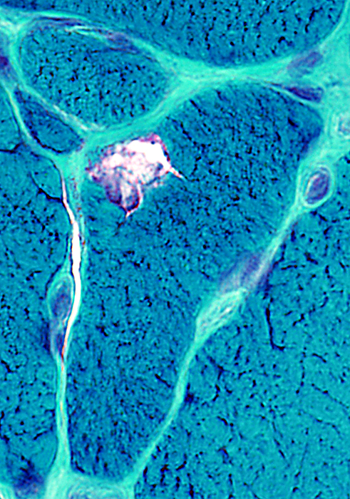
 VvG stain |
 VvG stain |
 VvG stain |
 Gomori trichrome stain |
 Gomori trichrome stain |
 Gomori trichrome stain |
 Gomori trichrome stain |
 Congo red stain |
 Congo red stain |
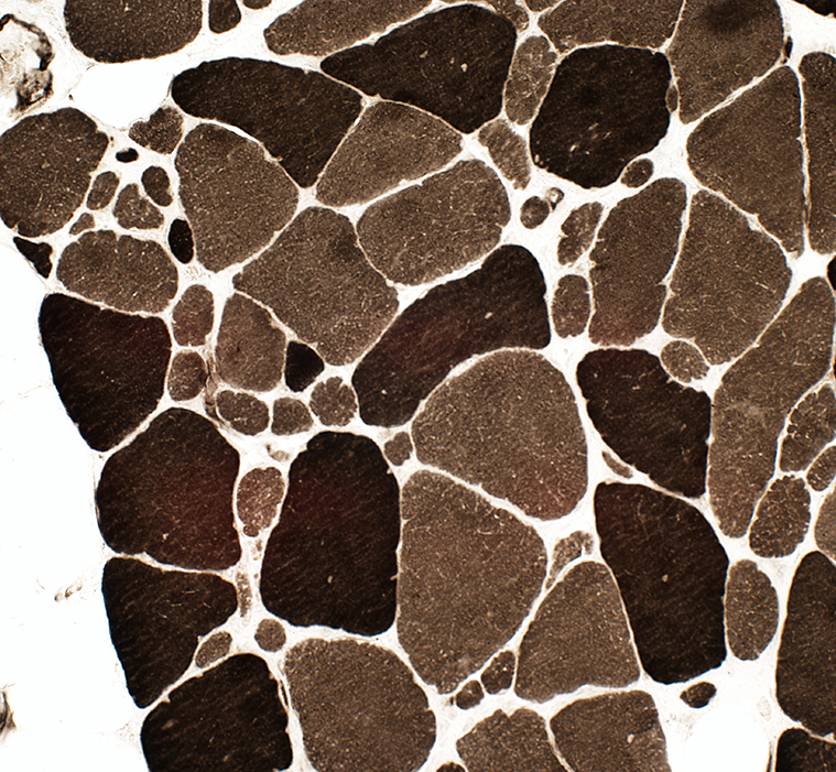
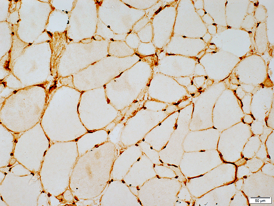
 AMPDA stain |
Fiber types
 ATPase pH 9.4 stain |
Intermediate-stained muscle fibers at ATPase pH 4.3
 ATPase pH 4.3 stain |
MHC Class I: Upregulated on muscle fiber surface
 MHC Class I stain |
 Esterase stain |
 Esterase stain |
Return to Distal myopathy
Return to SQSTM1 myopathy
Return to Muscle biopsies
Return to Biopsy illustrations
Return to Neuromuscular home page
6/2/2021