|
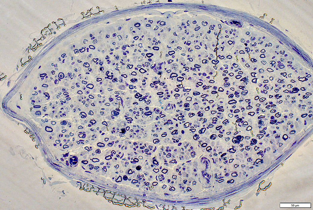
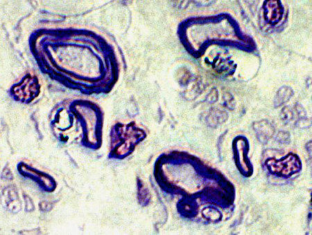
Metachromatic Leukodystrophy (MLD)
Schwann cells
Contain cytoplasmic debris, particulate & clear
Axons surrounded by
Thin myelin sheaths & Onion bulbs, small
|
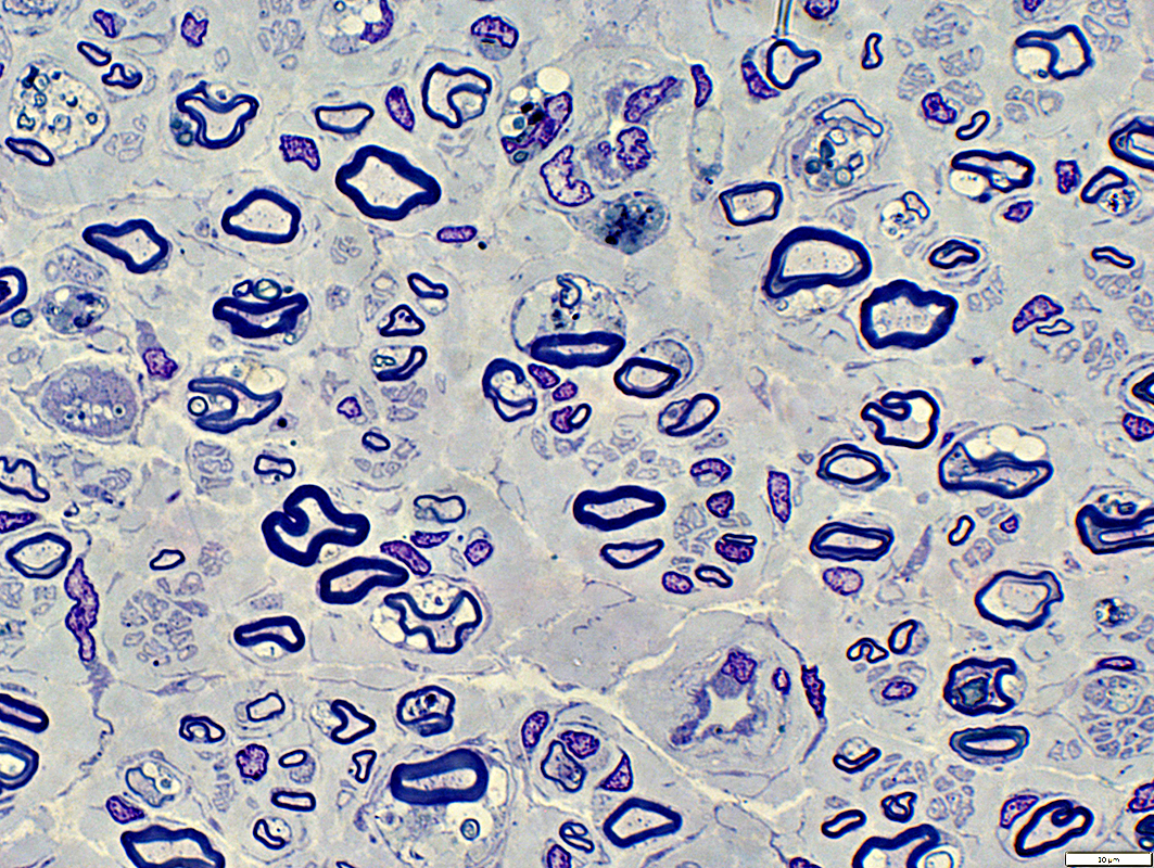
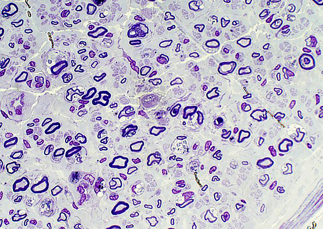
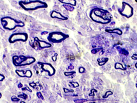
Toluidine blue stain
|
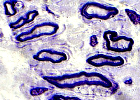
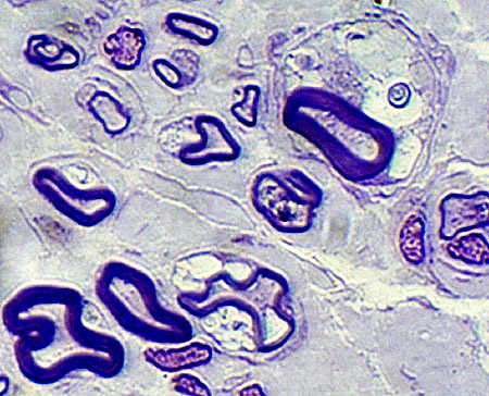
Myelinating Schwann cells
Contain particulate & clear debris in cytoplasm
Onion Bulbs: Small
|
|
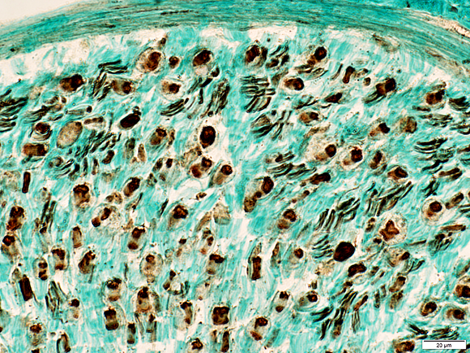
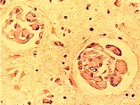
MLD Histochemistry
- Hypomyelination
- Lysosomal activation: Some endoneurial cells
- Storage material
- Most reliably assessed on frozen tissue
- Stains
- Brown-Yellow metachromatic material
- Hirsch-Peiffer (Acidified Cresyl Violet)
- Toluidine blue
- PAS positive
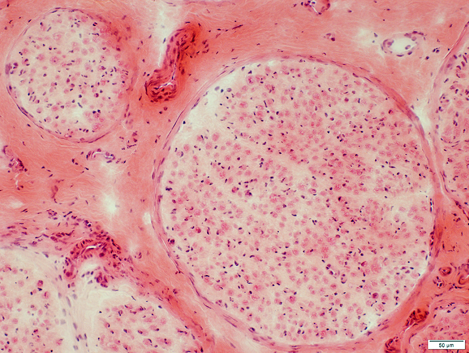
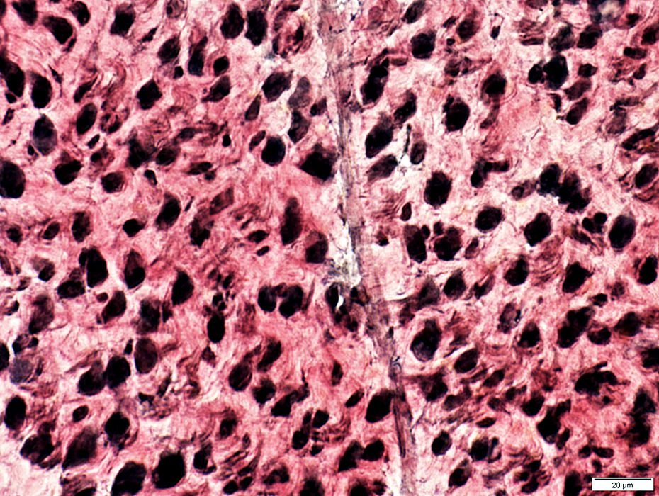
H&E stain
|
Endoneurium
Pale
Increased space between axons
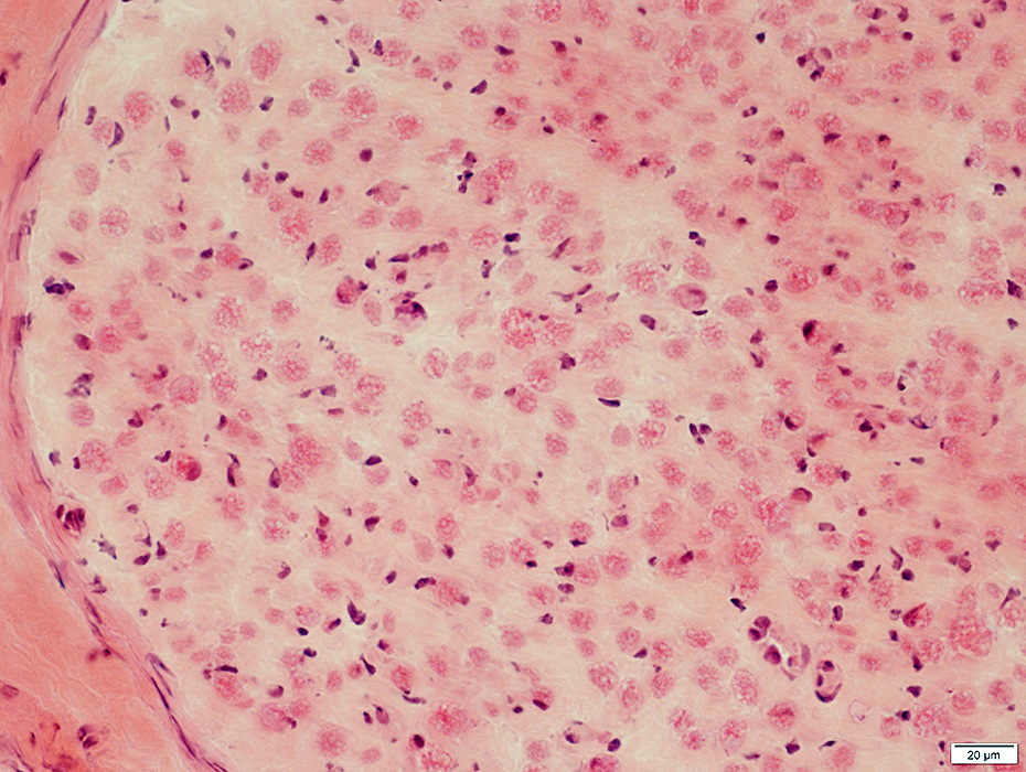
H&E stain
|
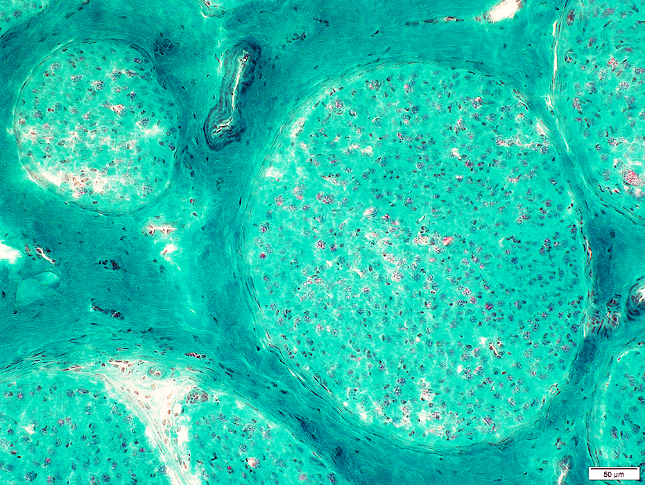
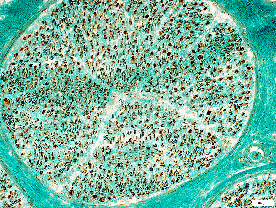
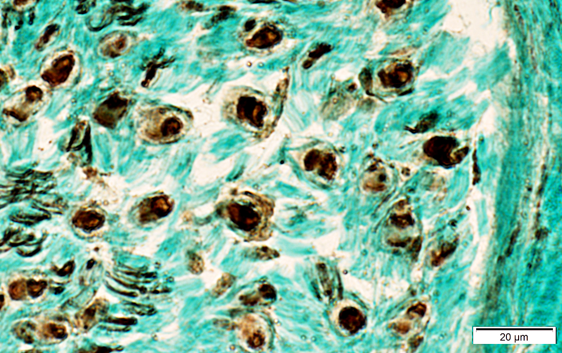
Gomori trichrome stain
|
Reduced staining for myelinated axons
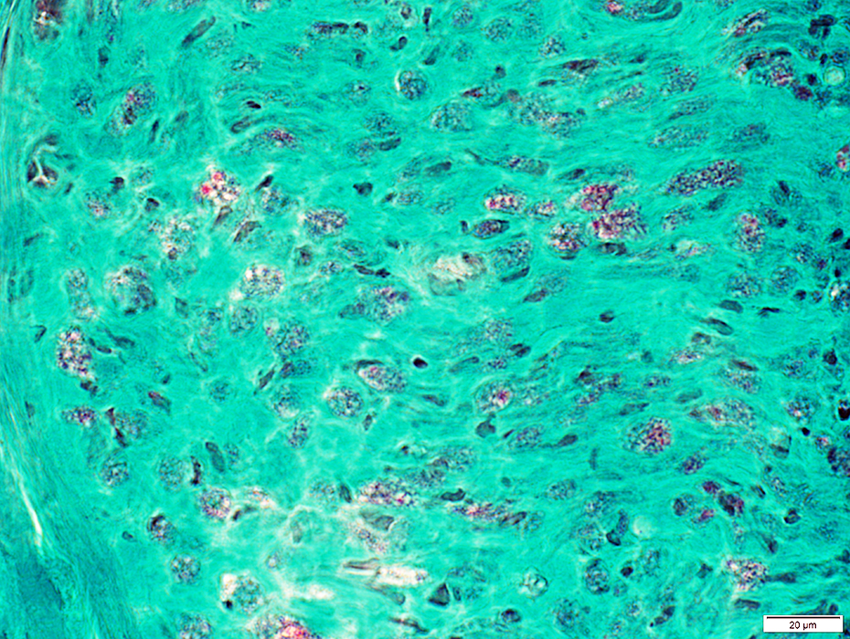
Gomori trichrome stain
|
Larger axons
Increased space between axons
VvG stain
|
Larger axons: Normal to mildly reduced numbers
Smaller axons: Reduced numbers in some endoneurial areas
Neurofilament stain
|
Lysosomal activity in some endoneurial cells
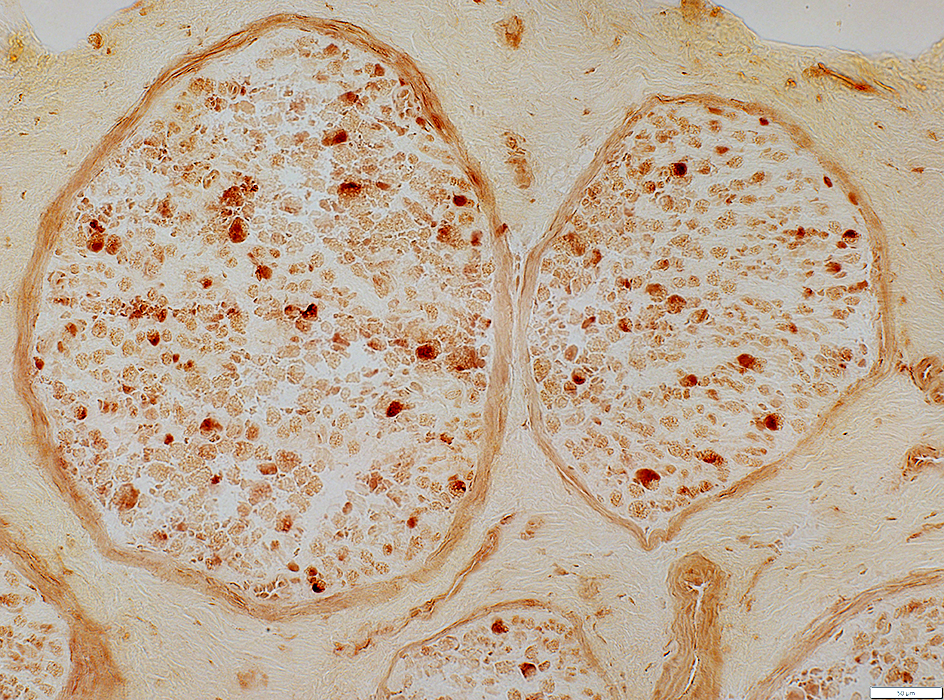
Acid phosphatase stain
|
Schwann Cell Types
Bungner Bands (Yellow)
Contain both P0 & NCAM
Scattered in endoneurium
P0(r).jpg)
NCAM stain (Green)
P0 stain (Red)
|
NCAM(r).jpg)
Neurofilament stain (Green)
NCAM stain (Red)
|
MBP(r).jpg)
Neurofilament stain (Green)
MBP stain (Red)
|
P0(r).jpg)
Neurofilament stain (Green)
P0 stain (Red)
|
MBP(r).jpg)
P0 stain (Green)
MBP stain (Red)
|
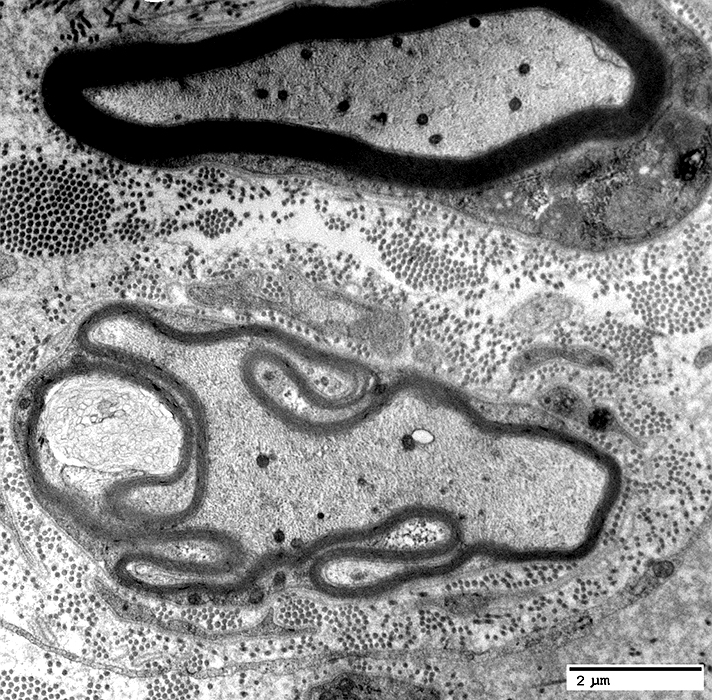
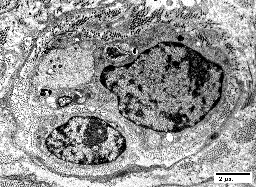
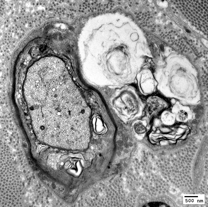
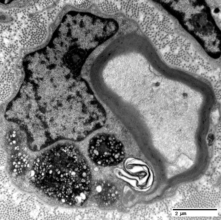
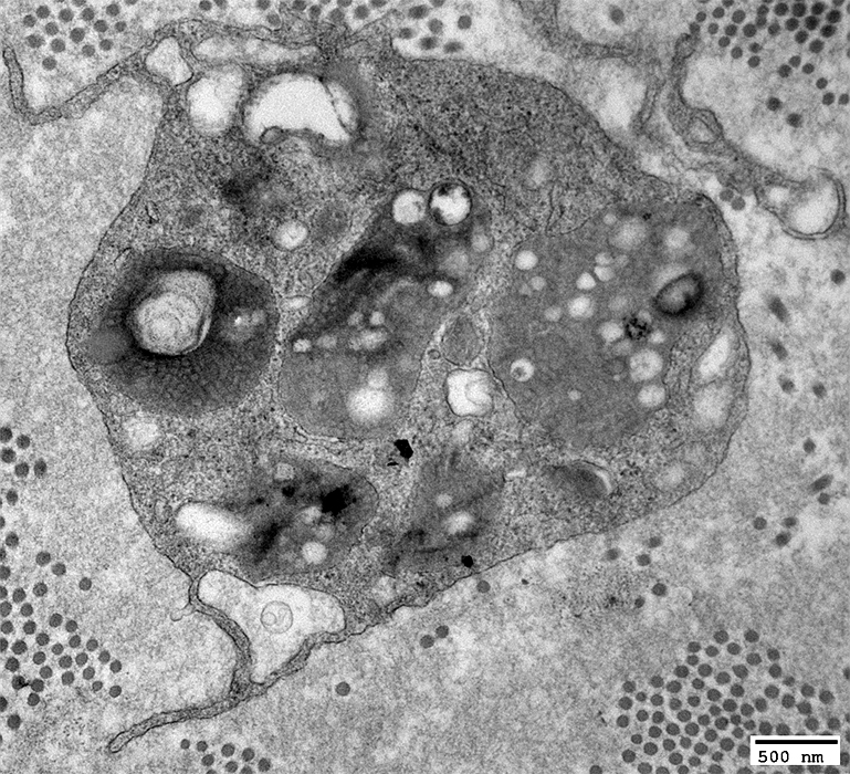
MLD: Reduced or Absent Myelin Sheath
Unmyelinated axons: Surrounded by Schwann cells
Axons with thin, or folded, myelin sheath
Onion bulb: Small, single layer of basal lamina around axons & Schwann cells
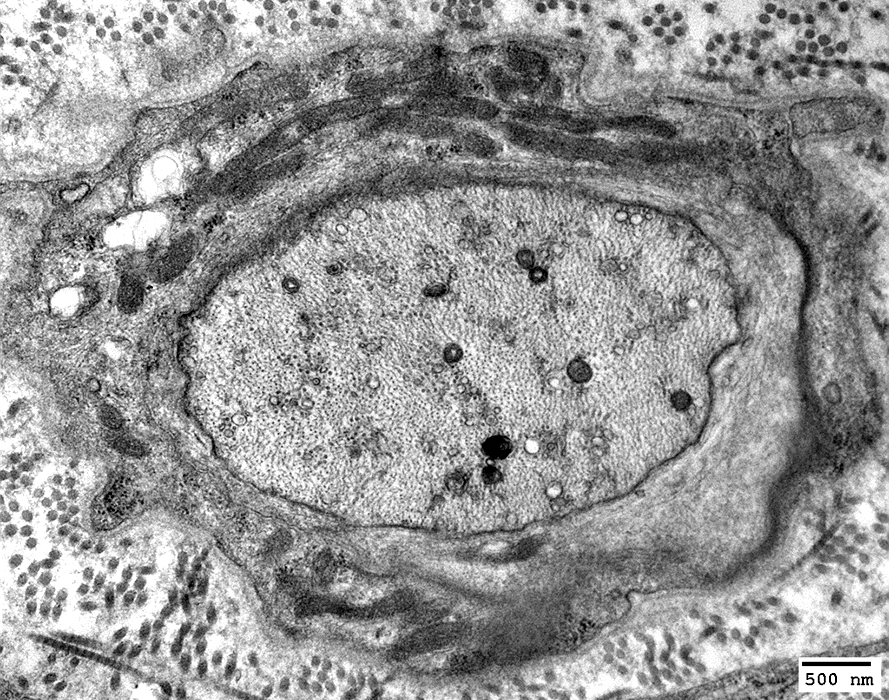
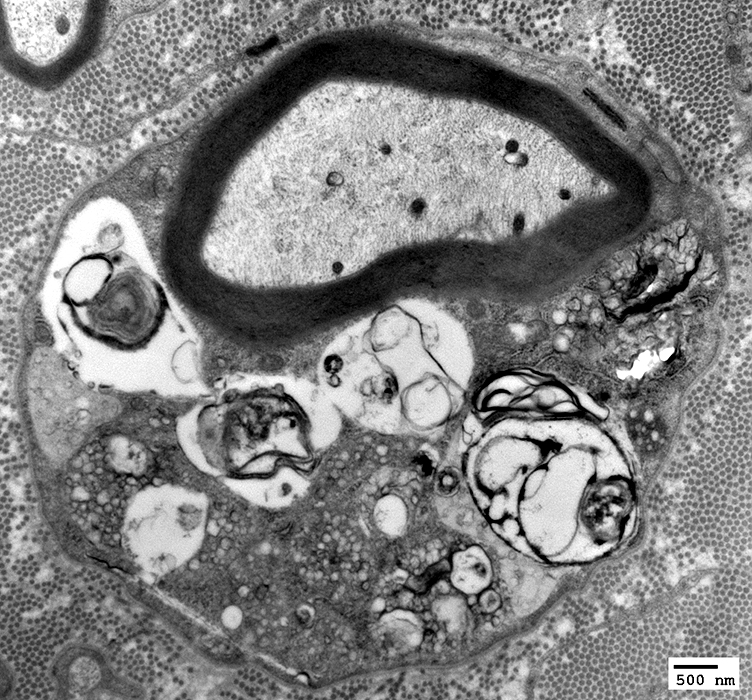
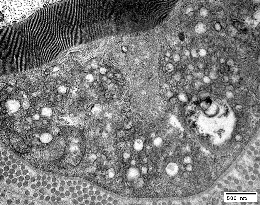
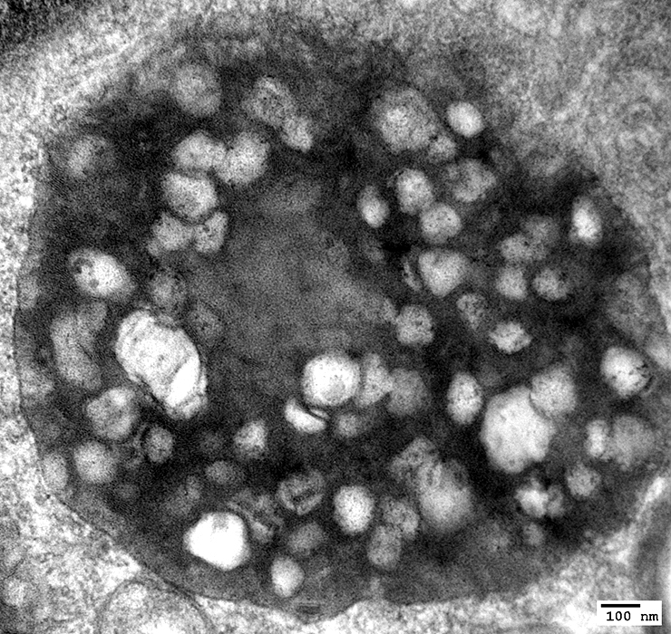
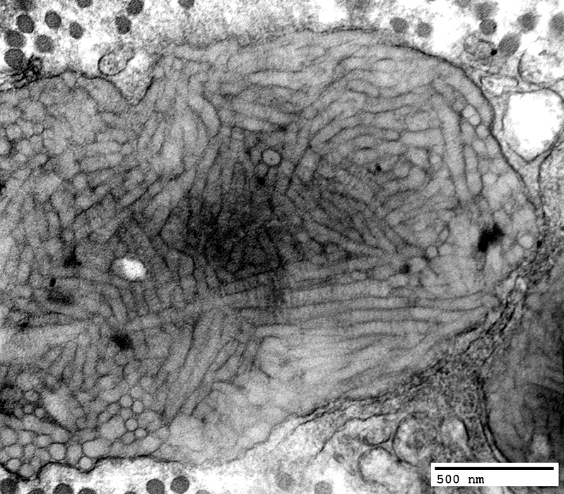
MLD: Schwann cell inclusions
Axons: Moderately or Severely thin myelin sheath
Schwann cells: Contain myeloid debris in secondary lysosomes
Schwann cells: Cytoplasm contains Tuffstone inclusions & Myeloid debris
Schwann cells with Cytoplasmic inclusions
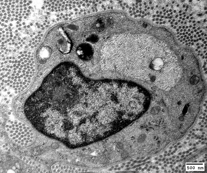
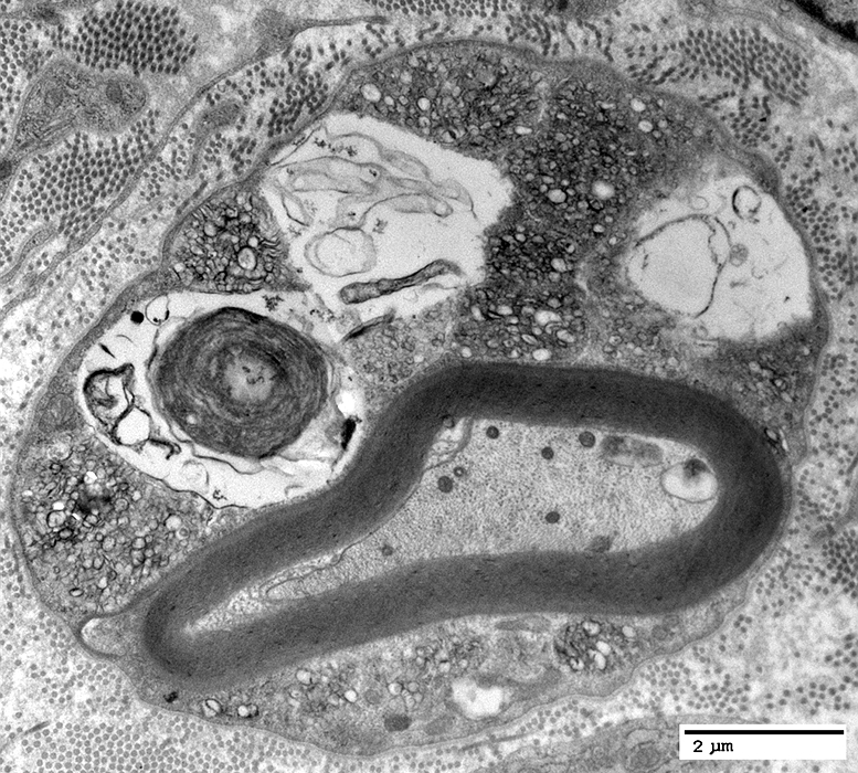
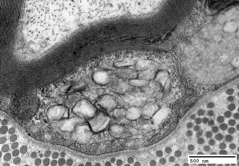
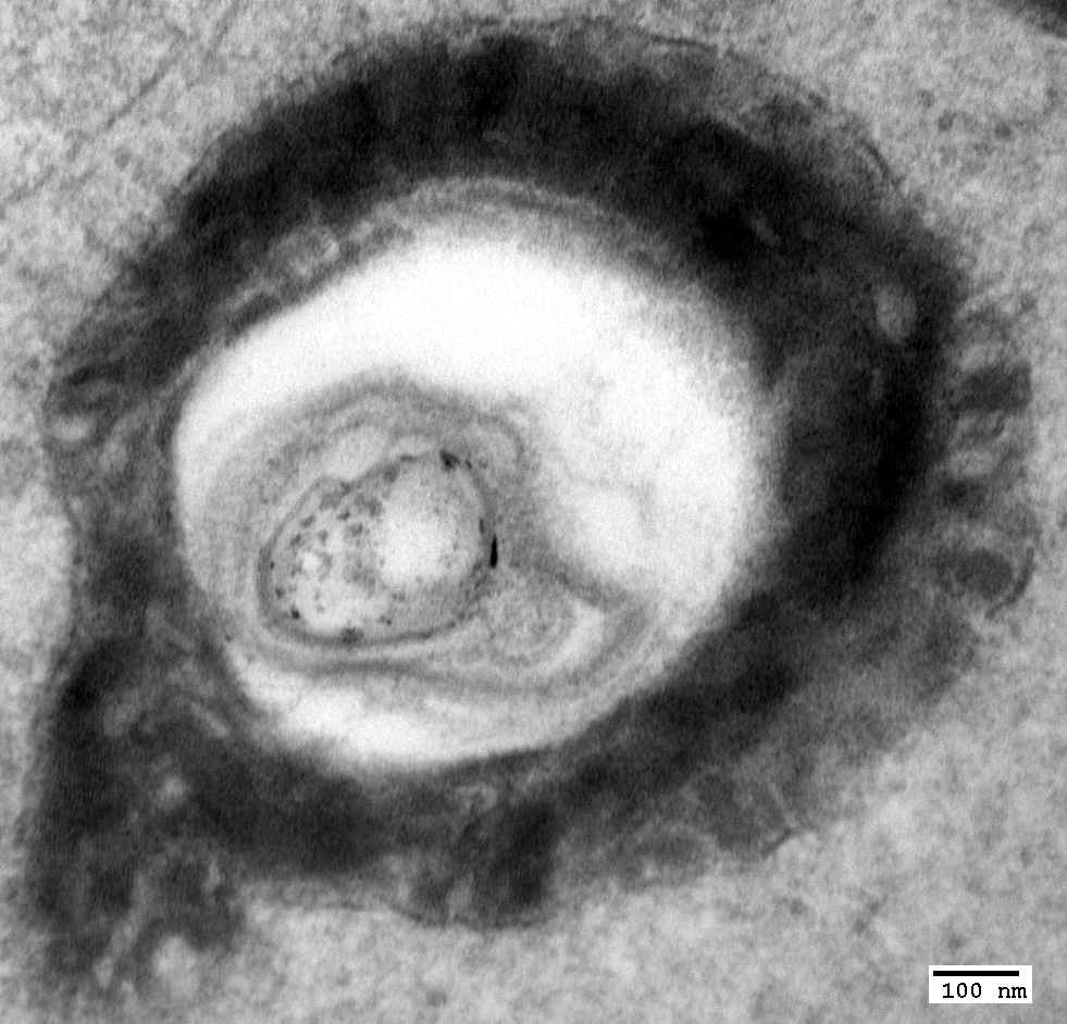
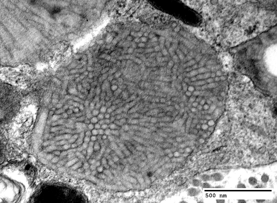
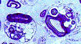
MLD: Tuffstone Bodies
Tuffstone bodies
Electron dense: Sharply demarcated
Most common in Schwann cells of myelinated & unmyelinated axons
Contain
Irregularly oriented granular material
Areas with 5.8 nm periodicity
Peripheral lamellae oriented perpendicular to surface
Vacuoles: Electon lucent
Tuffstone bodies
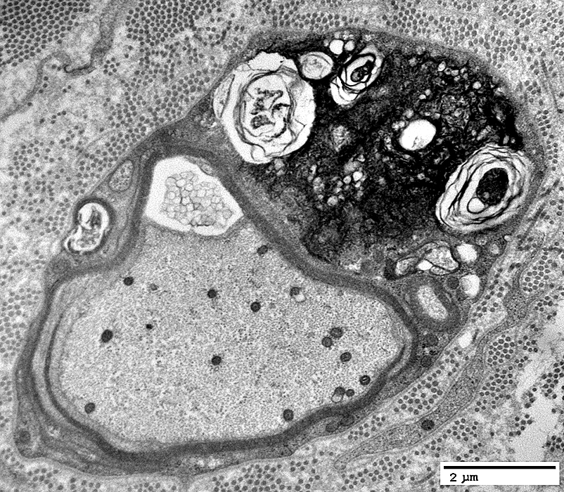
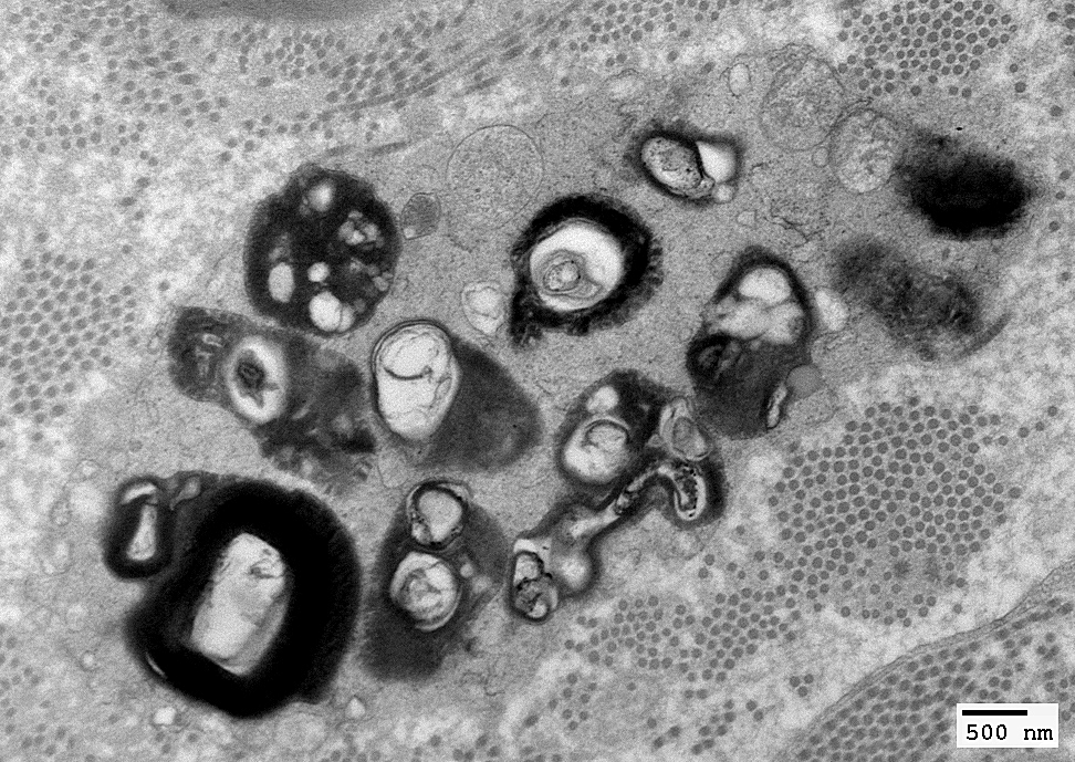
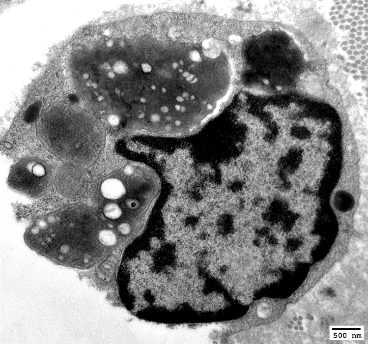
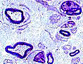
MLD: Macrophages
Prismatic ("Herringbone") inclusions
Commonly in endoneurial macrophages
Stacks of lamellar disks
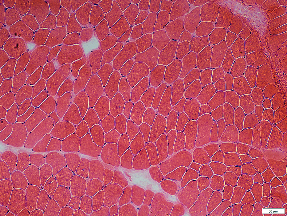
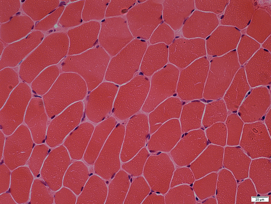
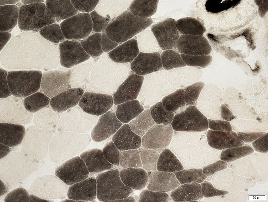
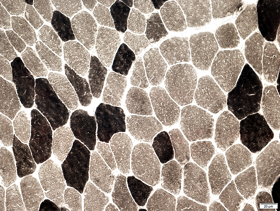
MLD: Muscle
Muscle fiber sizes: Mild variation; No denervation
Muscle fiber types
Fiber type grouping: Mild
Type 2C: Increased numbers
|
Krabbe disease: Globoid cells
|
Return to
Normal nerve biosies
Return to
Biopsy illustrations
Return to
Neuromuscular Home Page
Return to
Nerve biopsy
Return to
Demyelinating neuropathies
5/21/2021
|