Immune Myopathies: MDA5 antibody-associated
Myopathologic features 1,2
PerimysiumMacrophages: Scattered perimysial & endomysial; Some perivascular
Muscle fibers
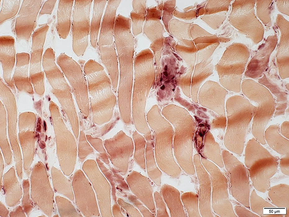
Necrosis & Regeneration: Few fibers; Scattered
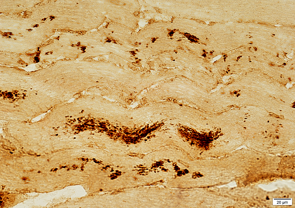
MHC Class I: Upregulated in some patients
LC3 aggregates
NOS2, HSP70, NCAM in sarcoplasm, especially in perifascicular fibers near inflammation
Interferon-related molecules: RIG-I; ISG15
Capillaries
Normal or Mildly reduced numbers
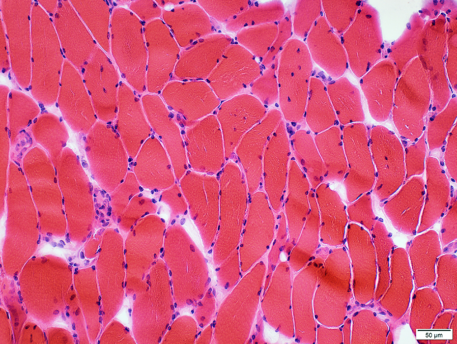 H&E stain |
Necrosis & Regeneration: Scattered
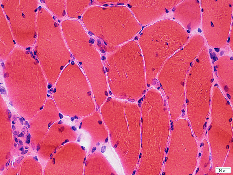 H&E stain |
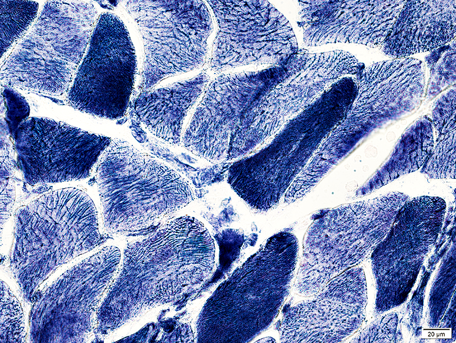
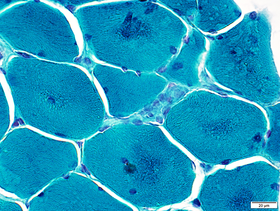 Gomori trichrome stain |
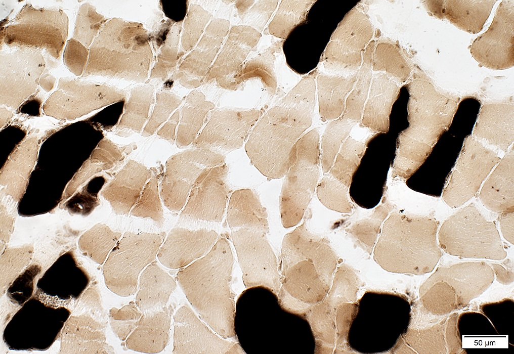
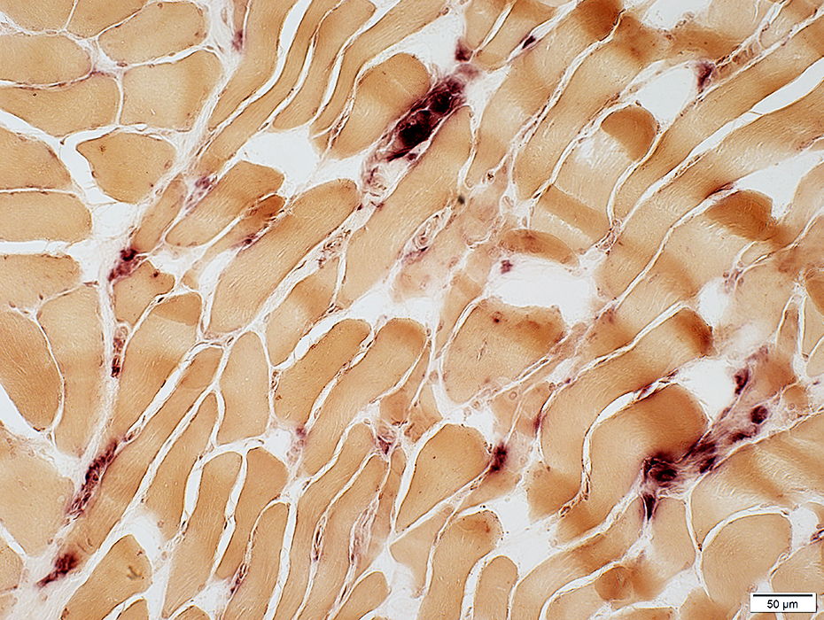 Acid phosphatase stain |
Scattered in endomysium
Replacing necrotic muscle fibers
 Esterase stain |
 ATPase pH 4.3 stain |
 NADH stain |
 LC3 stain |
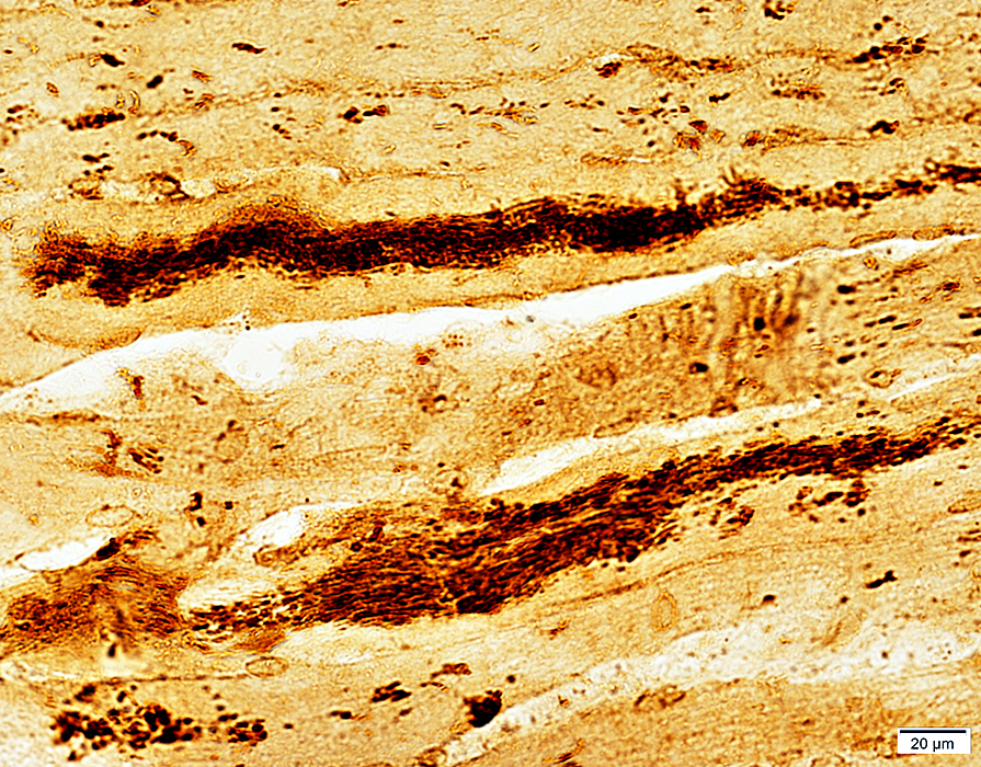 LC3 stain |
 MHC Class I stain |
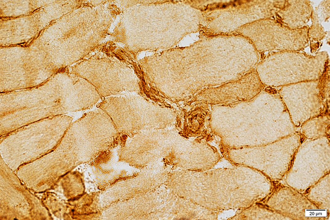 MHC Class I stain |
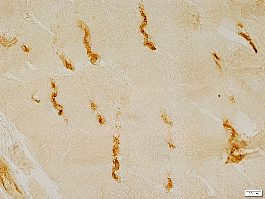 Ulex stain |
Return to Inflammatory myopathies
Return to IMPP
Return to Pathology index
Return to MDA5
References
1. Curr Opin Rheumatol 2011;23:595-604
2. Am J Pathol 2016;186:691-700
9/24/2019