Herpes Zoster
|
Clinical Drawings DRG pathology Early Hemorrhage Late Nerve |
From: Head H, Campbell AW. The pathology of herpes zoster and its bearing on sensory localization. Brain 1900;23:353-529
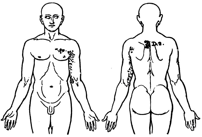
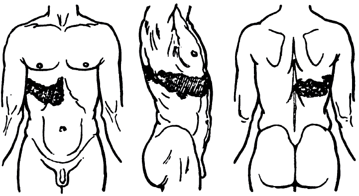
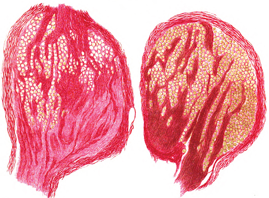
Radicular Rash: T2 root
|
Radicular Rash: T7 root
|
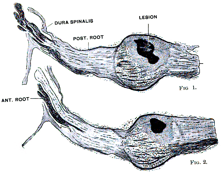
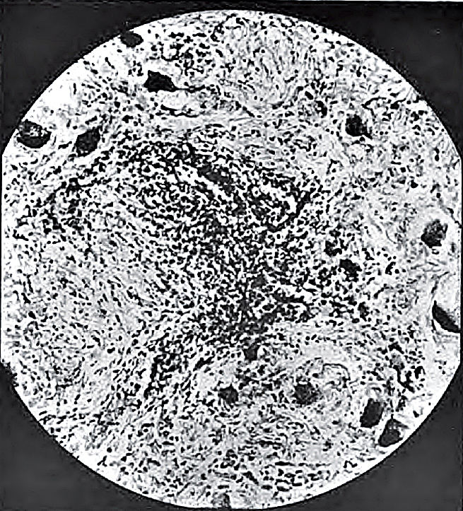
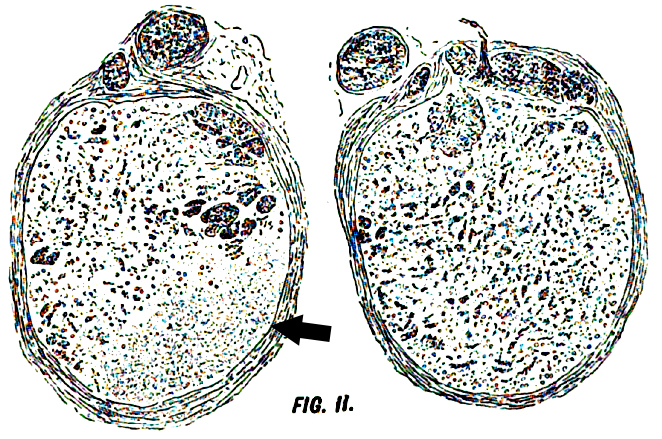
Zoster-related Dorsal Root Ganglion: Early Pathology
|
i.e., in that portion opposed to the anterior root. . . .
In the centre of these haemorrhagic foci the ganglion cells are absolutely destroyed. . . .
But [elsewhere] in the affected ganglia many cells are usually seen which show no noticeable alteration,
and in no instance of spontaneous zoster have we been able to see those minor chromatolytic changes
that are so commonly present in degenerative diseases of the nervous system"
|
|
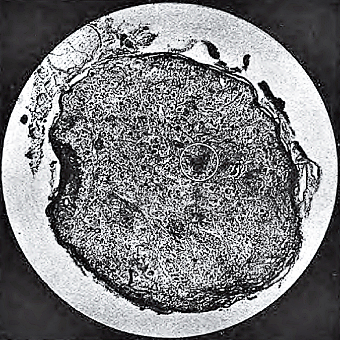
Hemorrhage &
Cellular inflammation Affecting & Around Ganglion cells
|
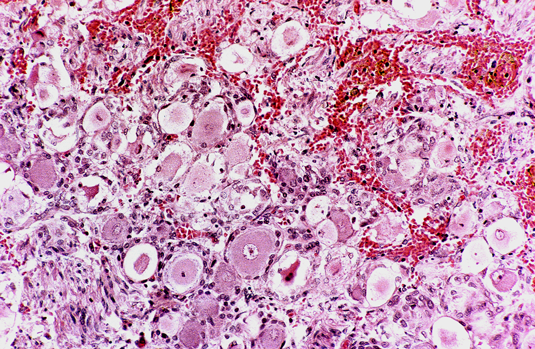 H&E stain From: R Schmidt |
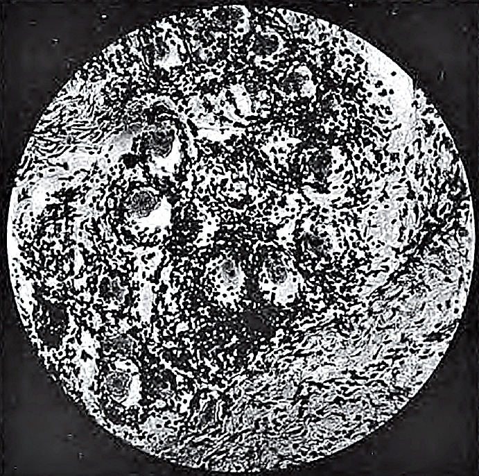
Dorsal Root Ganglion: Unaffected Control (Opposite side)
|
Degenerating ganglion cells & Satellite cell inclusions
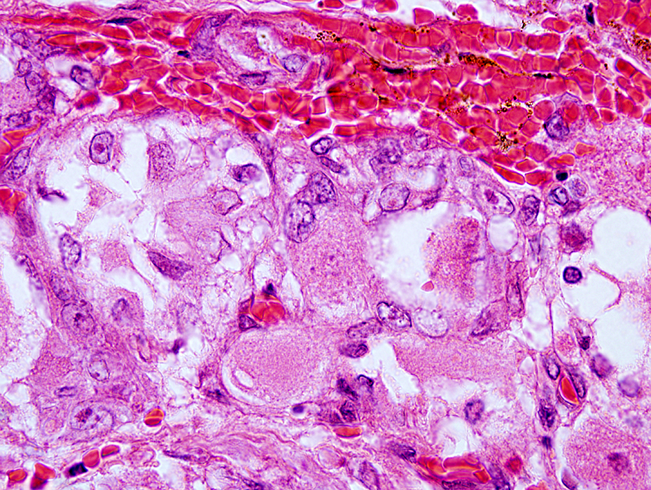 H&E stain From: R Schmidt |
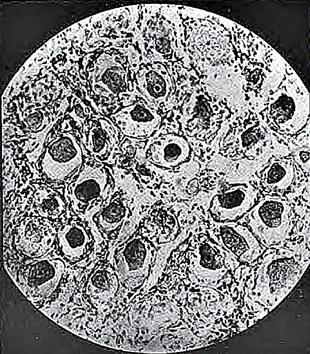
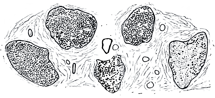
Zoster-related Dorsal Root Ganglion: Late Pathology
|
Within this area all ganglion cells and nerve-fibres are destroyed,
and no structure remains to show where they once existed.
But in several cases a few healthy ganglion cells, and still more often a few healthy nerve-fibres,
can be seen scattered in the scar tissue
|
Zoster-related Nerve: Axon loss, Differential fascicular
|
Return to Neuromuscular Home Page
Return to Muscle autoantibodies
9/18/2023