Hypokalemic Periodic Paralysis
|
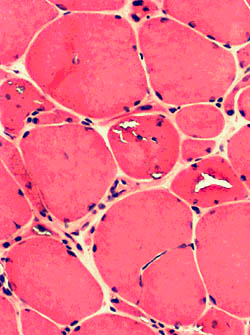
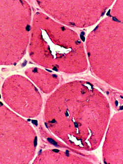
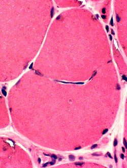
General Pathology: Early; During attacks of weakness
|
Pathology: Early
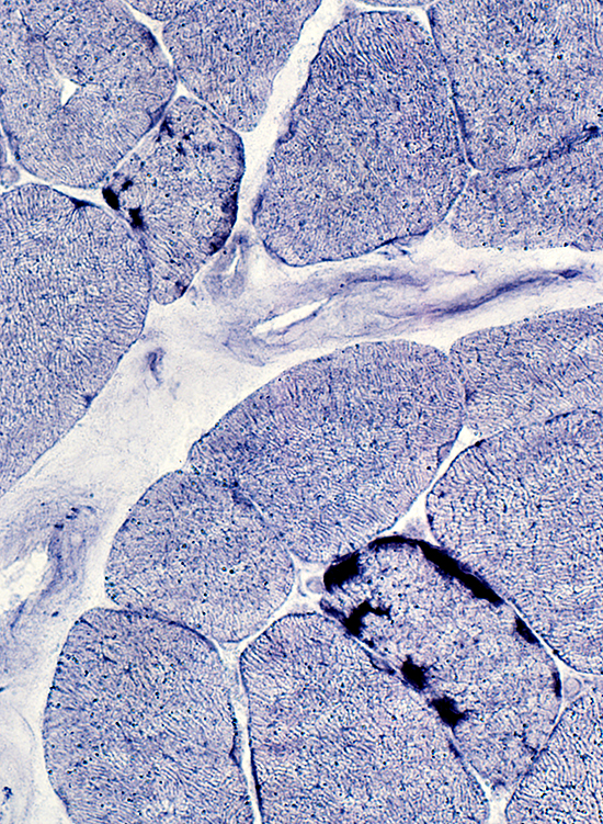
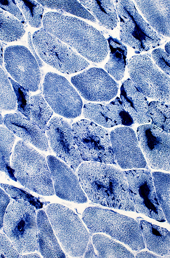
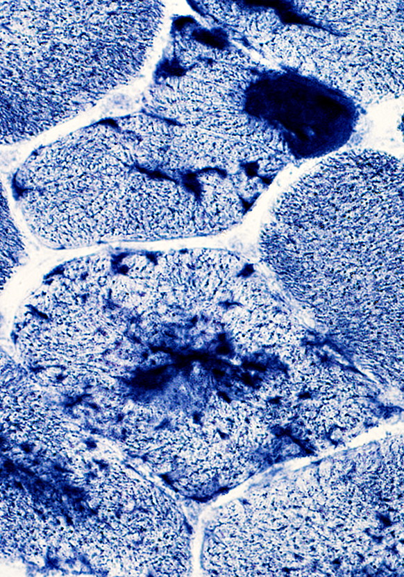
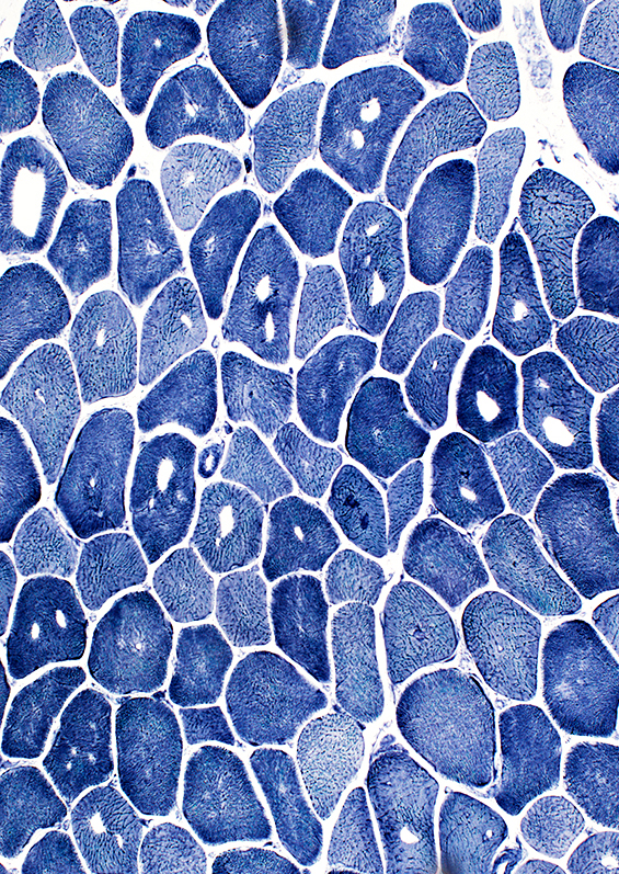 NADH stain |
 NADH stain |
Tubular aggregates: Stain for NADH
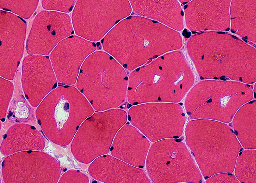 H & E stain |
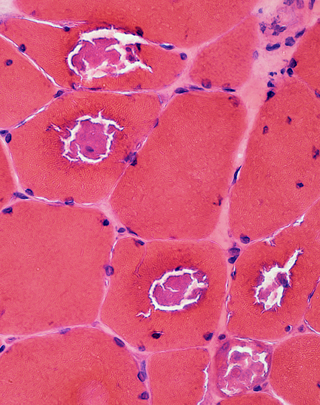
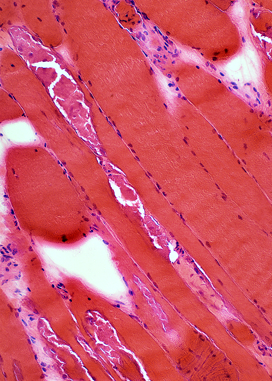
 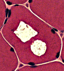 H & E stain |
 H & E stain |
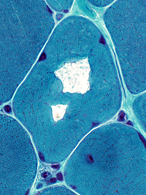 Gomori trichrome stain |
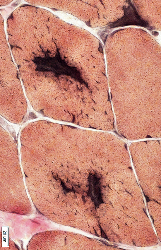
AMPDA: Stains tubular aggregates but not contents within vacuoles
 AMPDA stain |
 AMPDA stain |
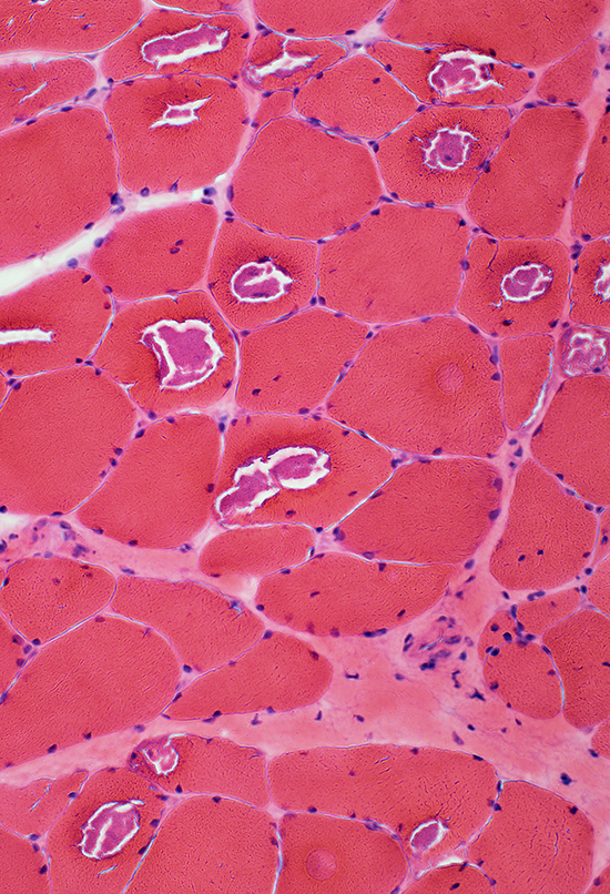
Pathology: Late; During permanent weakness
- Internal architecture changes
- Vacuoles
- Tubular aggregates
- Chronic myopathic changes
- Muscle fibers
- Size: Varied; Small & Large
- Internal nuclei
- Split fibers
- Endomysial connective tissue: Increased
- Muscle fibers
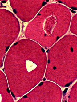
 H & E stain |
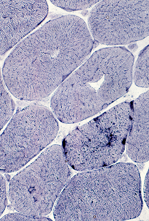
Hypokalemic Periodic Paralysis: Large Tubular Aggregates
 H & E stain |
 H & E stain |
 H & E stain |
 VvG stain |
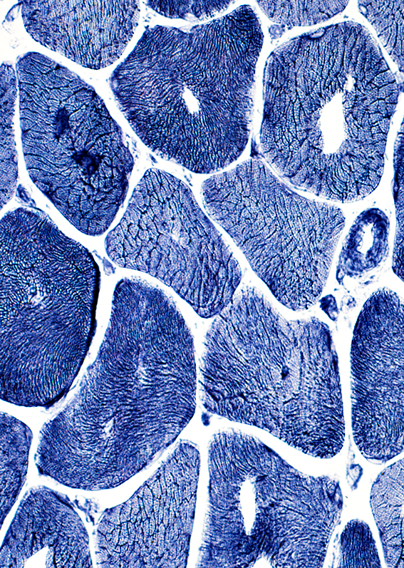
 NADH stain |
 NADH stain |
Return to Hypokalemic periodic paralysis
6/22/2015