FACIOSCAPULOHUMERAL (FSH) DYSTROPHY
|
FSH Dystrophy Adult Mild (Early) Moderate Late Congenital Muscle MRI Not consistently useful to identify myopathy |
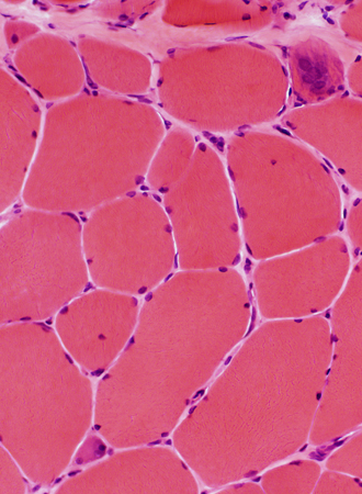
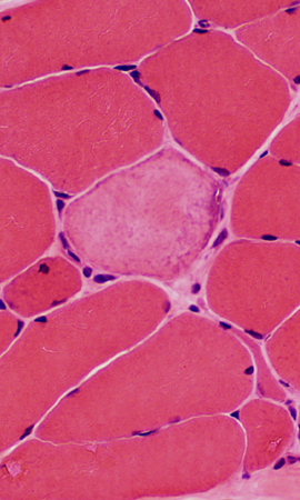
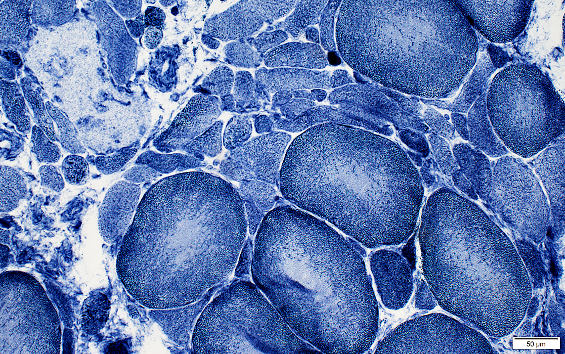
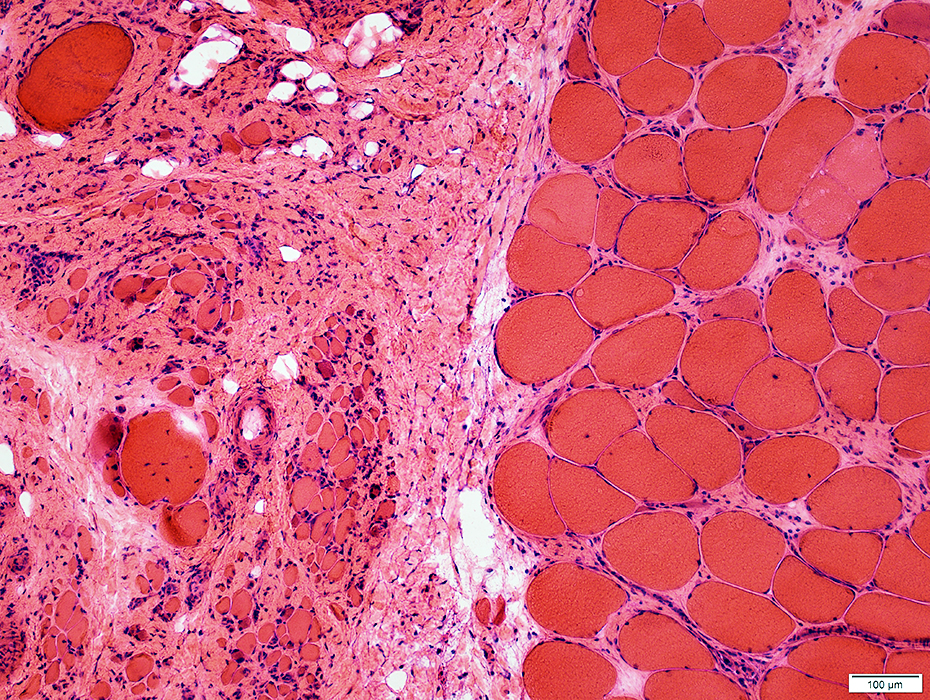
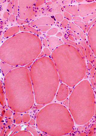
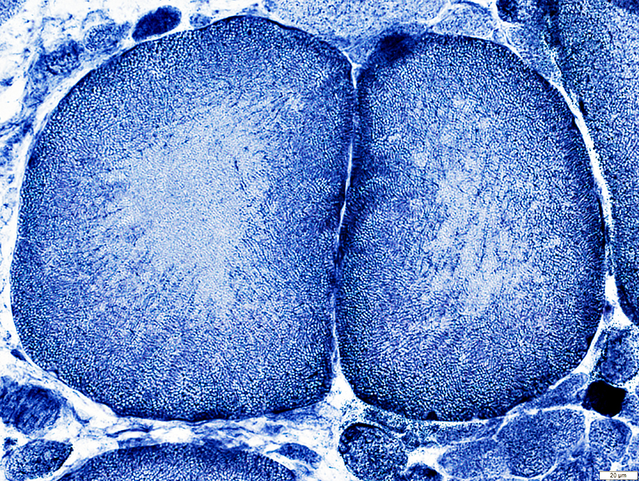
FSH: Adult, Moderate severity
|
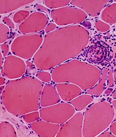
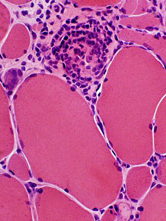
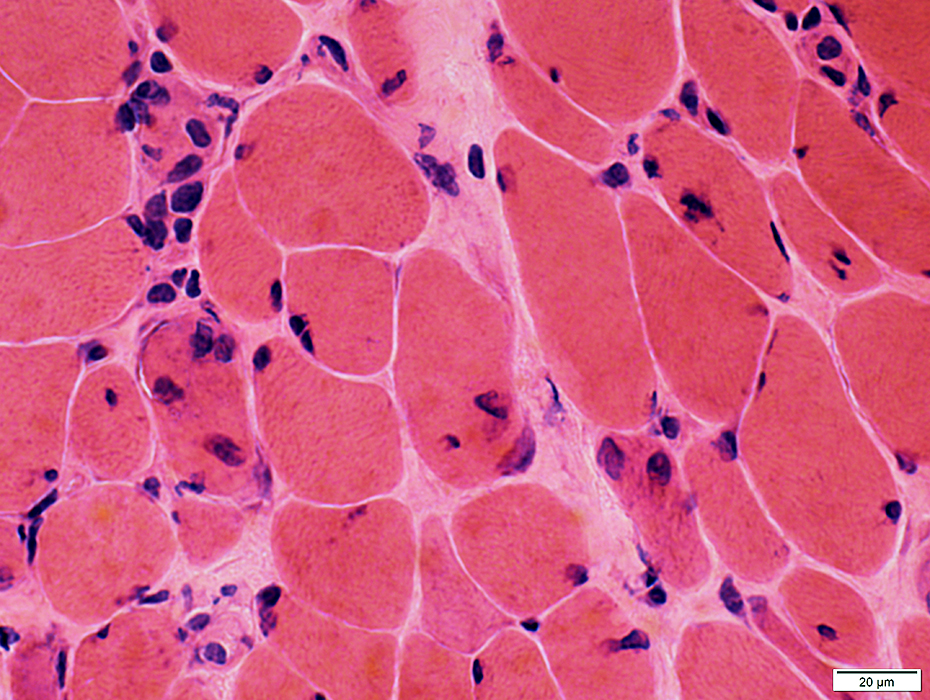
Endomysial Capillaries: May be Large Misoriented ATPase+ Inflammation: Lymphocyte foci Muscle fibers Fiber types MHC-I: Upregulation Myopathy: Hypertrophy & Atrophy Fiber pathology Necrosis |
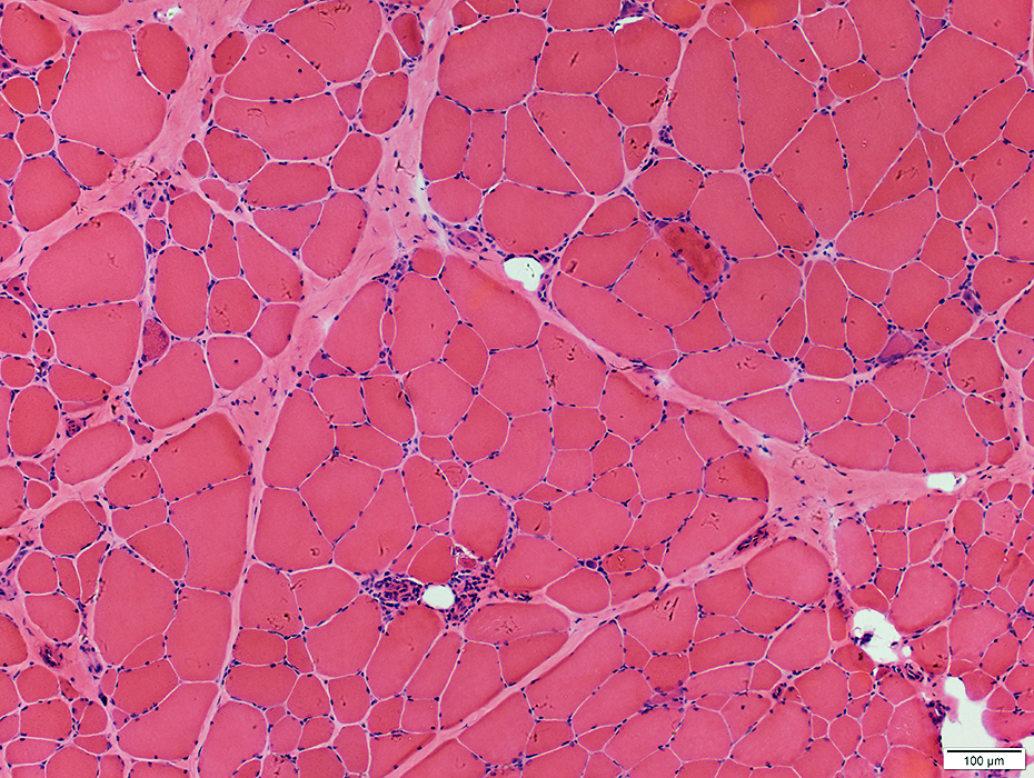 H&E stain |
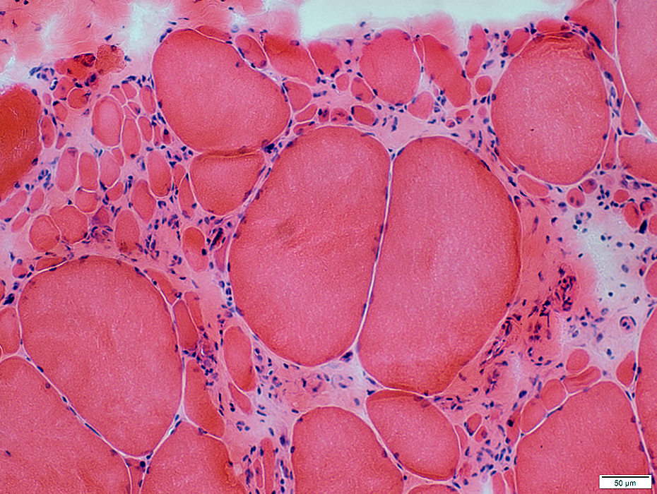
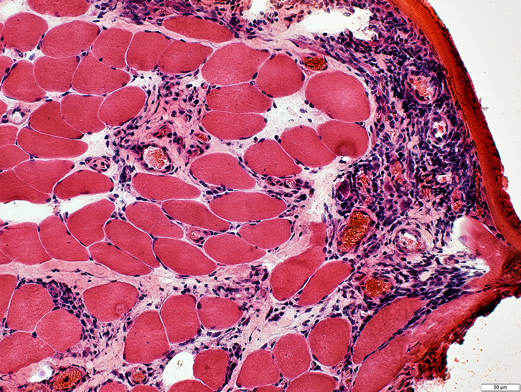
Size: Varied; Atrophy & Hypertrophy
Pathology: Necrosis, Regeneration & Persistent immaturity
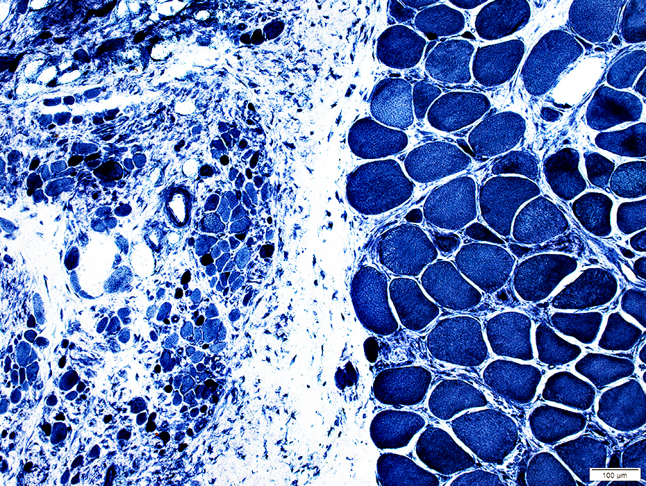
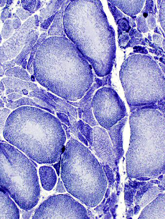
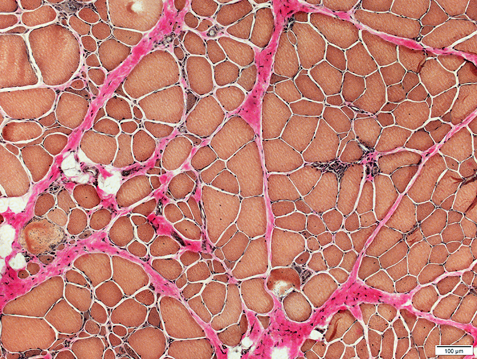 VvG stain |
 H&E stain |
 H&E stain |
 H&E stain |
|
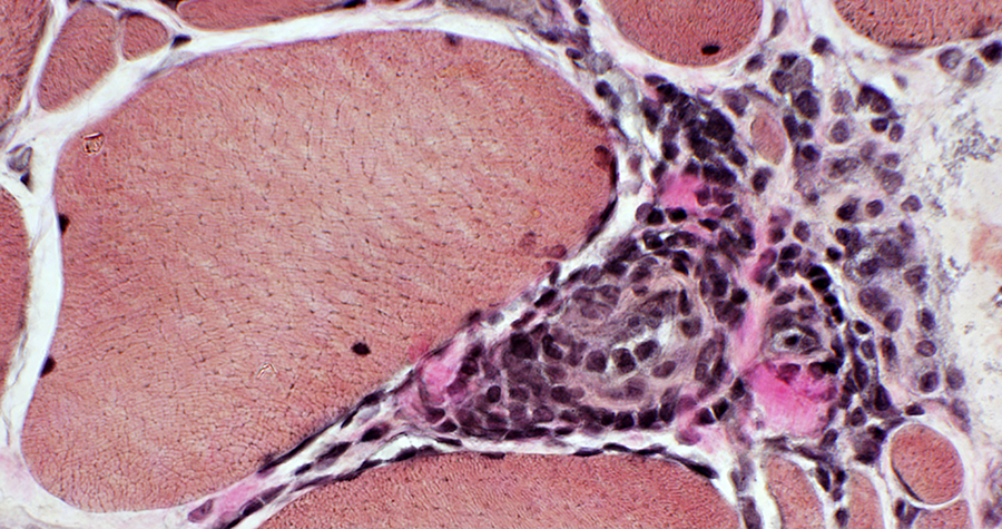
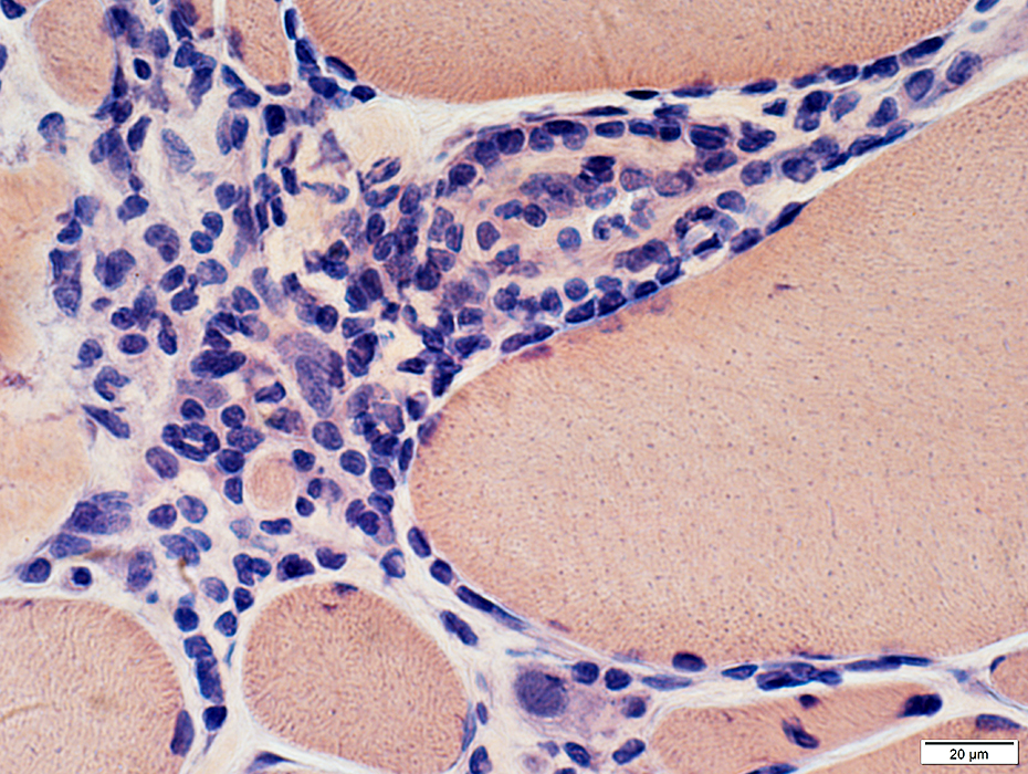
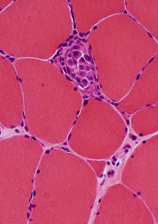
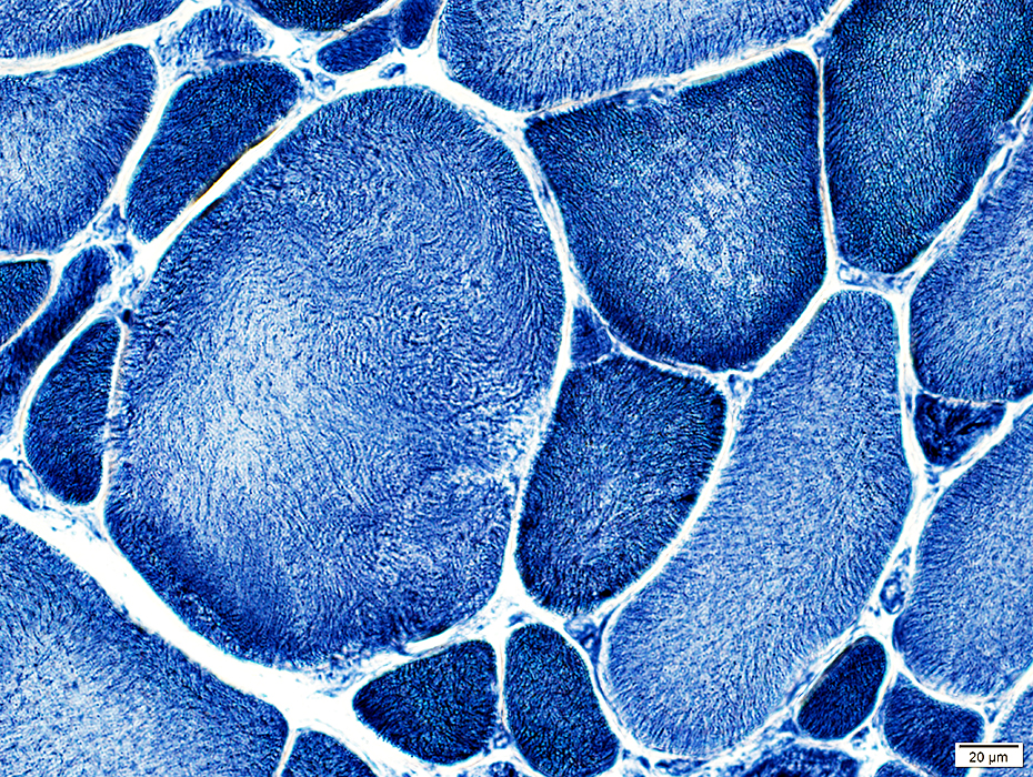
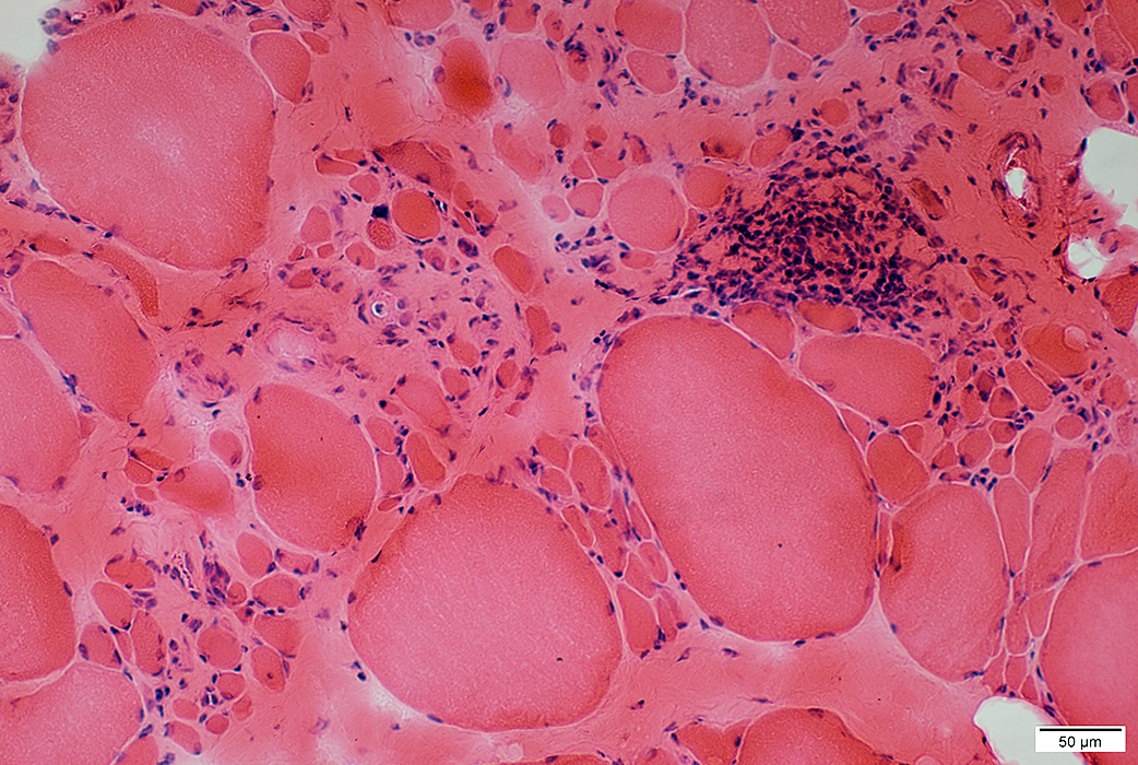
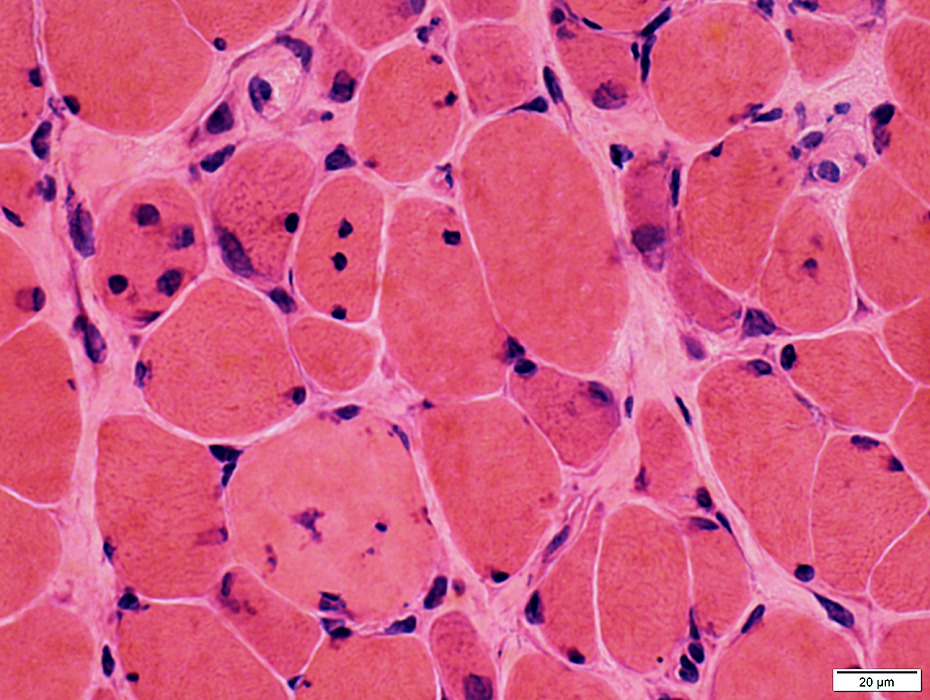
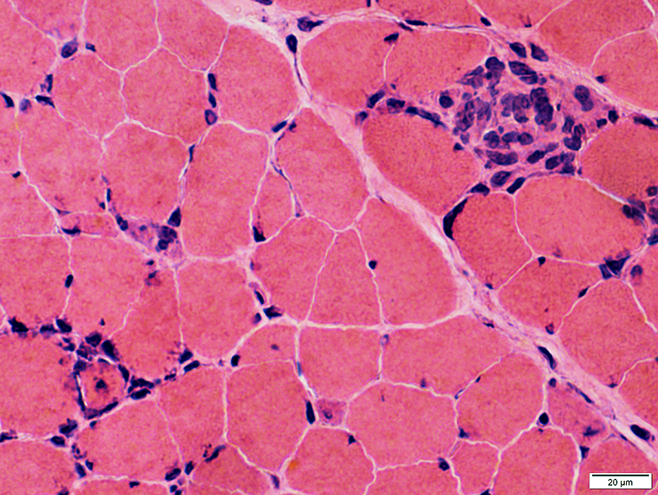
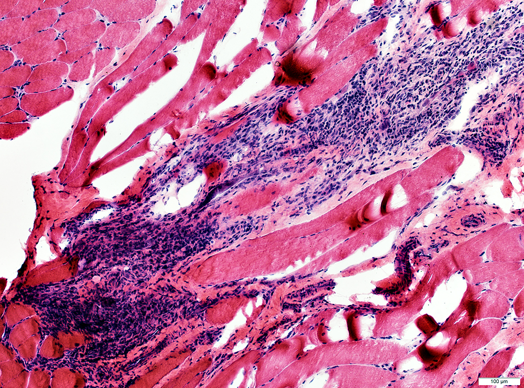
Muscle fibers Size: Varied; Atrophy & Hypertrophy Regeneration: Basophilic muscle fibers Endomysial connective tissue: Moderately increased Inflammation: Around smaller, intermediate-sized vessels, not arteries or veins |
||
 VvG stain |
Location
Around small perimysial vessels with no elastin in their wall (Above)
Not usually around arteries or veins (Below)
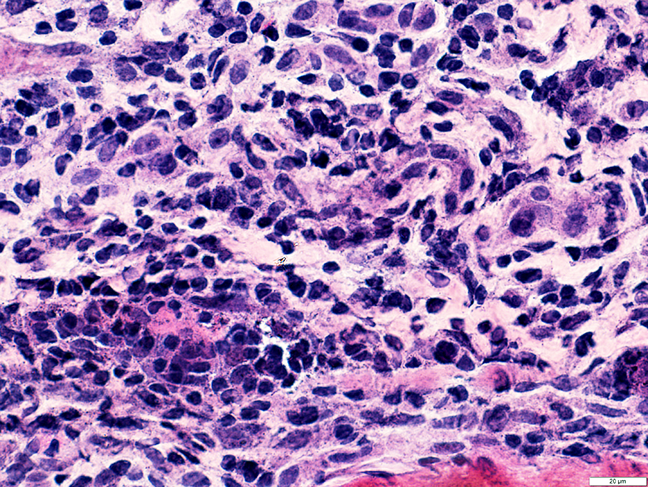
Cell types
Mononuclear: Especially CD4 & CD8
Large with large nuclei: ? Muscle regeneration
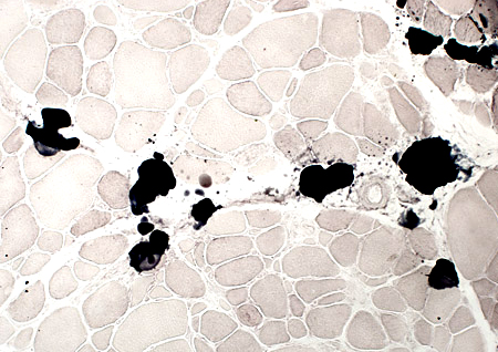
 Congo red stain |
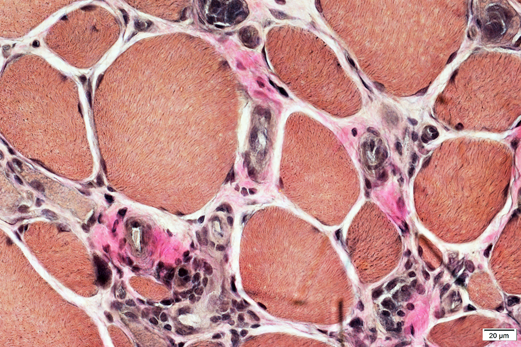
Capillaries
 VvG stain |
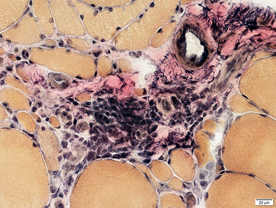
Multiple large endomysial capillaries
 VvG stain |
Capillary pathology
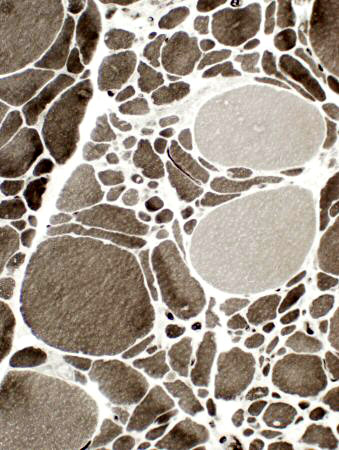
 ATPase pH 4.3 stain |
Dark, abnormal staining for ATPase
 ATPase pH 4.3 stain |
 ATPase pH 4.3 stain |
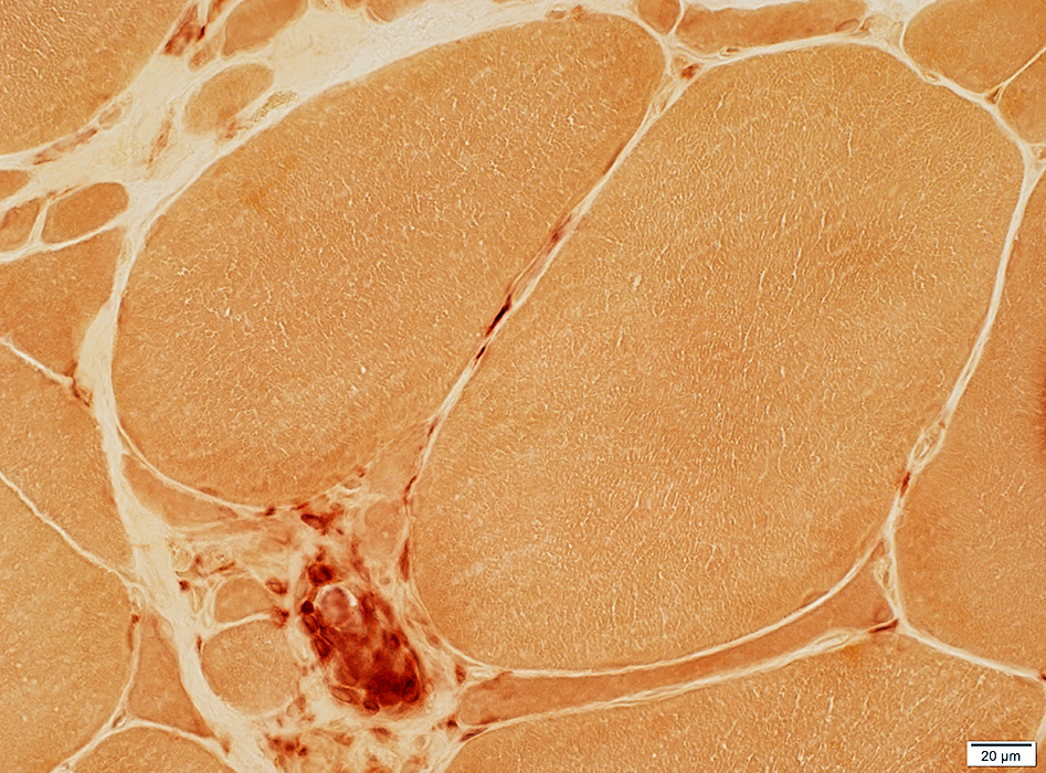
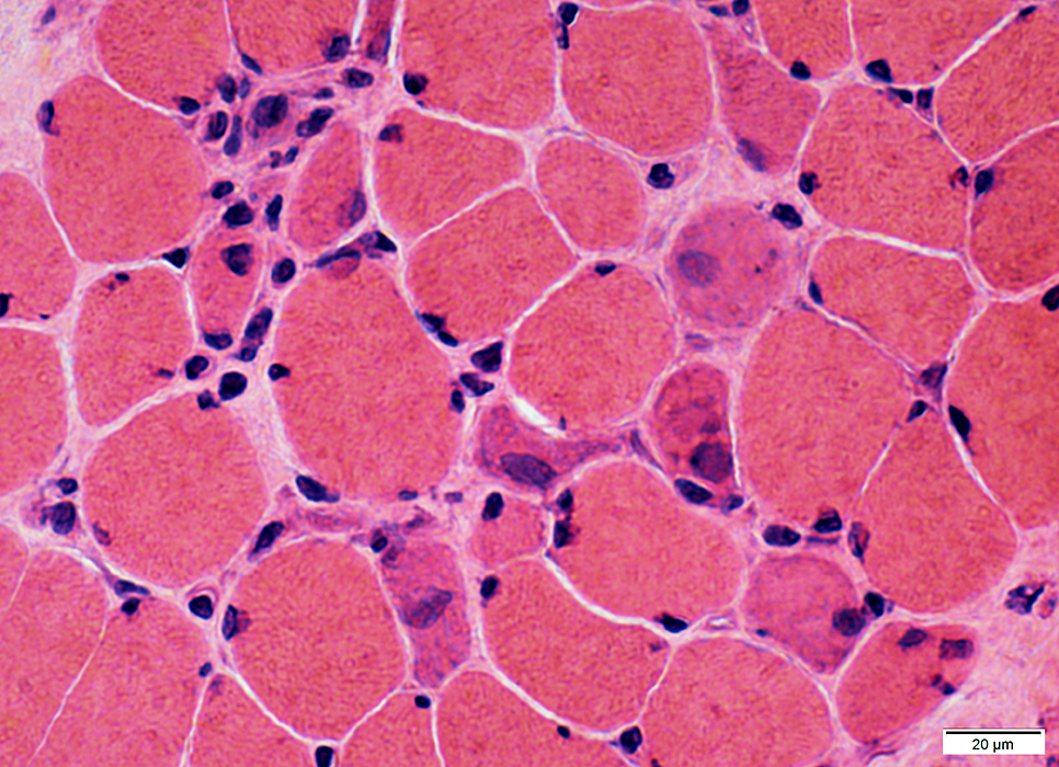
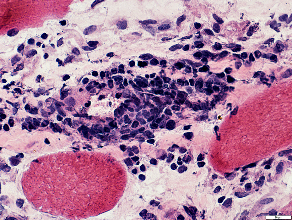
Necrotic muscle fibers
Scattered
Histiocytic cells slow to migrate away from necrotic fiber
More common in muscles with less fibrosis
 H&E stain Necrosis: Early Pale muscle fiber cytoplasm |
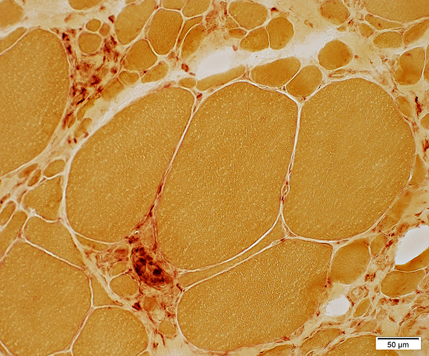
 H&E stain Necrosis Muscle fibers replaced by histiocytic cells |
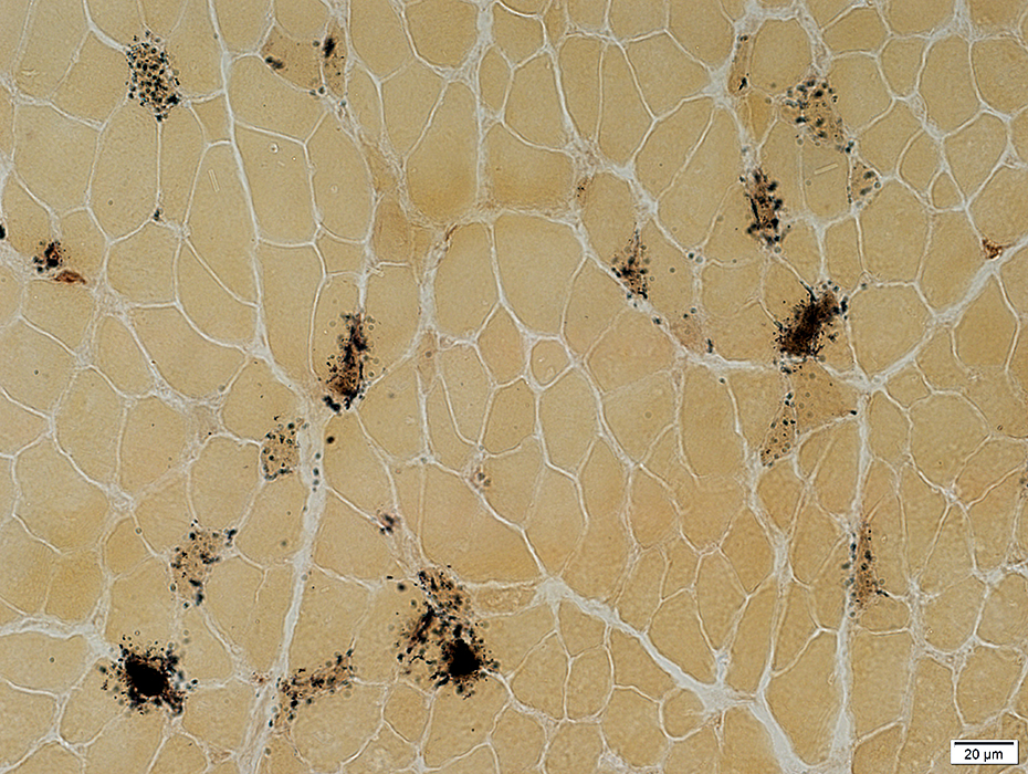
 Acid phosphatase stain |
 Congo red stain |
Replaced by esterase-positive histiocytic cells
 Esterase stain |
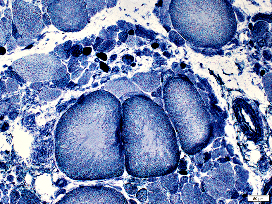
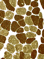
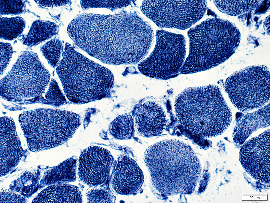
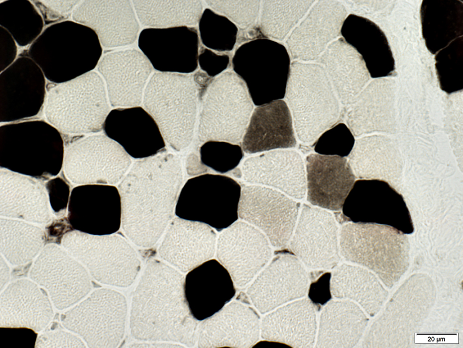
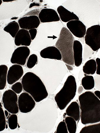 ATPase, pH 4.3 stain Small & large fibers are both types.
A few immature, type 2C, fibers are present. (Intermediate staining; Arrow) |
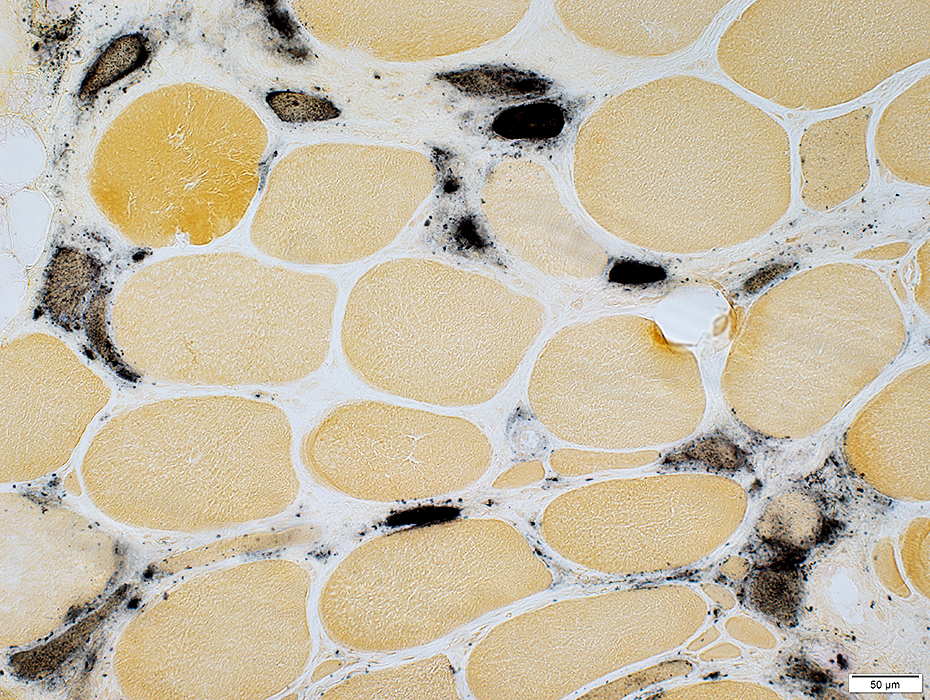
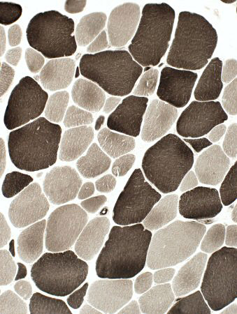 ATPase pH 9.4 stain, Patient 1 Large fibers: Types I & II.
Small fibers: Types I & II. |
 ATPase, pH 9.4 stain | |
FSH: Fiber pathology
 H&E stain
|
|
 Gomori trichrome stain |
 H&E stain |
 NADH stain |
Larger fibers
Pale centers (Above
Whorled or Irregular (Below)
Small fibers: Dark stained
Necrotic muscle fiber: Pale stained (Top left)
 NADH stain |
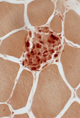
MHC-1: Upregulation by muscle fibers
On the rim of all muscle fibers
In the cytoplasm of smaller, immature fibers.
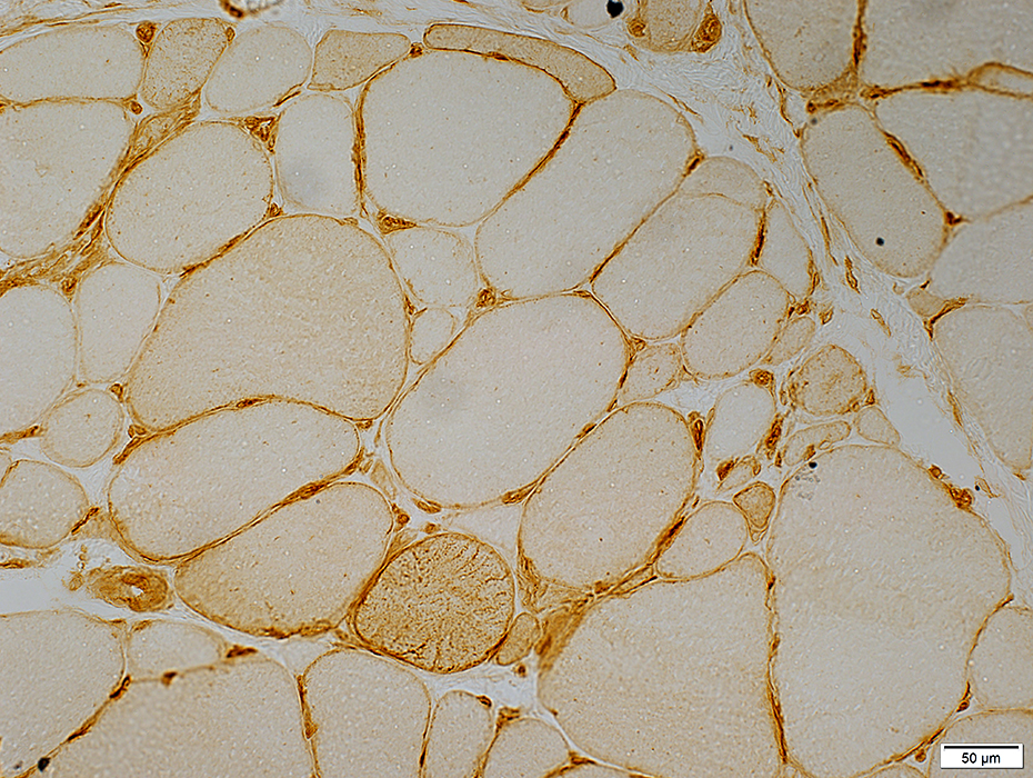 MHC-1 stain |
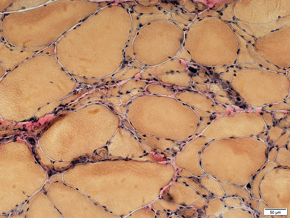
 Sudan black stain Fat replaces perimysial connective tissue |
FSH: Later stage
|
Myopathy Active Chronic Muscle fibers Inflammation |
 H&E stain |
 NADH stain |
Endomysial connective tissue: Increased between muscle fibers
Inflammation: Foci surrounding smaller perimysial vessels
 H&E stain |
 H&E stain |
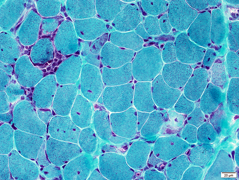
 H&E stain Varied fiber size: Marked
Hypertrophic & Very small fibers |
 NADH stain Hypertrophic fibers: Often have pale centers.
Small fibers: Dark & Pale stained |
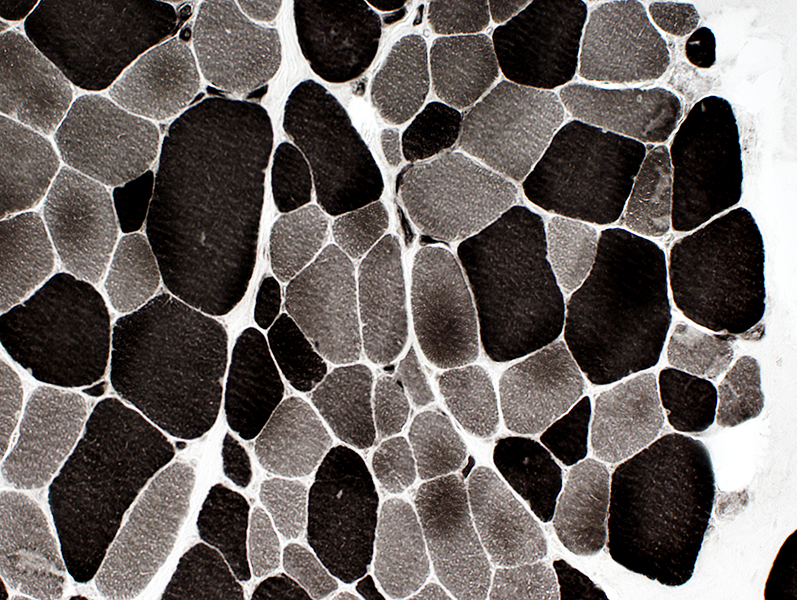
 ATPase pH 9.4 stain, Patient 2 Large fibers: Types I & II.
Small fibers: Often type II. |
 Gomori trichrome stain |
Small muscle fibers: Round; Dark stained on NADH
Hypertrophied muscle fibers: Very large; clear centers on NADH
 NADH stain |
Varied muscle fiber sizes
Small rounded muscle fibers
Vary large, hypertrophied muscle fibers
 VvG stain |
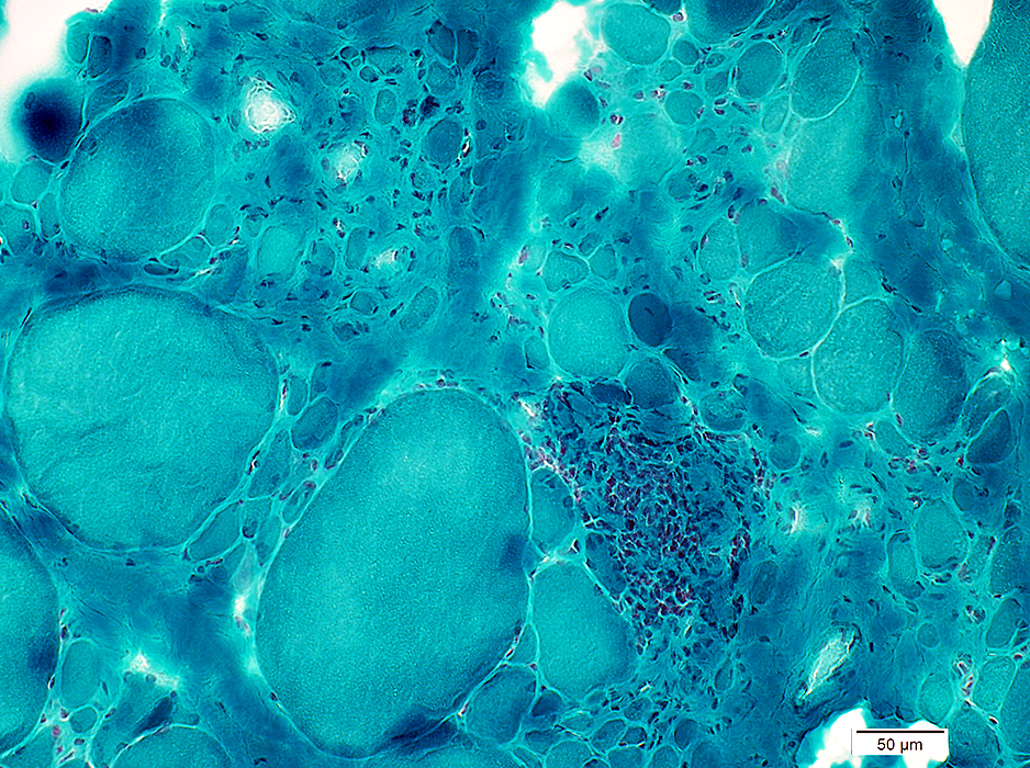
Inflammation: Foci surrounding smaller perimysial vessels
 H&E stain |
Inflammation: Foci surrounding smaller perimysial vessels
Endomysial connective tissue: Increased betwen muscle fibers
 Gomori trichrome stain |
Inflammation: Perimysial focus
 Congo red stain |
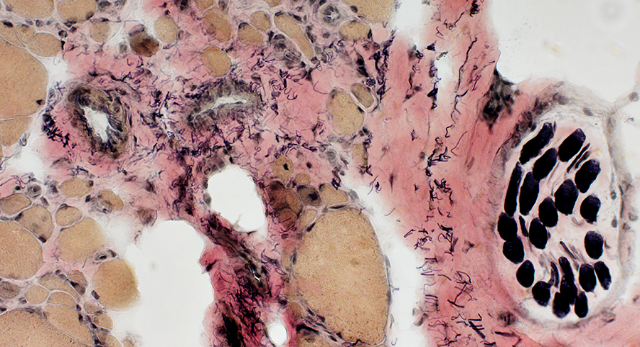
 Esterase stain |
 VvG stain |
Larger vessels: Normal
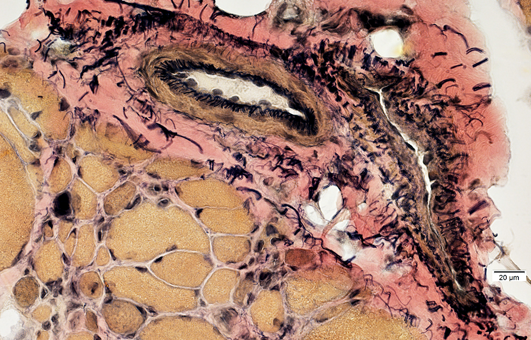 VvG stain |
Intermediate-Sized vessels & Intramuscular Nerves: Normal in FSH
 VvG stain |
 H&E stain |
 H&E stain |
 VvG stain |
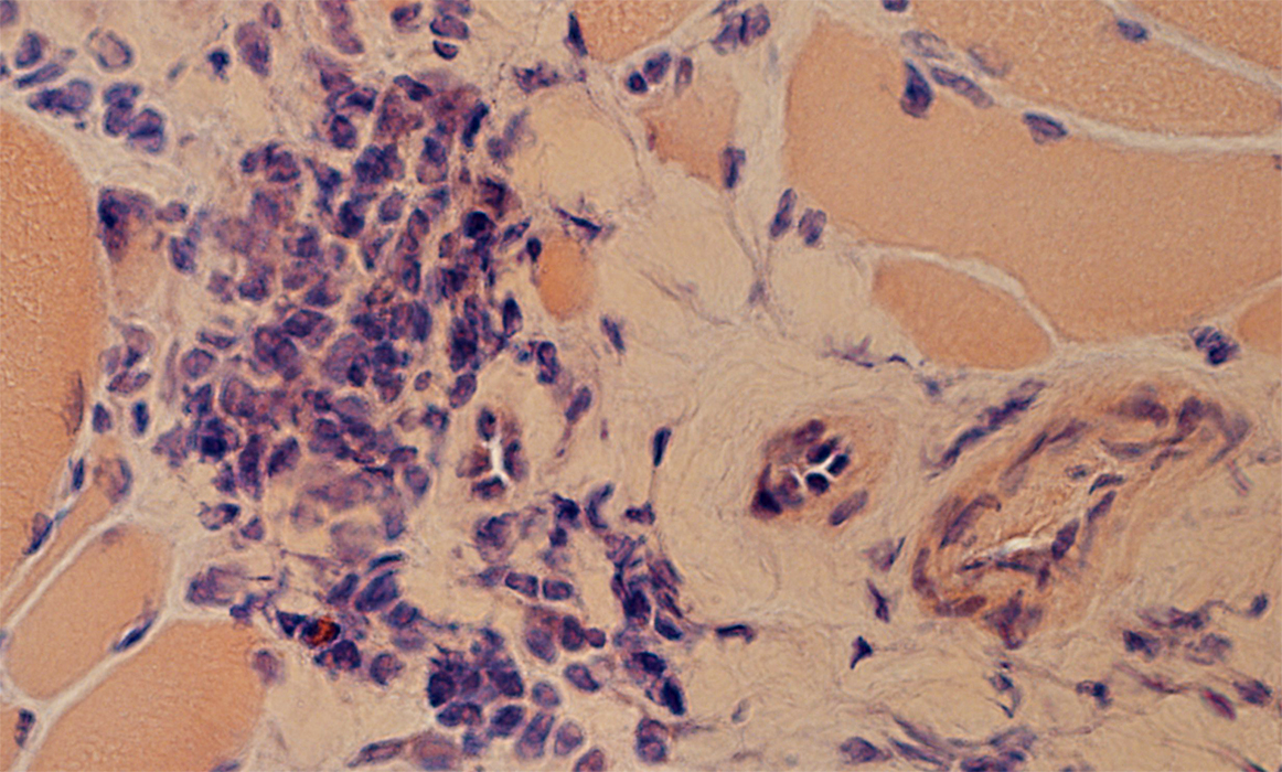
Necrotic muscle fibers
Immature, regenerating muscle fibers: Darker stained, rounded, small muscle fibers
Acid phosphatase positive cells scattered in endomysium
 Acid phosphatase stain |
 Esterase stain |
 Acid phosphatase stain |
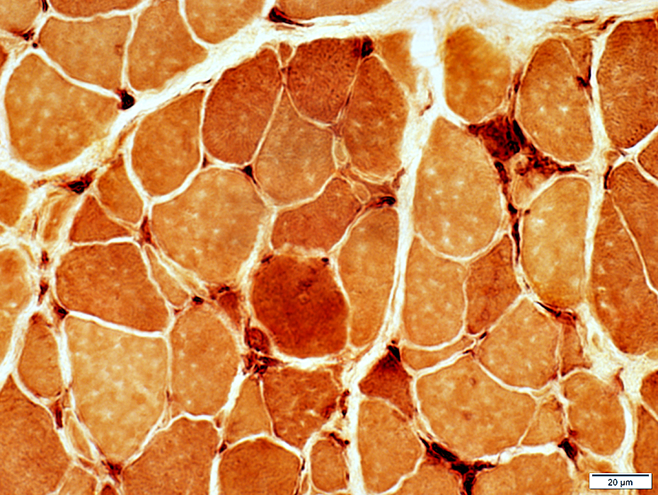
 NADH stain |
Small muscle fibers: Varied staining, from normal to dark
Hypertrophied muscle fibers: Often have clear central regions
 Alkaline phosphatase stain |
Small, rounded
Cytoplasm: Stains for alkaline phosphatase
 Alkaline phosphatase stain |
Small, rounded
Cytoplasm: Darker staining on VvG
|
VvG stain |
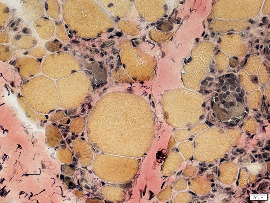
 ATPase pH 9.4 stain |
Large fibers: Types I & II
Small fibers: More commonly type II
 ATPase pH 4.3 stain |
Congenital FSH
|
Inflammatory cell foci Myopathic features |
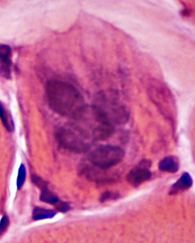
FSHD, Congenital: Myopathy
2 year old female
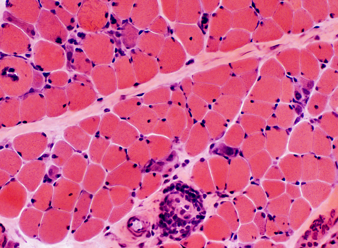 H&E stain Muscle fiber size: Varied Many small basophilic fibers Mononuclear inflammatory cells Around smaller, intermediate sized perimysial vessel (Above) In endomysium (Below) |
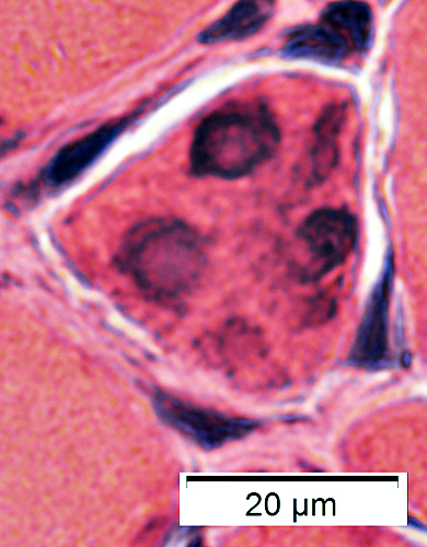
 H&E stain Varied muscle fiber size. Basophilic (immature) fibers. No hypertrophy. Endomysial inflammation. |
 ATPase pH 9.4 stain Smaller fibers are Type I & II |
 Gomori trichrome stain |
Myopathology patterns
 H&E stain |
Varied
Small fibers: Rounded; Some basophilic
Nuclei: Irregular shapes; Large; One or several internal
 H&E stain |
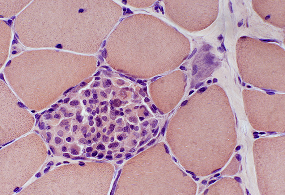
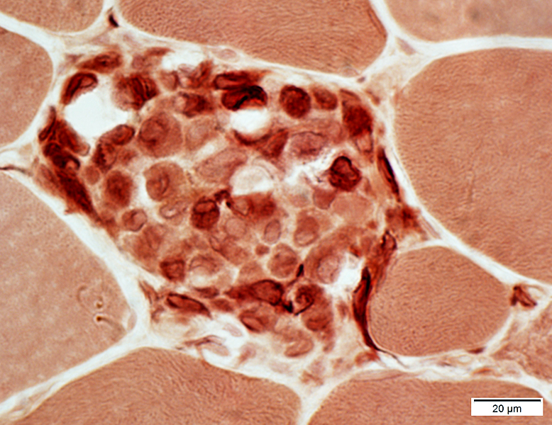
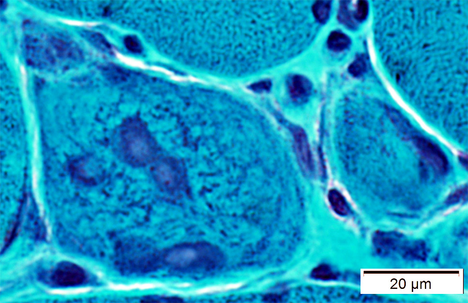
Endomysial cell clusters
 H&E stain |
Small muscle fibers: Basophilic cytoplasm; Large nuclei
 H&E stain |
Internal architecture: Coarse; Immature
 NADH stain |
 ATPase pH 4.3 stain |
Type 2C: Intermediate color on ATPase pH 4.3
Alkaline phosphatase positive
Scattered; Small
 Alkaline phosphatase stain |
Endomysial cells
Histiocytic; Scattered
Common pattern with MHC-I upregulatioon by muscle fibers
 Esterase stain |
MHC-1: Abnormal upregulation by muscle fibers
 MHC-1 stain |
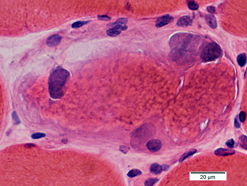
FSHD, Congenital: Inflammation 4 year old female
 H&E stain |
 H&E stain |
 H&E stain |
 H&E stain |
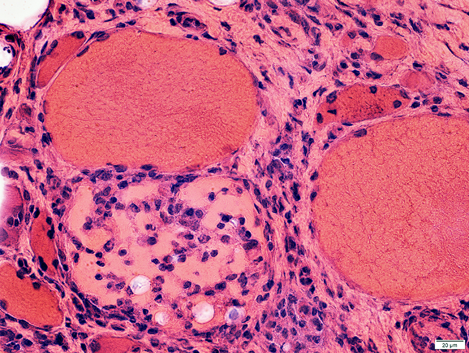
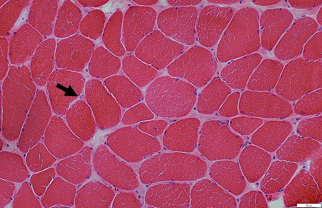
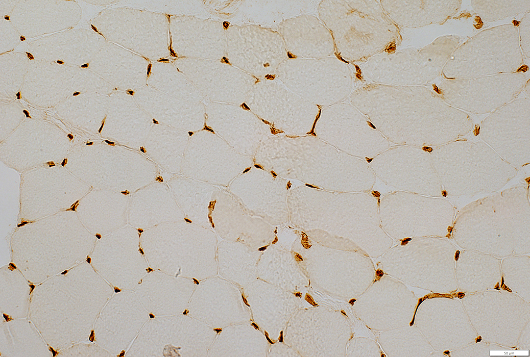
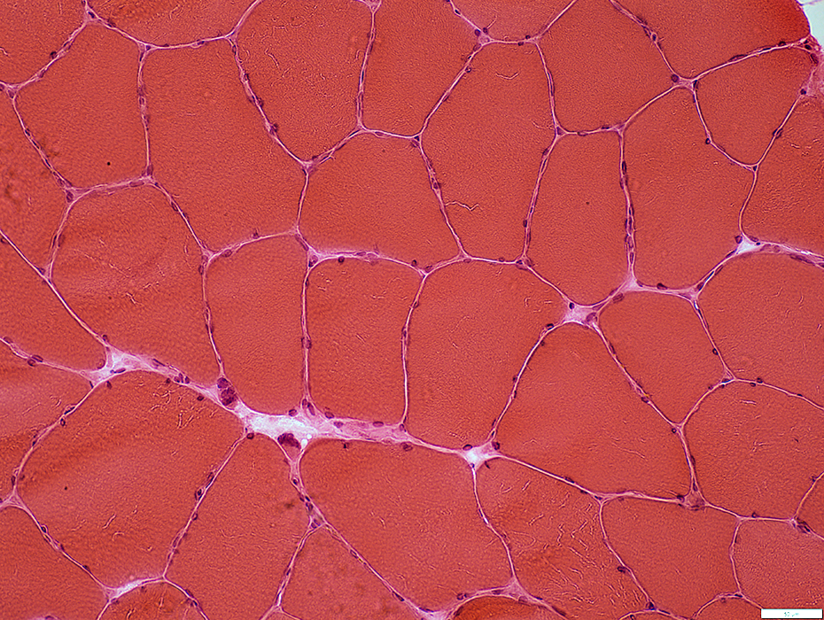
FSHD: Early in course
 H&E stain |
Muscle fibers
Sizes: Mild variation; Few small fibers
Endomysial capillaries
Large (Arrow; Above)
Misoriented (Below): Often circumferential around muscle fibers
 H&E stain |
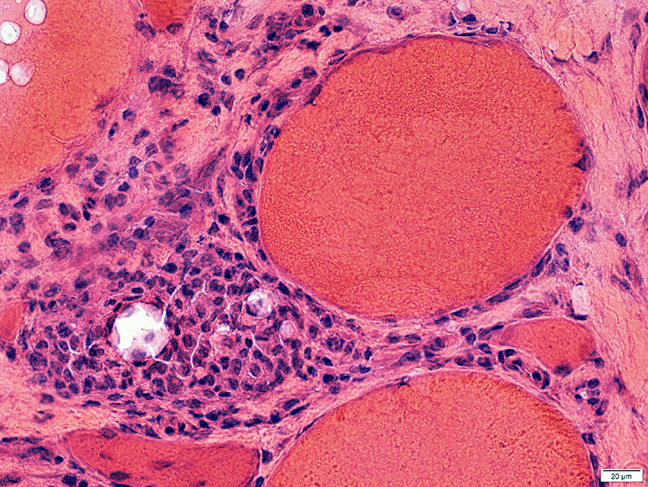
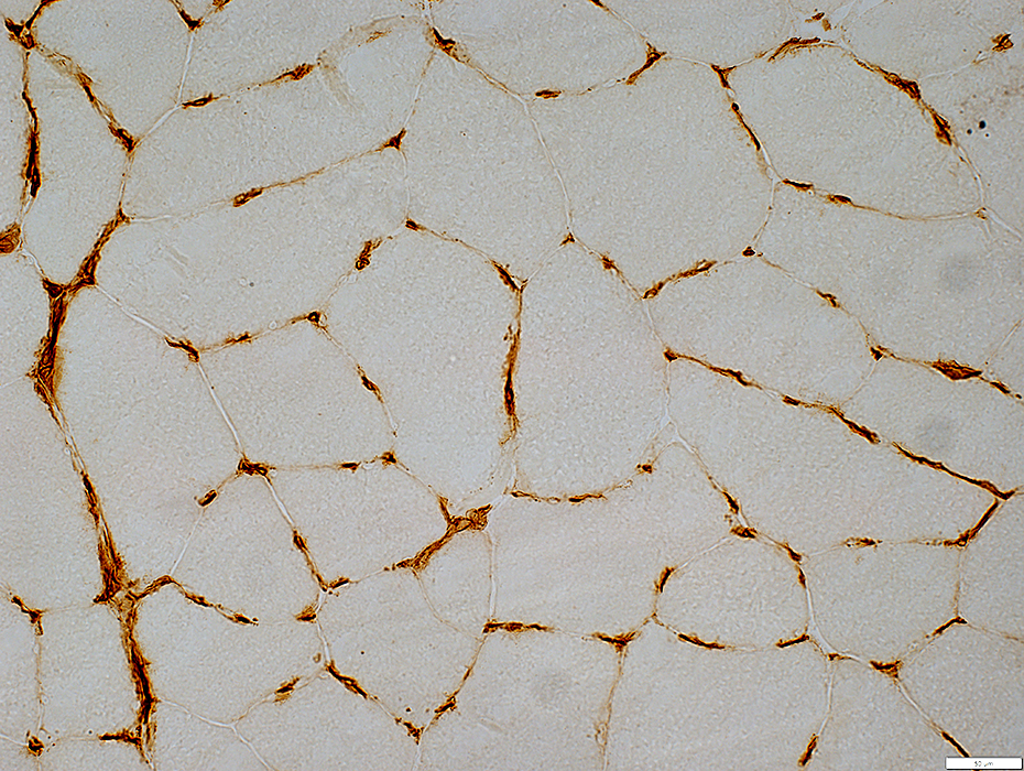
 MHC1 stain |
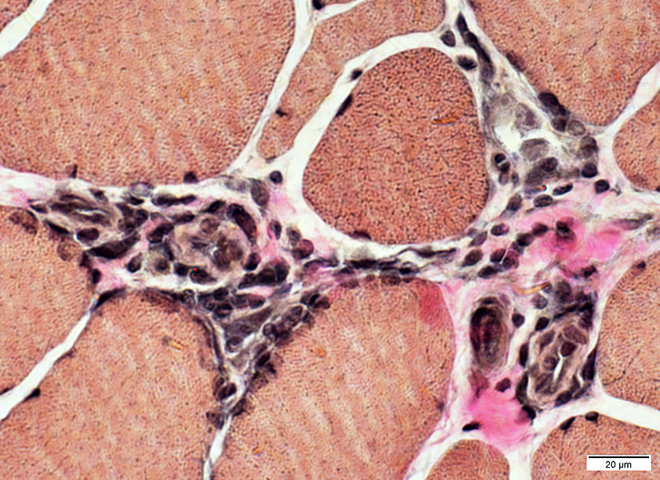
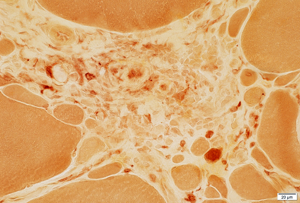
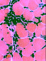
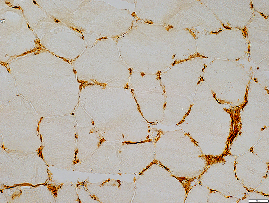
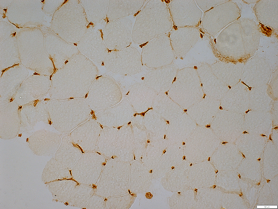
Endomysial Capillaries
Large
Misoriented: Often circumferential around muscle fibers
 MHC1 stain |
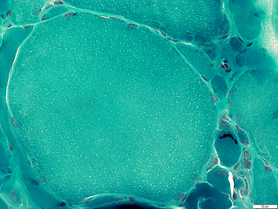
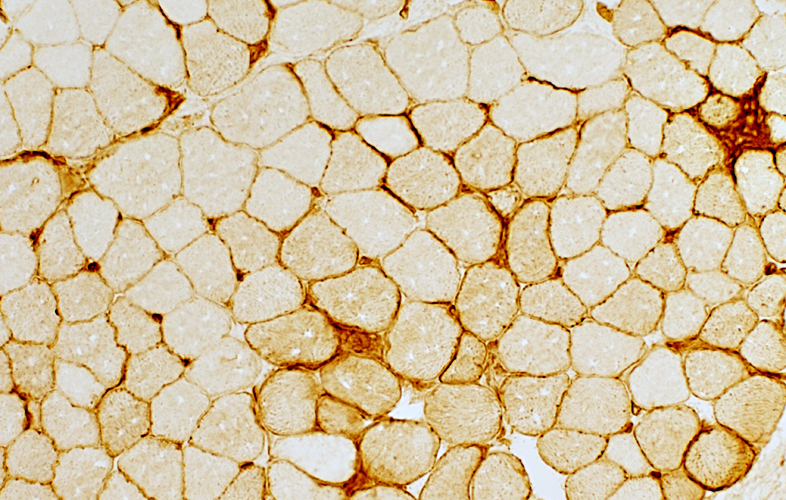
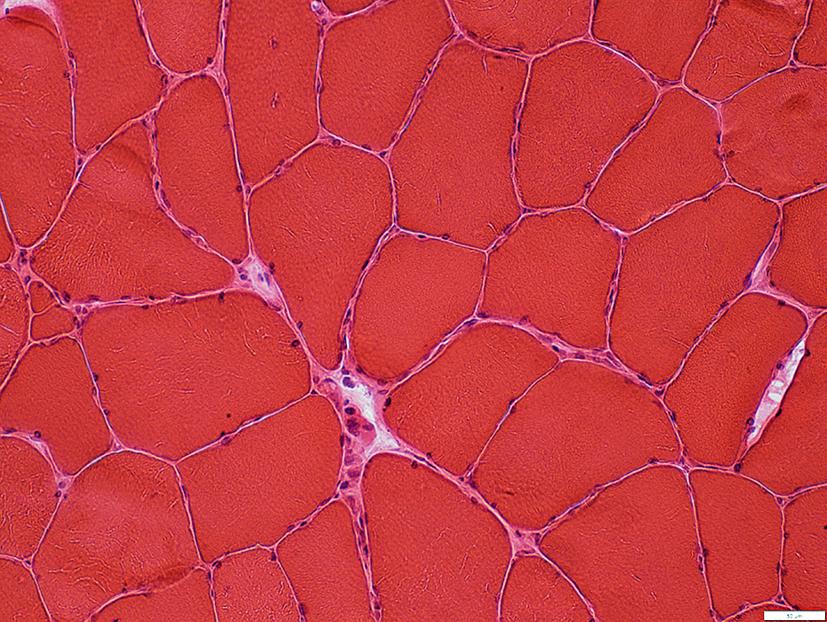
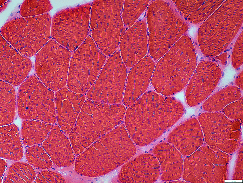
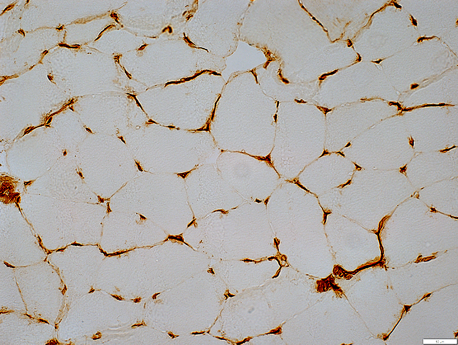
Control muscle
Capillaries: Most parallel to length of muscle fibers; Cut in cross-section
 MHC1 stain |
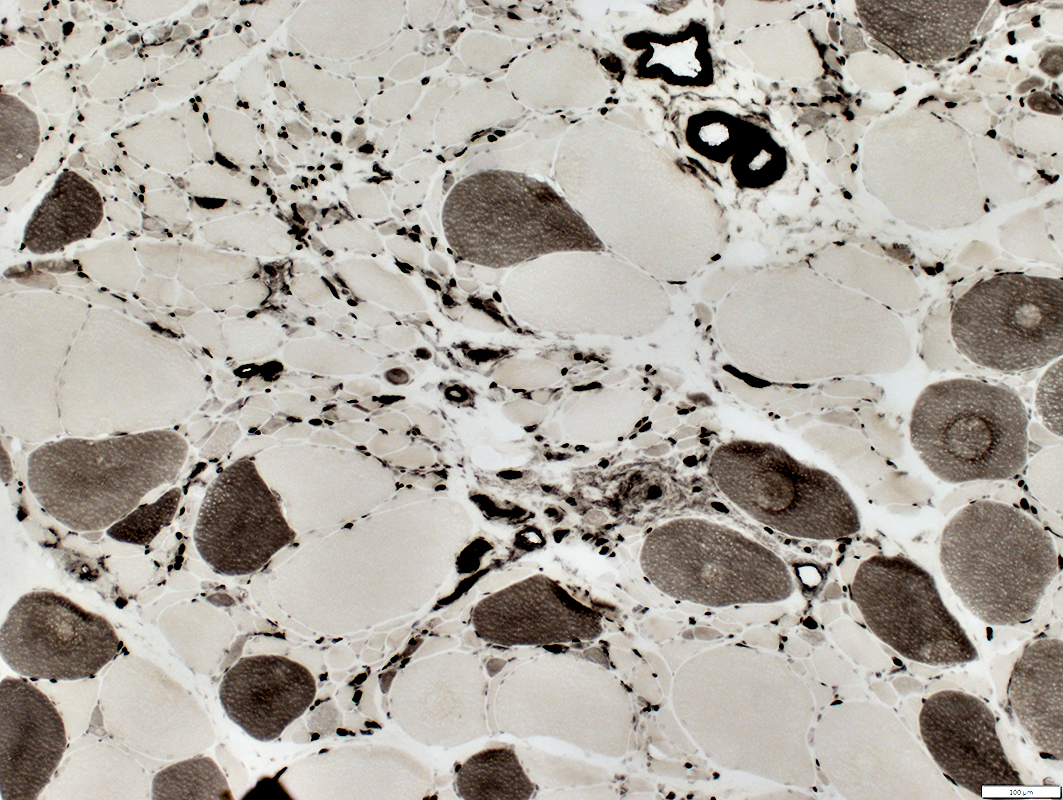
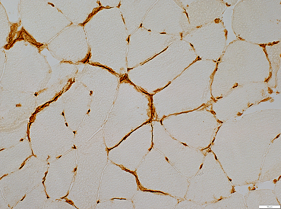
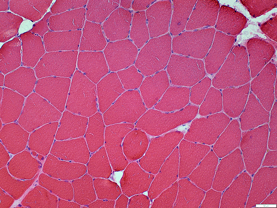
6 FSHD Muscle Biopsies: Identified by MRI as "Active Myopathy"
MRI does not consistently identify ongoing myopathy
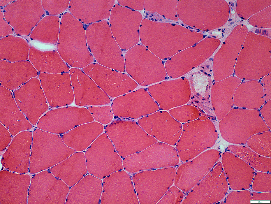
 H&E stain |
Fibers: Varied sizes; Hypertrophy
Endomysial capillaries: Misoriented; Increased numbers
 MHC1 stain |
 H&E stain |
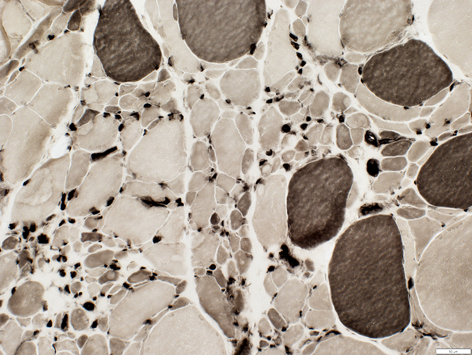
Fibers: Varied sizes; Hypertrophy; Few small fibers
Endomysial capillaries: Increased numbers, moderate
 MHC1 stain |
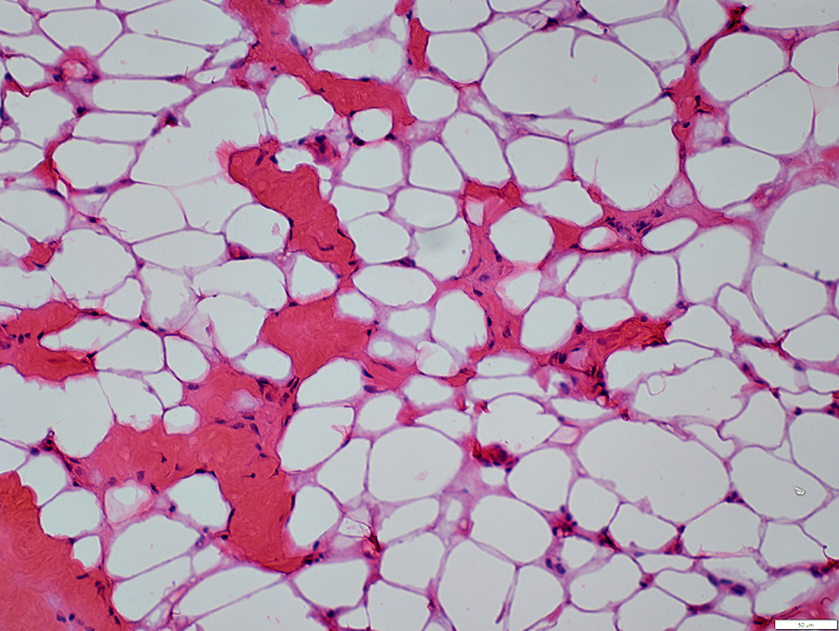
 H&E stain |
Fibers: Varied sizes; Hypertrophy
Endomysial capillaries: Misoriented; Increased numbers
 MHC1 stain |
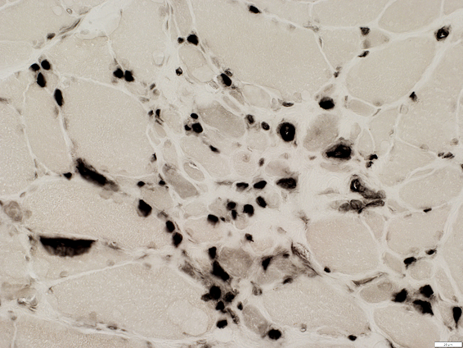
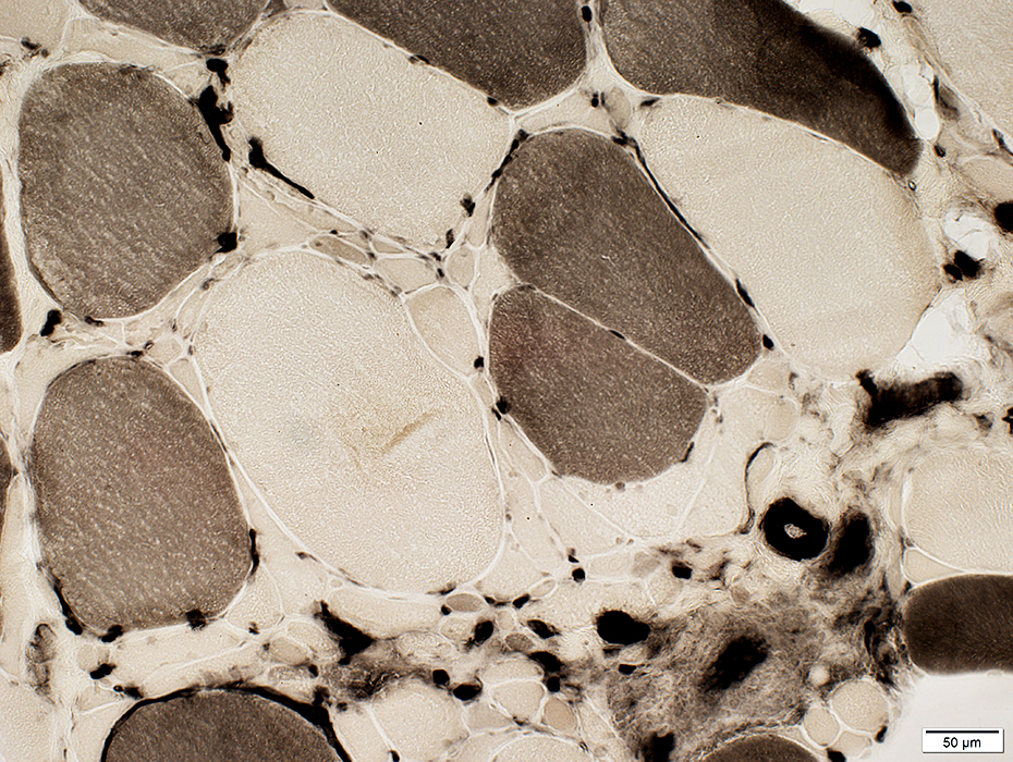
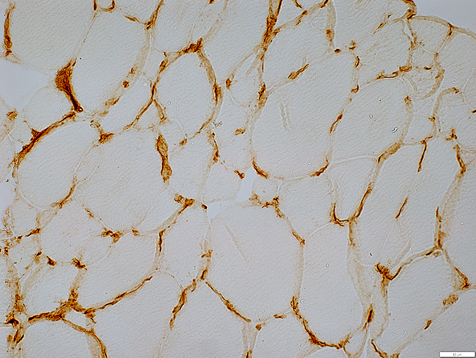
 H&E stain |
Fibers: Varied sizes; Hypertrophy, Few small fibers
Endomysial capillaries: Misoriented; Increased numbers
 MHC1 stain |
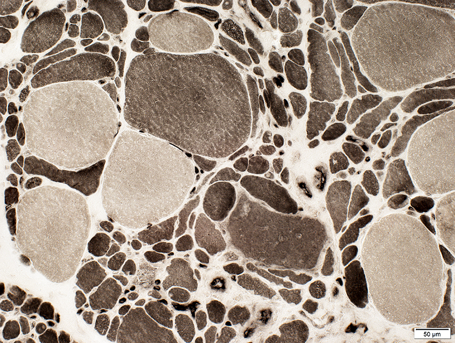
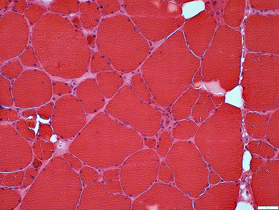 H&E stain |
Fibers: Bimodal sizes; Hypertrophy, Small & intermediate-sized fibers fibers
Endomysial capillaries: Misoriented; Increased numbers; Large
 MHC1 stain |
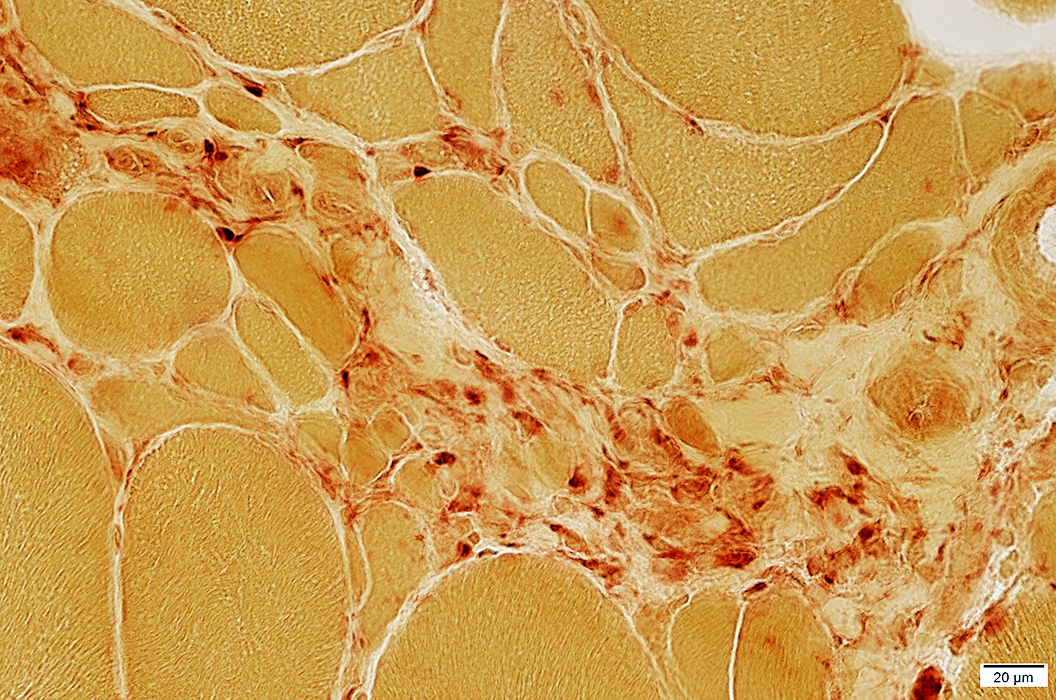
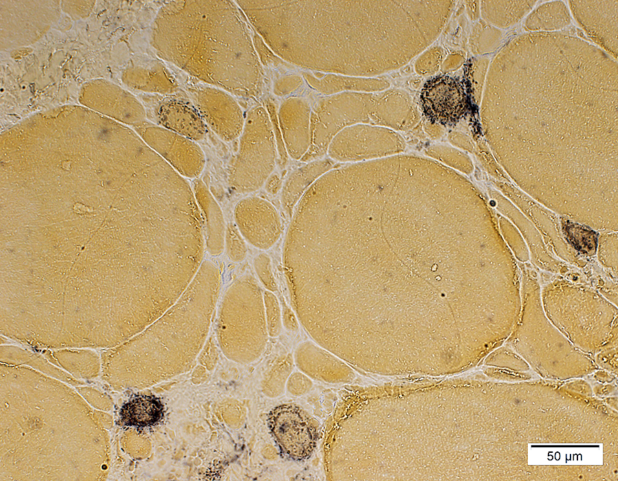
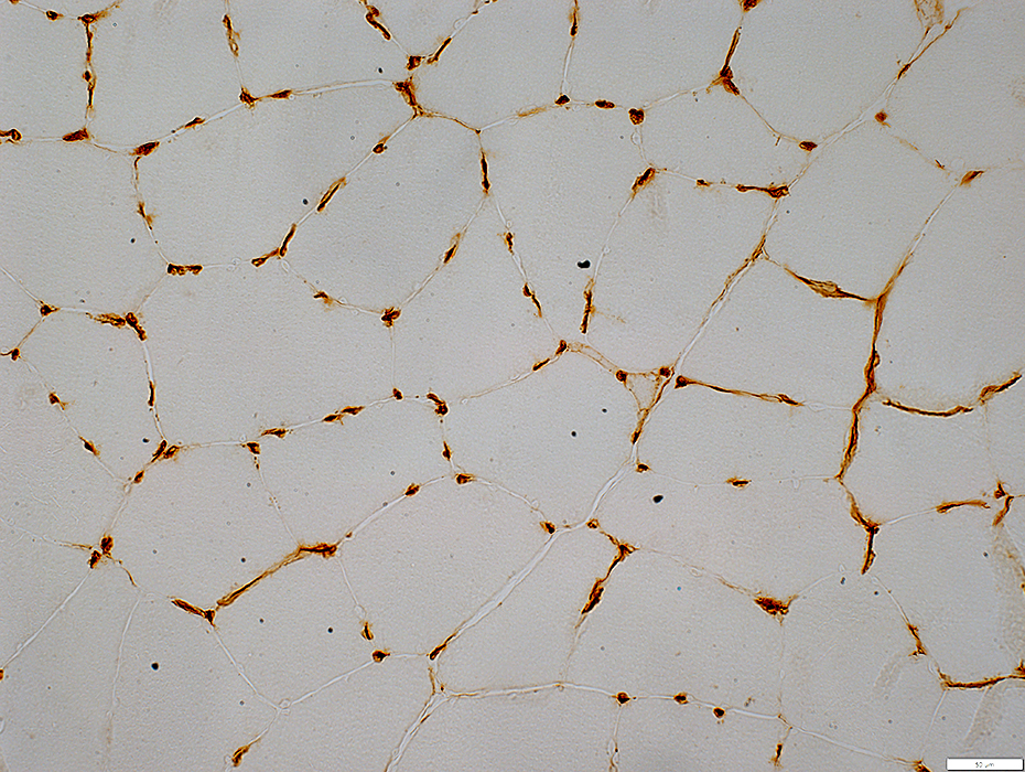
 H&E stain |
Fibers: None
Capillaries amid fat: Large
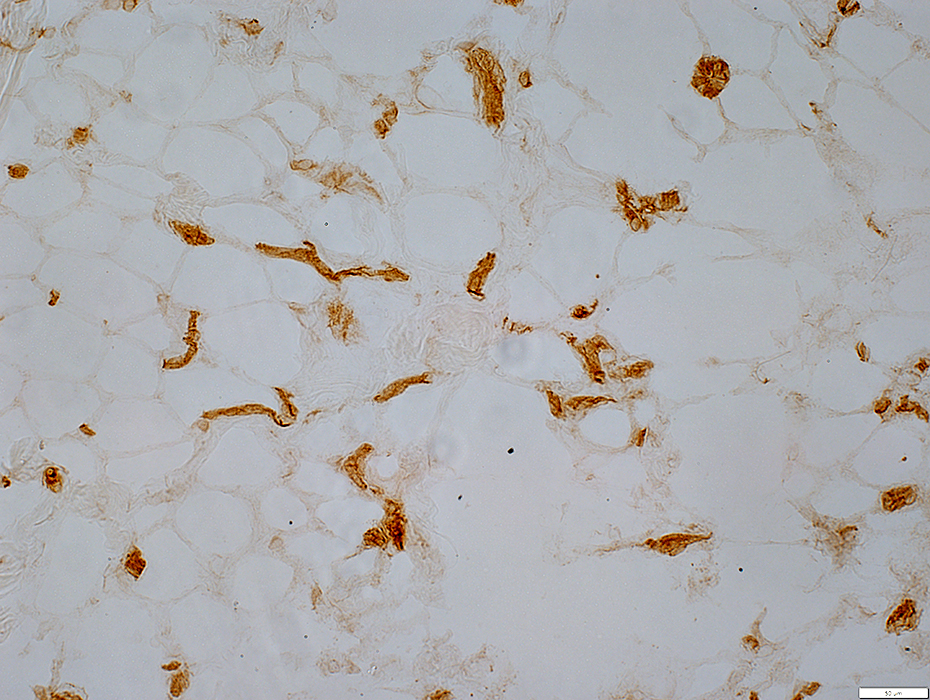 MHC1 stain |
Return to Neuromuscular Home Page
Return to FSH dystrophy
8/24/2025