Cryptococcus
|
Cryptococci: Organisms Budding Encapsulated Unencapsulated Ultrastructure Muscle Cryptococci Multifocal involvement Histiocyte association Foci (Cryptococcomas) Perimysium Capillaries Muscle fibers Atrophy Mitochondrial Δ Nerve |
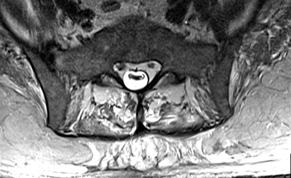 Muscle MRI: T2 Cryptococcal Myopathy
Patchy involvement of paraspinous muscles |
Muscle
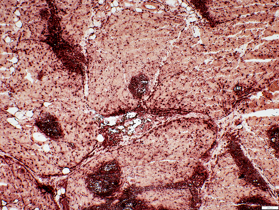
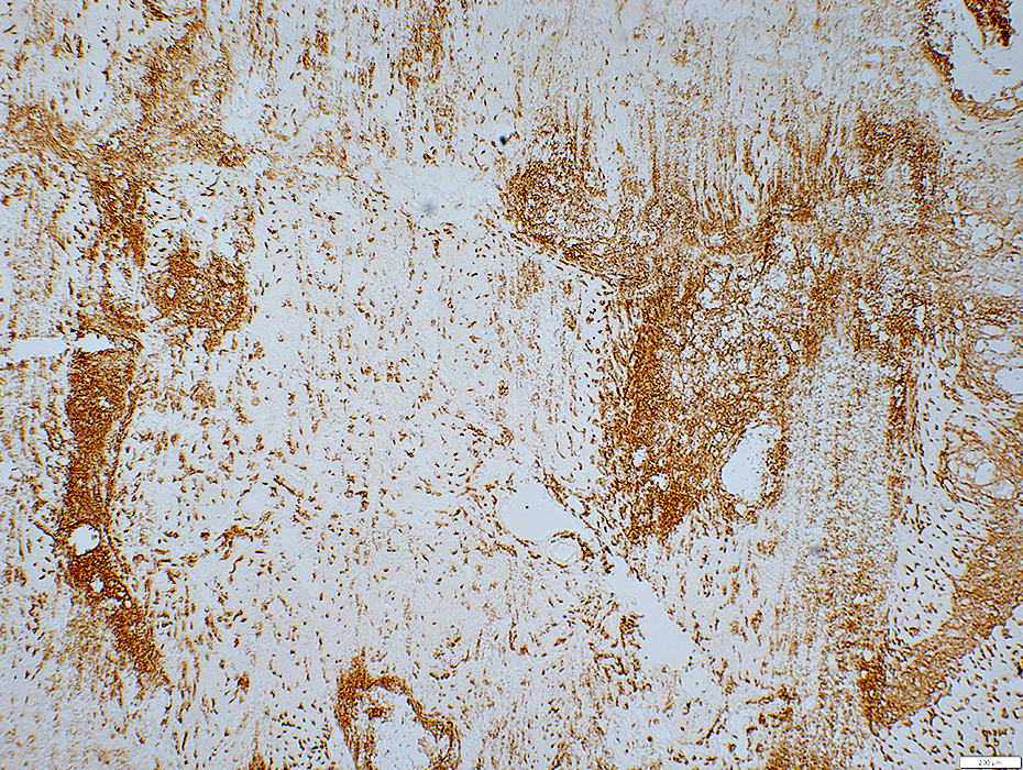
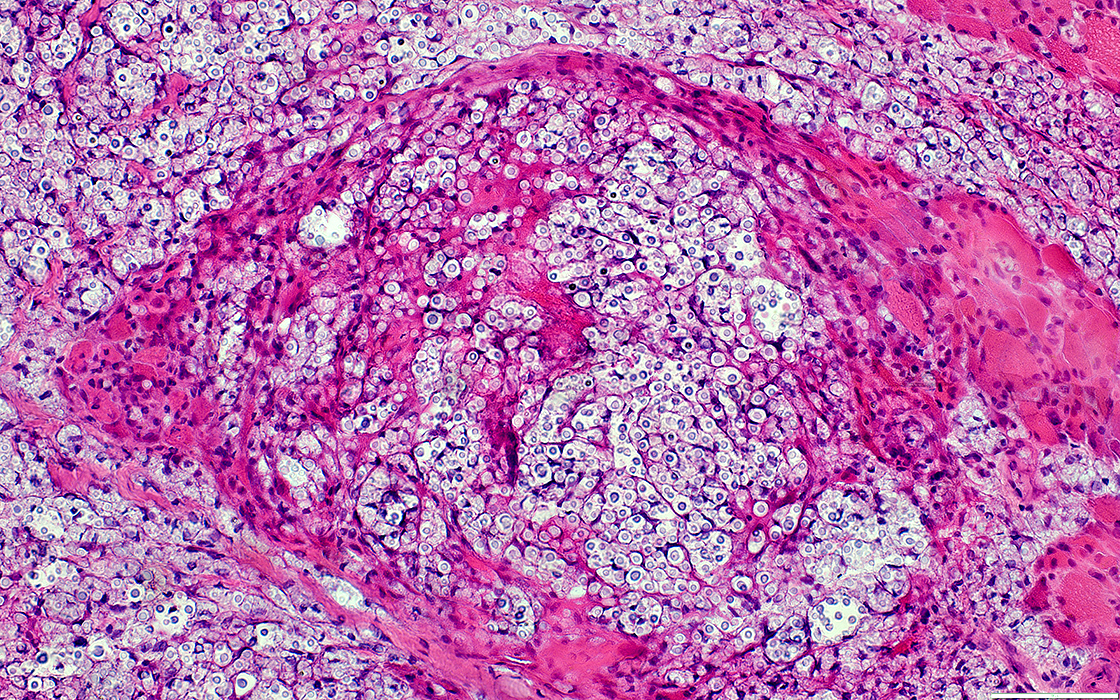
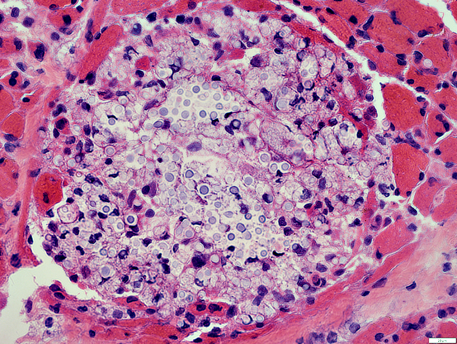
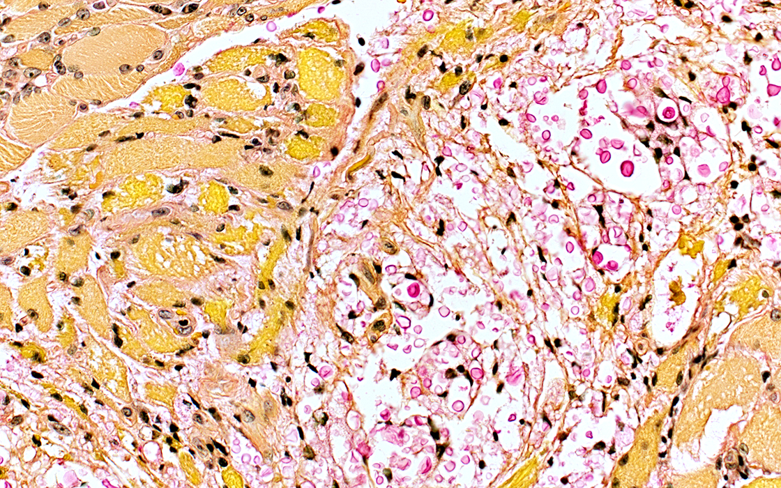
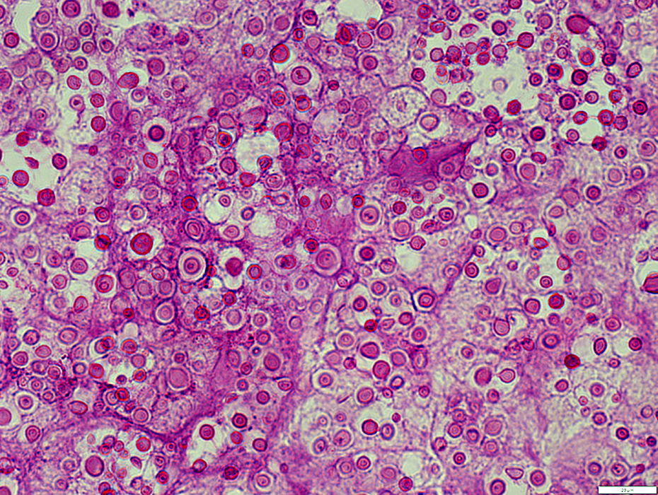
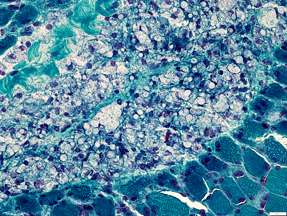
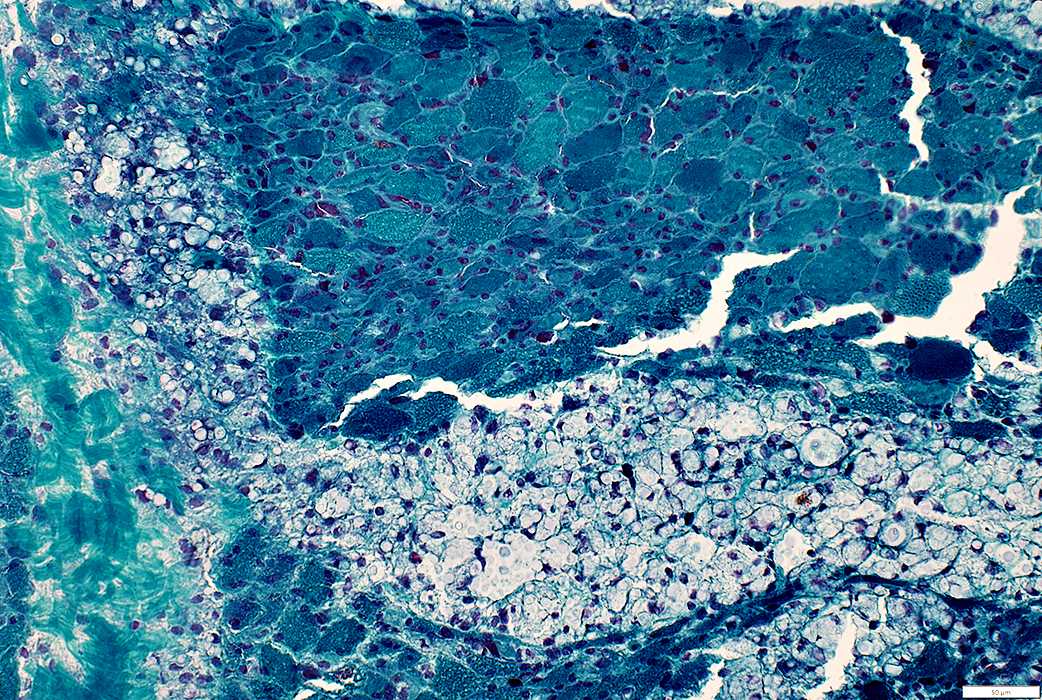
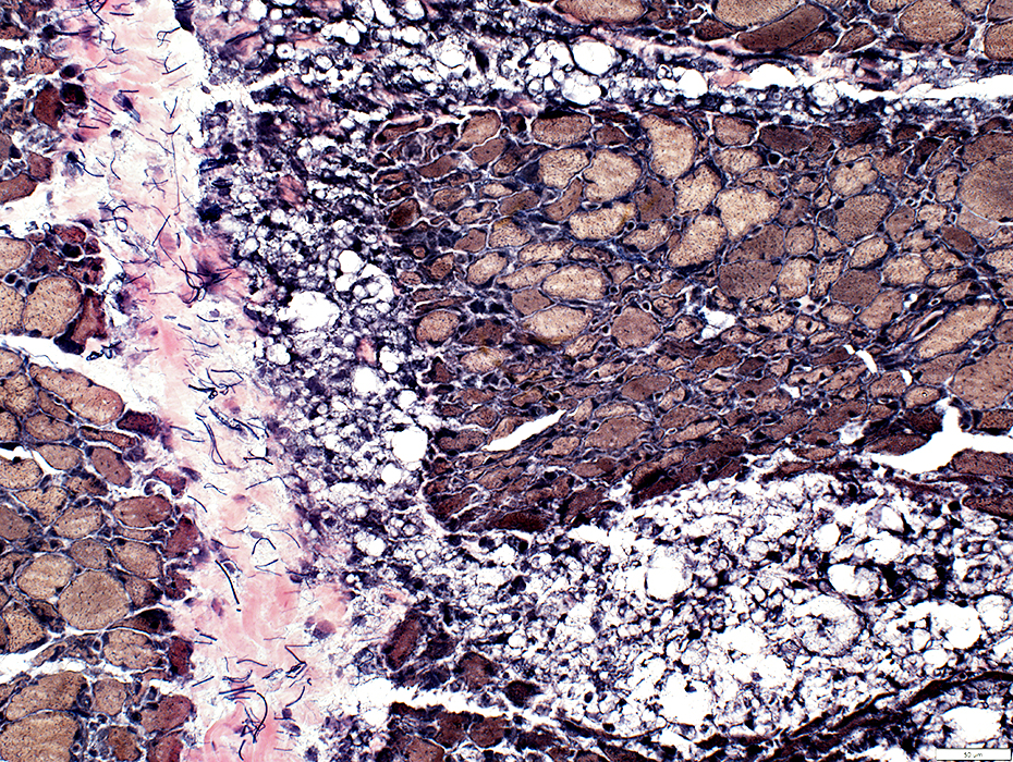
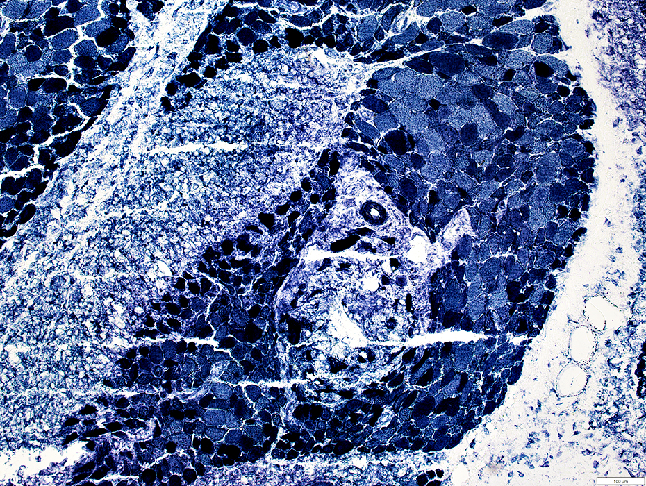
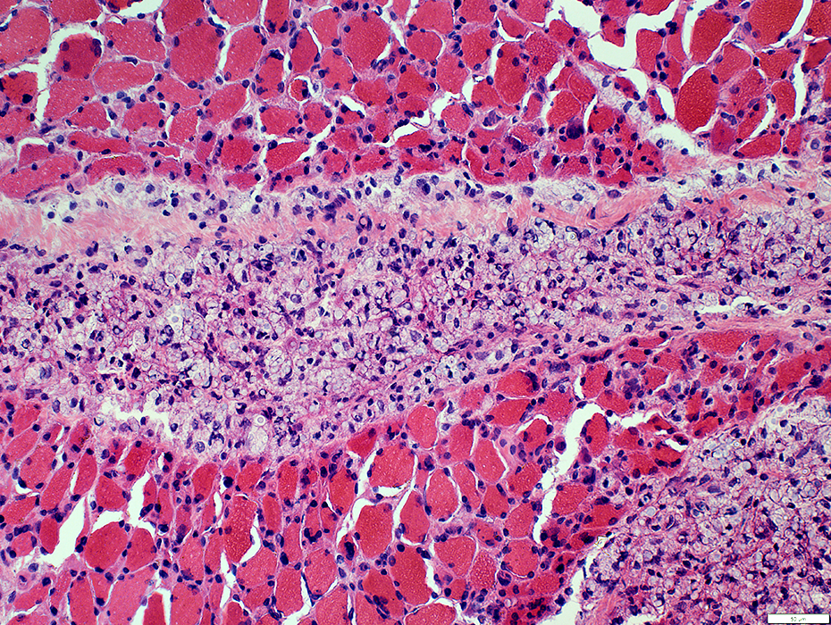
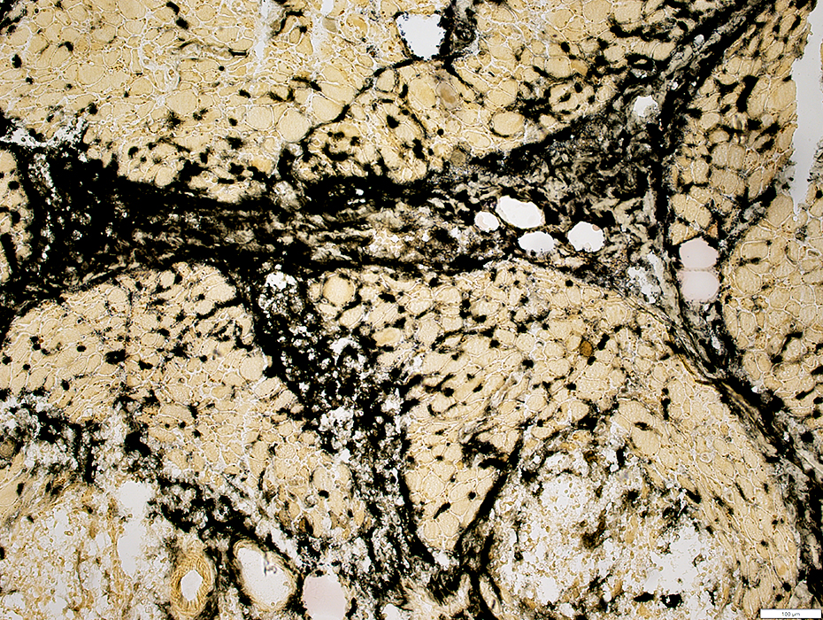
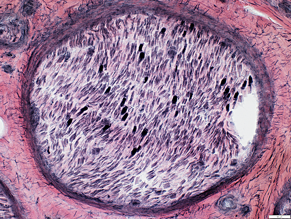
Cryptococcus: Multifocal Involvement of Muscle
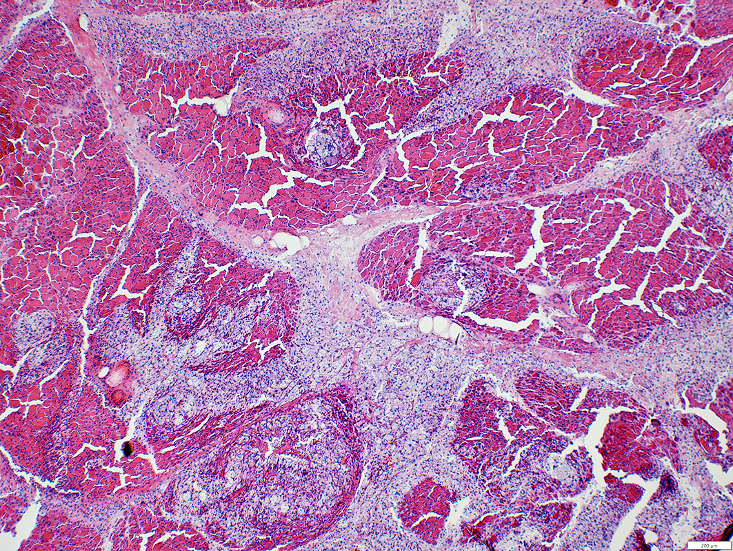 H&E stain |
Multifocal collections of organisms: Small cryptococcomas
Locations: Endomysium & Perimysium
Other cell components: Histiocytes
 H&E stain |
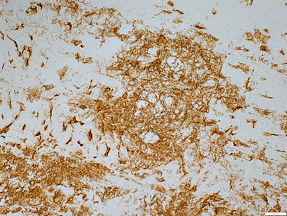
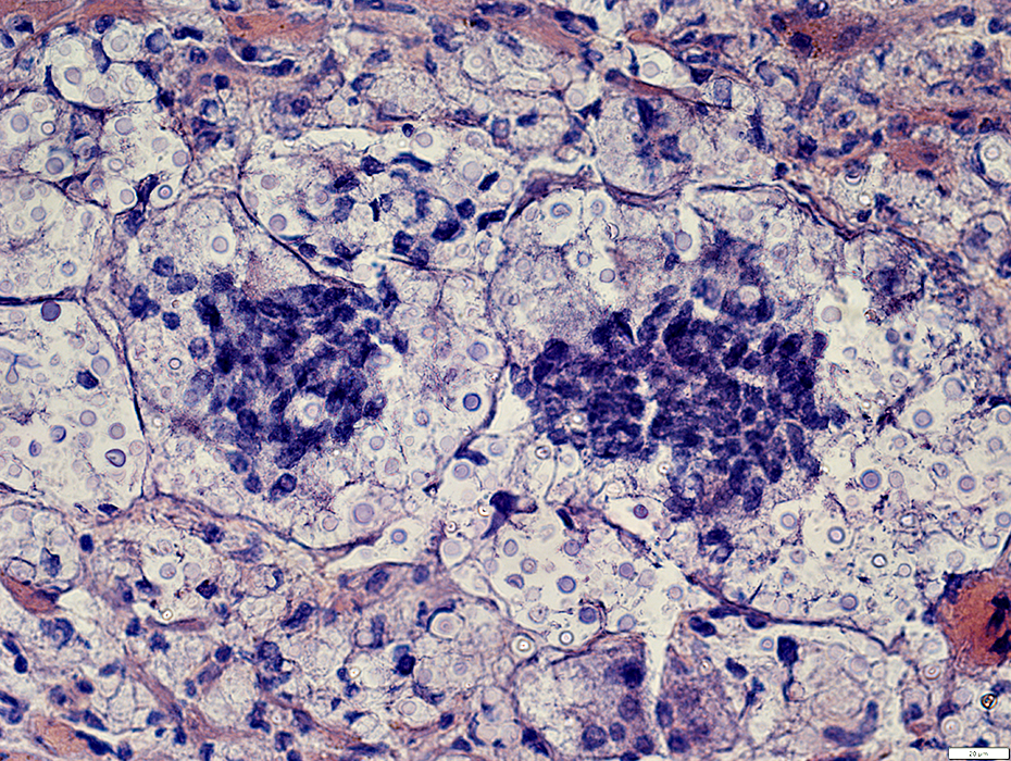
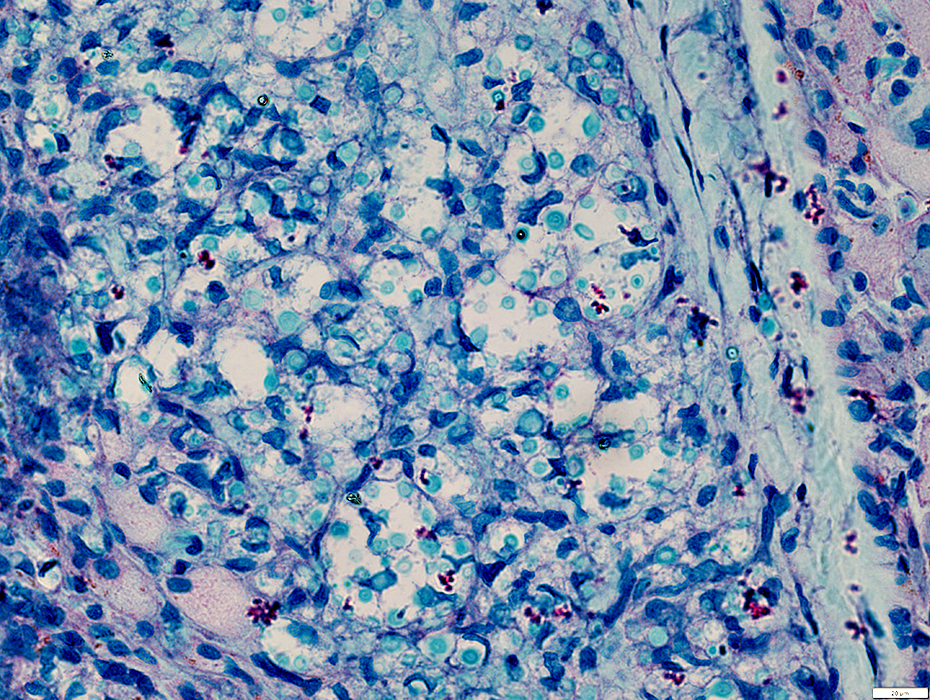
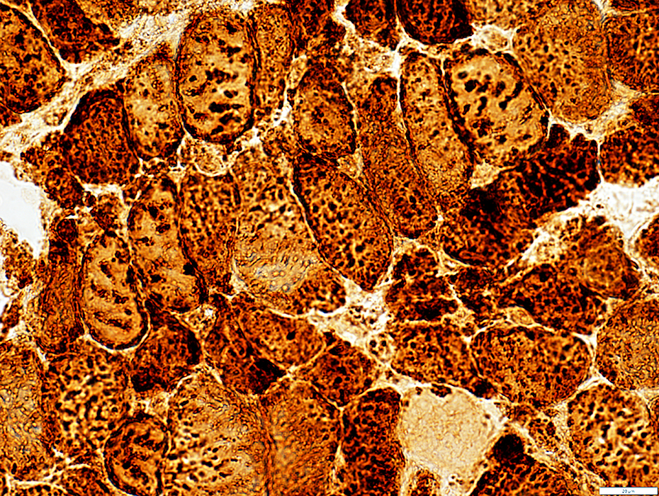
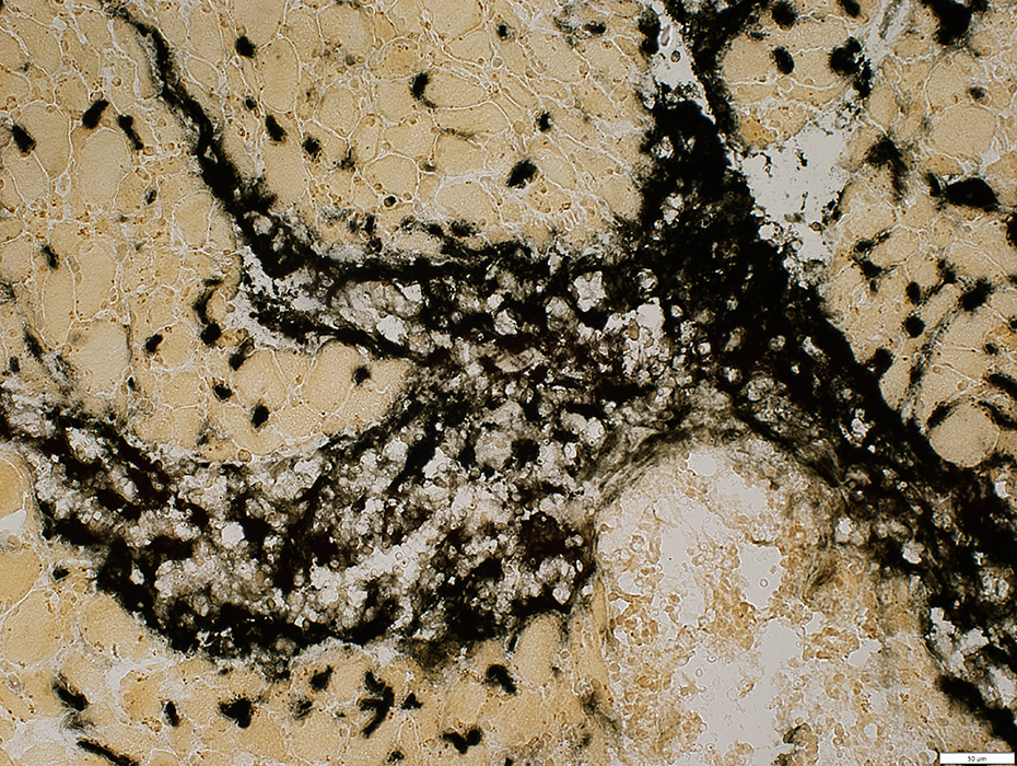
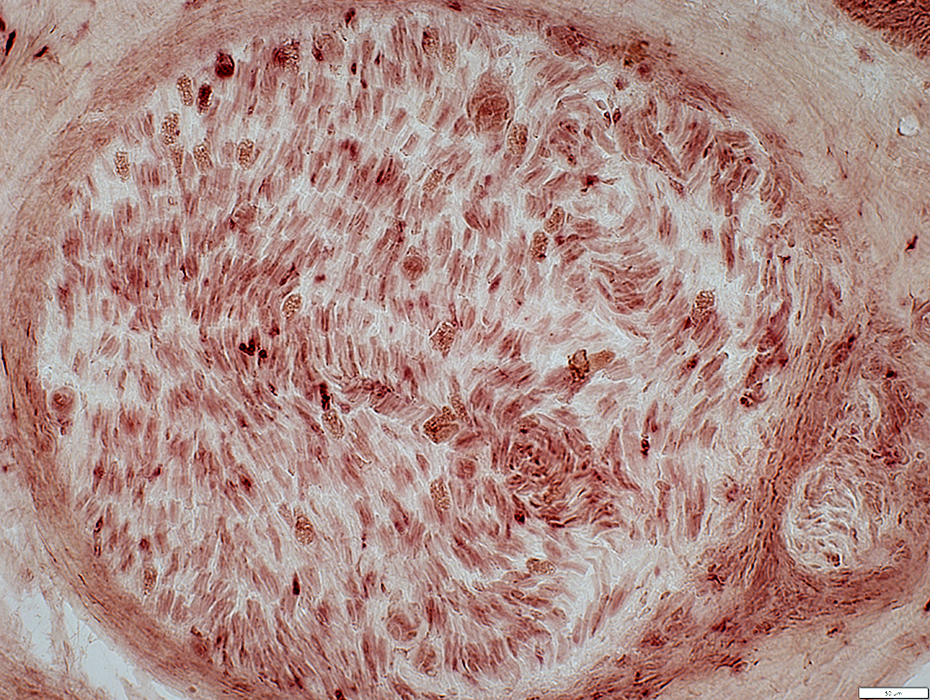
Cryptococcus: Histiocytic Cell & Cryptococcal foci
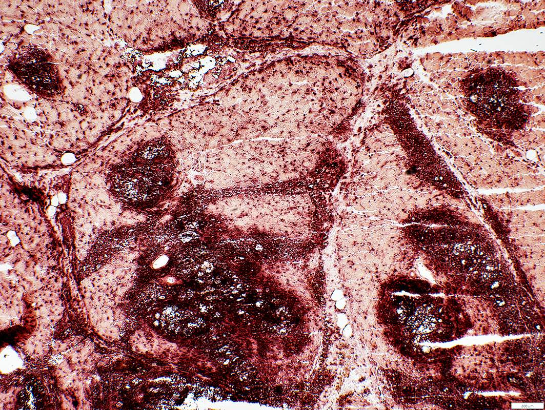 Acid phosphatase stain |
Histiocytic cells
Associated with cryptococcal clusters
Scattered in endomysium: Probably near capillaries
Locations: Endomysium; Perimysium
 Acid phosphatase stain |
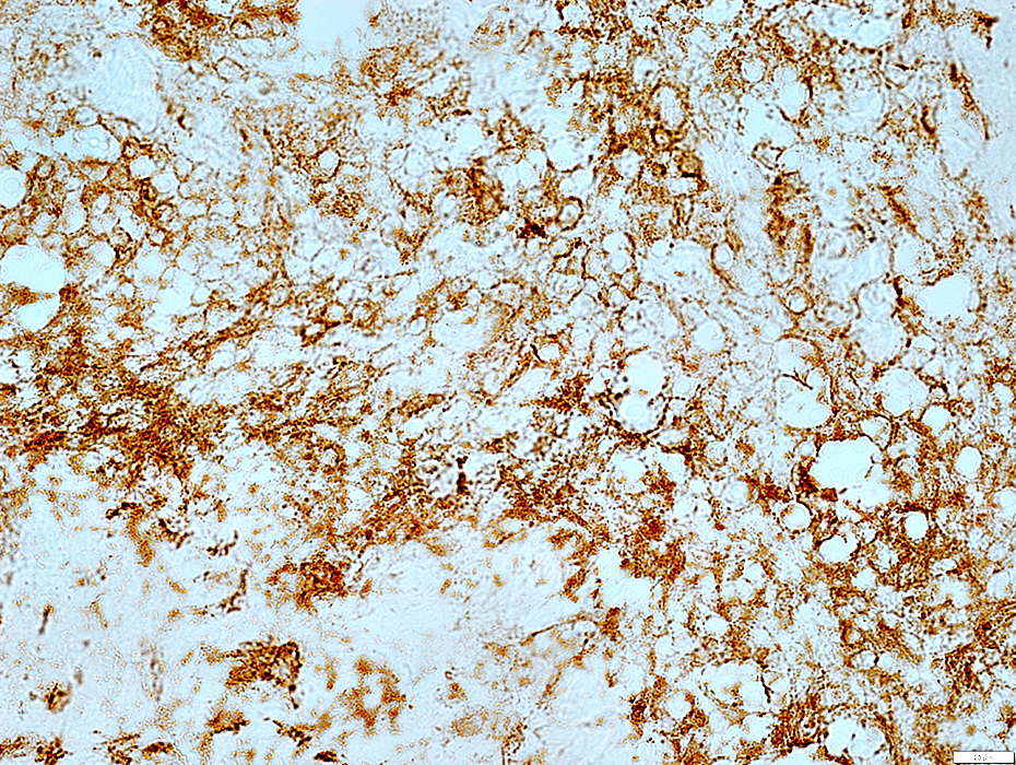
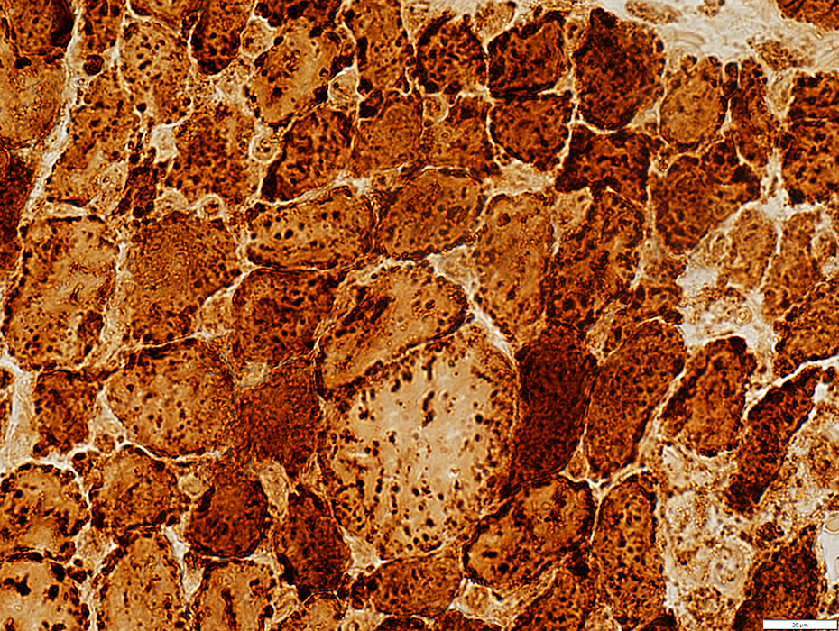
 Acid phosphatase stain |
Cell wall (Rim of cryptococci inside capsule)
Confluent through focus
May obscure cryptococcal organisms
Similar to granulomas associated with foreign bodies or IMAM
 Acid phosphatase stain |
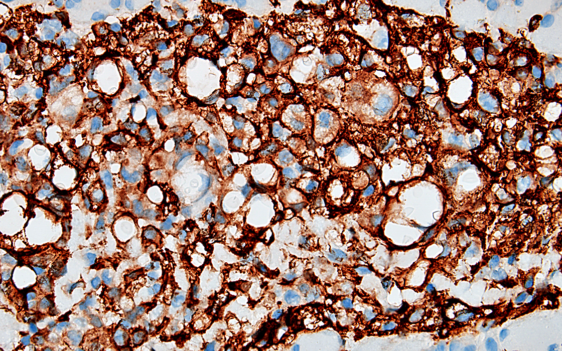
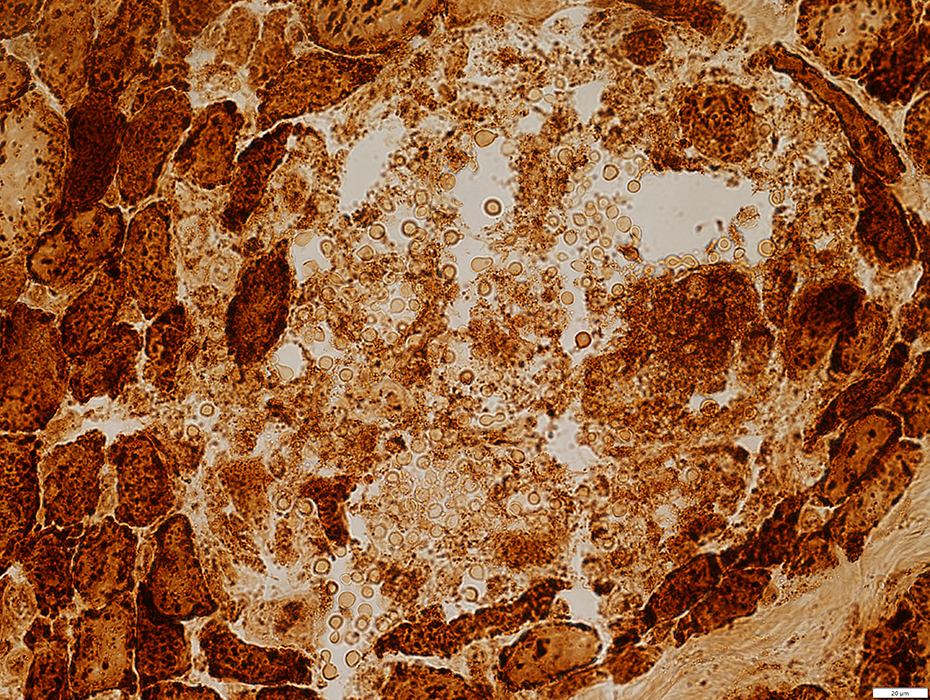
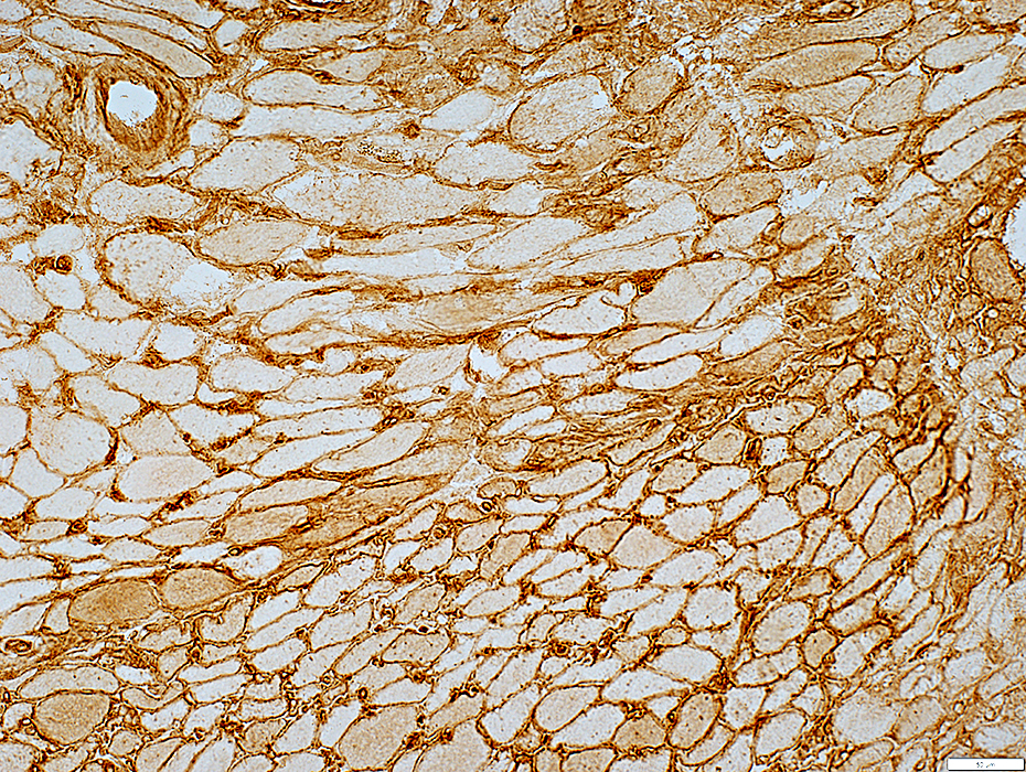
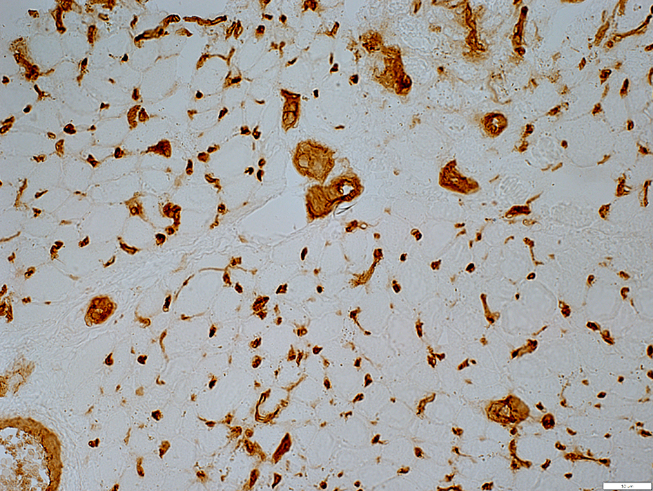
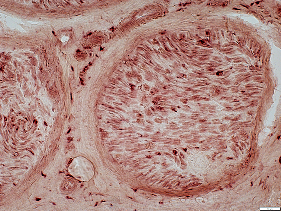
 CD 163 stain |
Foci: Present in clusters along with (around) cryptococci
Scattered: In muscle endymysium near capillaries
 CD 163 stain |
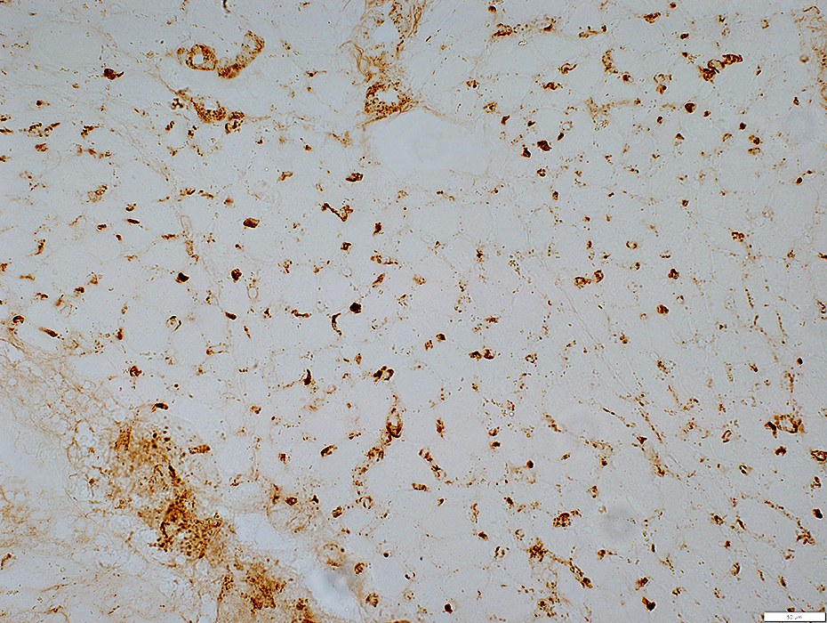
 CD 163 stain |
Foci: Present in clusters with processes around cryptococci
Cryptococci: Often intacellular within Histiocyte cytoplasm
 CD 163 stain |
 CD 163 stain |
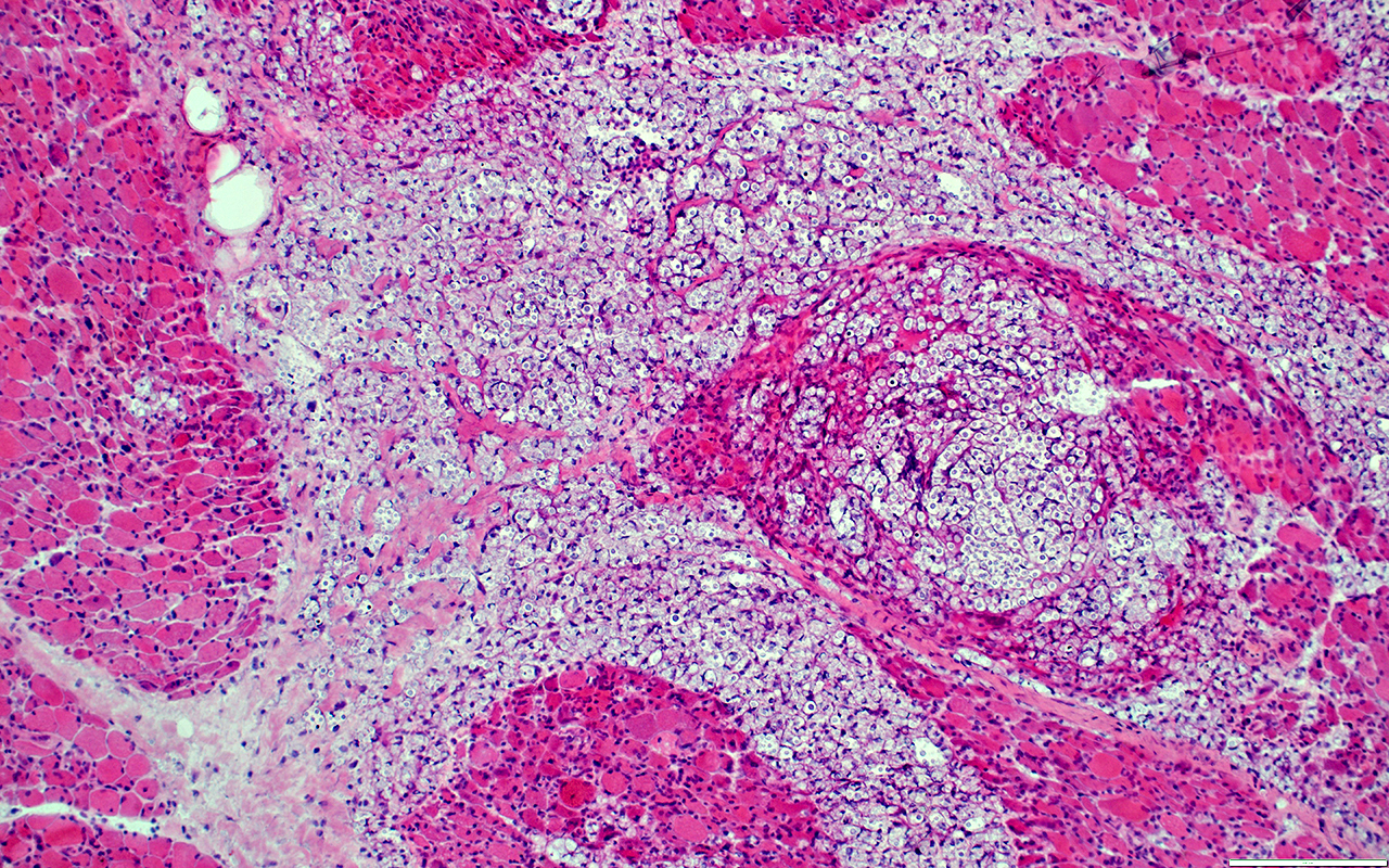
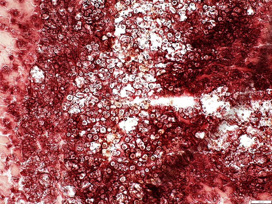
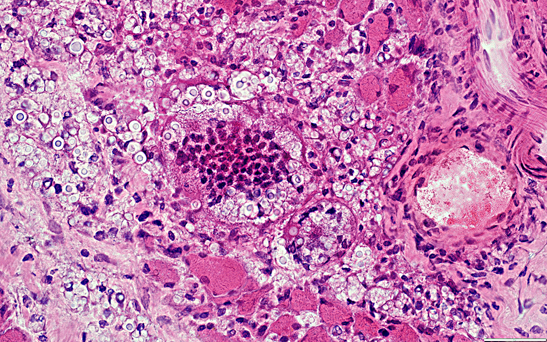
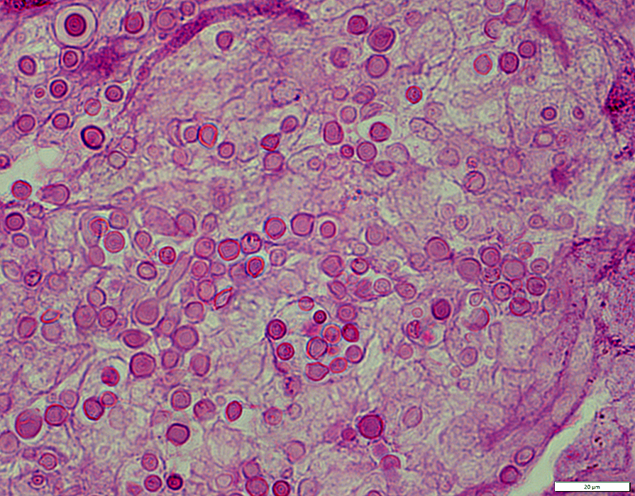
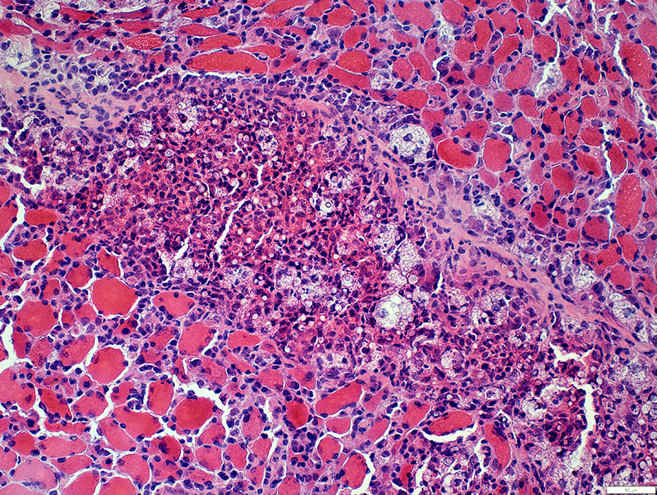
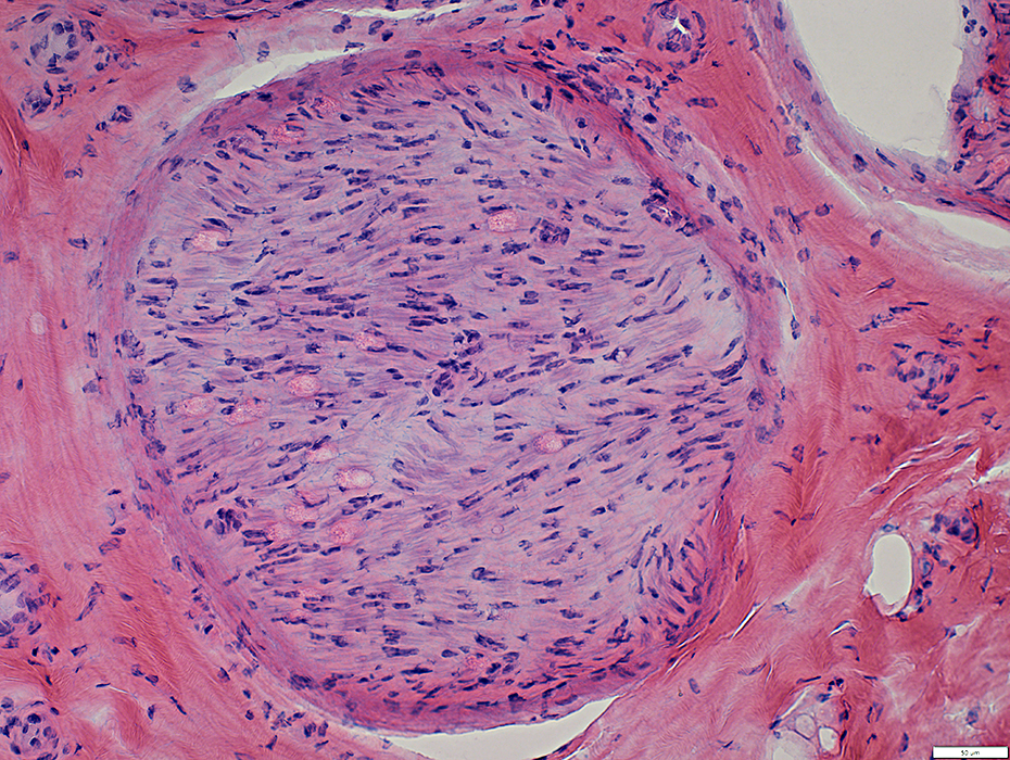
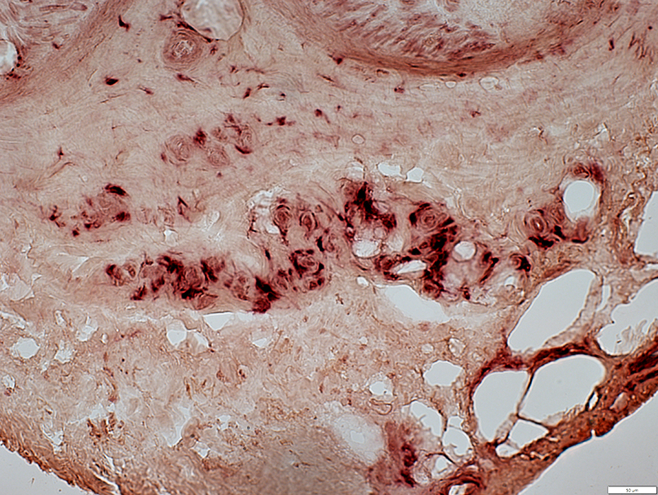
Cryptococcal Foci
 H&E stain |
Locations
Clusters (foci) occur in: Endomysium or Perimysium
Contents
Some foci contain mainly cryptococci, connective tissue & indistinct cells that stain for acid phosphatase (Above).
A few foci contain clusters of lymphocyte-like cells (Below)
Neighboring muscle fibers
Often smaller than in other areas
 H&E stain |
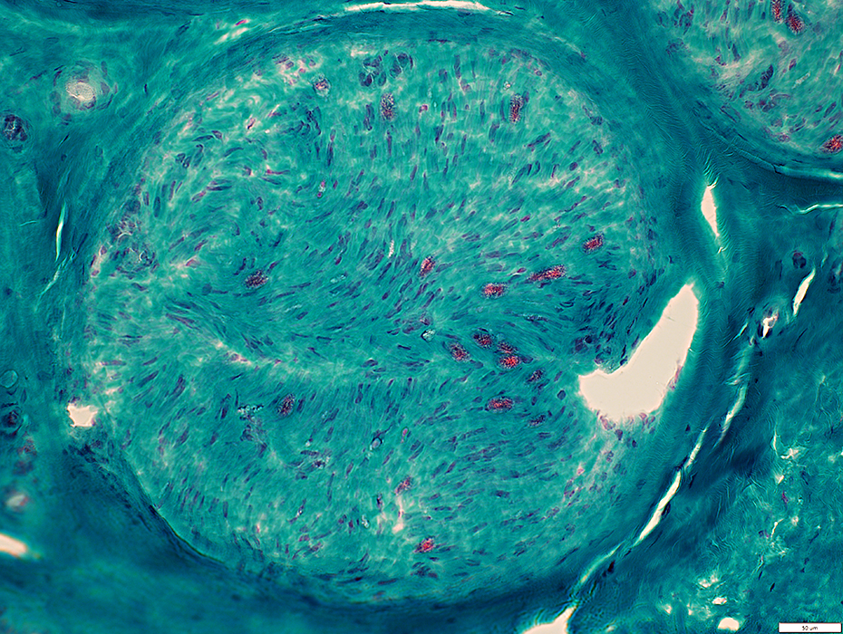
 Congo red stain |
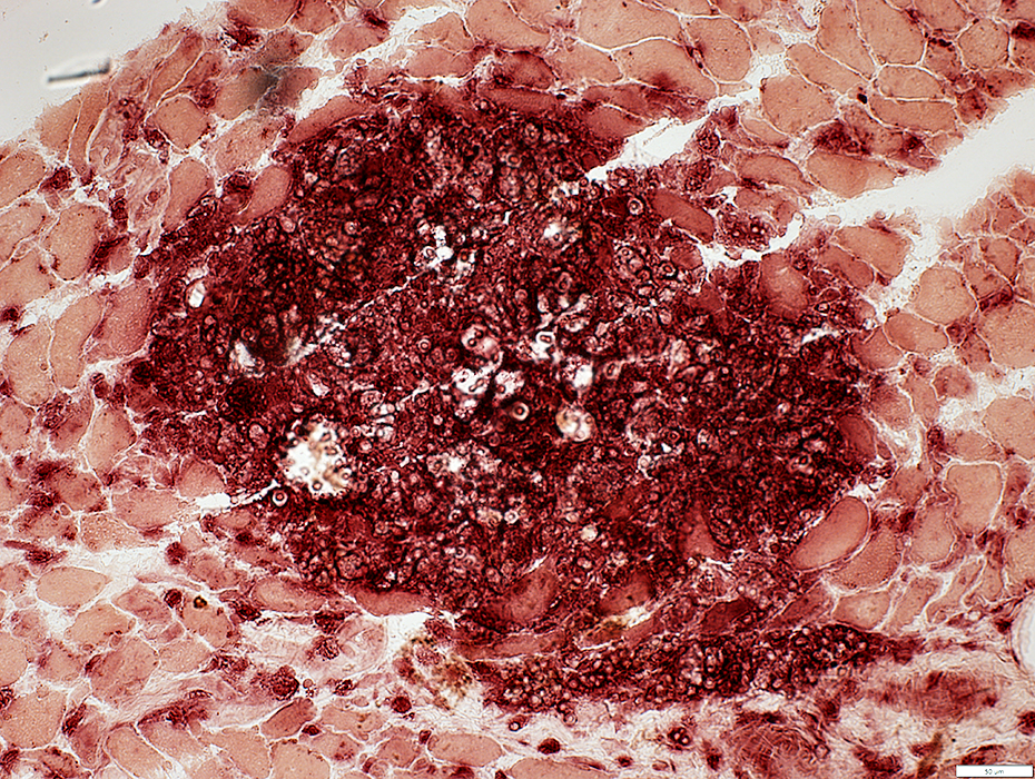
Cryptococcal focus
Fewer cryptococci
More other cells & tissue
 H&E stain |
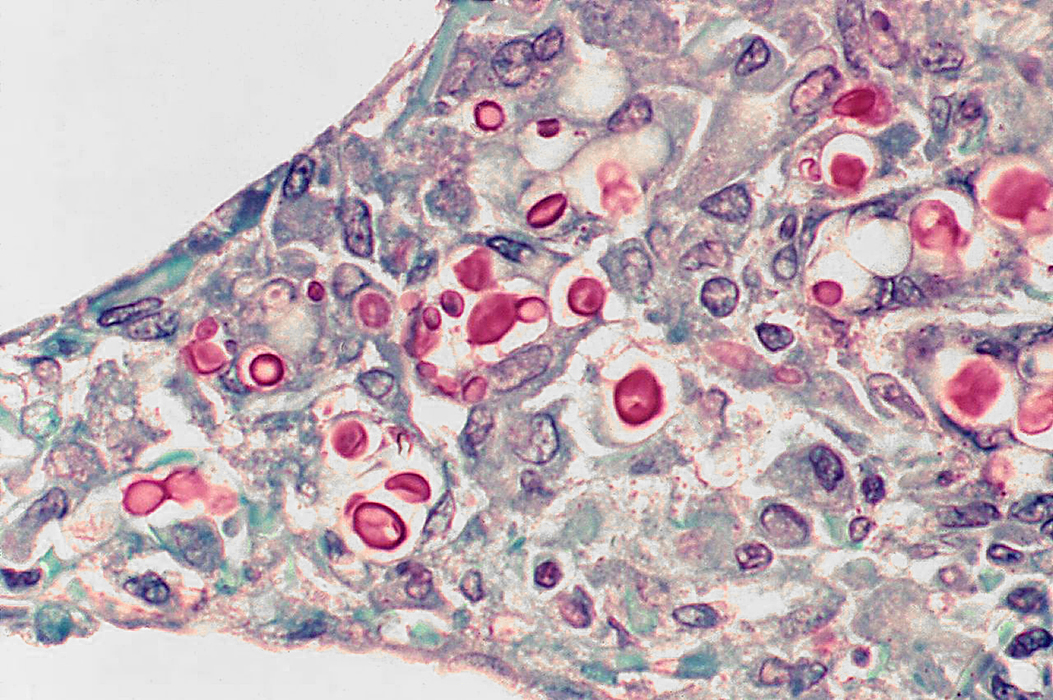
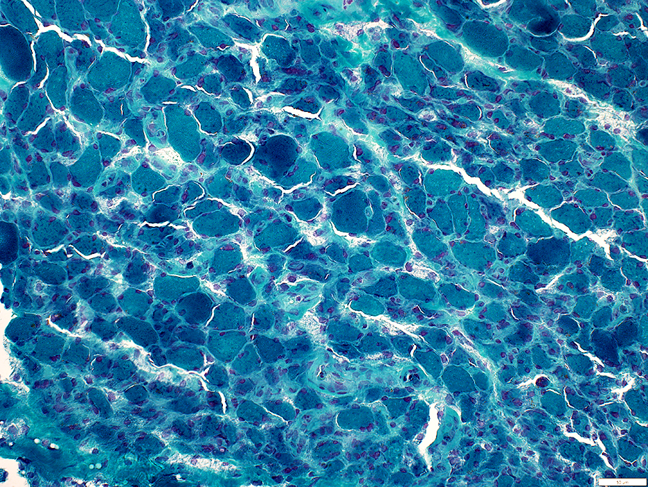
Cryptococci: Organisms
Cryptococci: Many budding
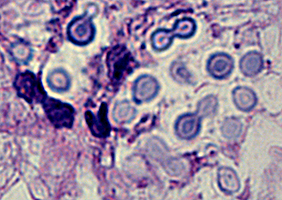 H&E stain |
 H&E stain |
Sizes: Varied
Structure
Peripheral: Clear capsule with varied thickness
Central
Rimmed circular region
A few show budding (Arrows, Below)
 H&E stain |
 Mucicarmine stain |
 Mucicarmine stain |
 Mucicarmine stain From: Wikimedia |
 Grocott methenamine silver (GMS) stain |
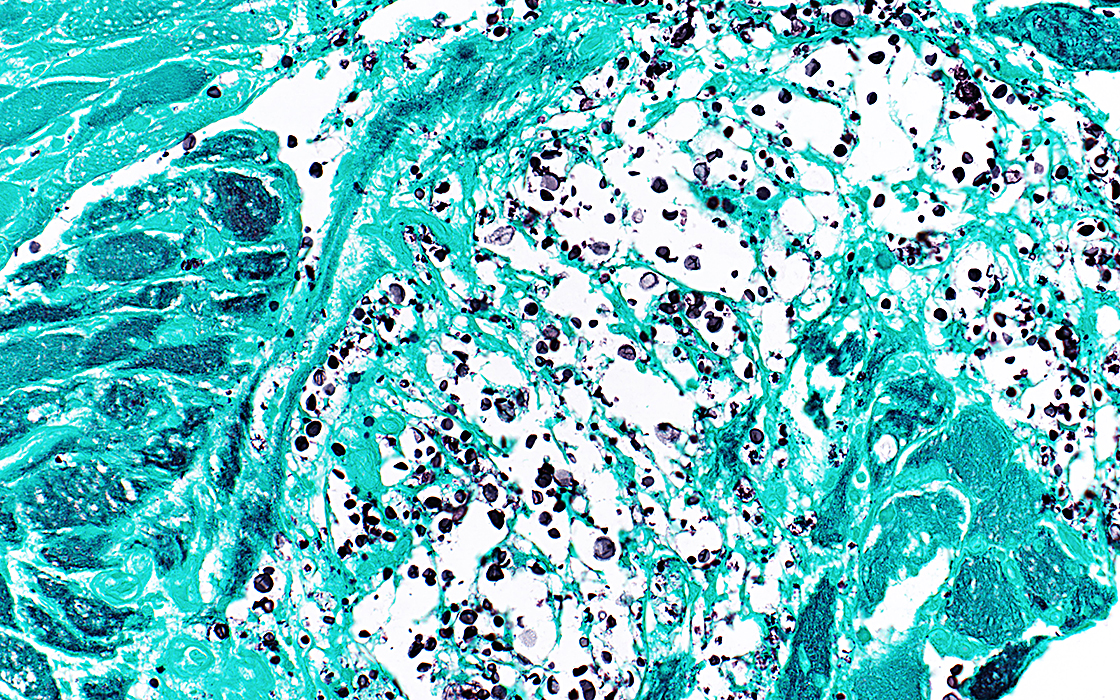 Grocott methenamine silver (GMS) stain |
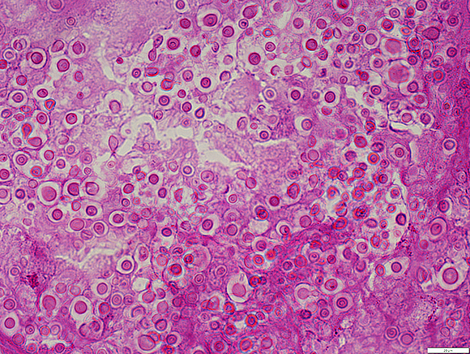 PAS stain |
Sizes: Varied
Structure
Peripheral: Clear capsule with varied thickness
Central: Rimmed circular region, PAS+
Clustered in endomysium or perimysium
 PAS stain |
Cryptococcal cluster: Most organisms in this cluster have thin, or no, capsules
 PAS stain |
Alcian blue: Stains central region of organisms
 Alcian blue stain |
Sudan black: Stains round area within central region of some organisms
 Sudan stain |
Thin layer of connective tissue around cryptococci
Stained by Gomori trichrome
 Gomori trichrome stain |
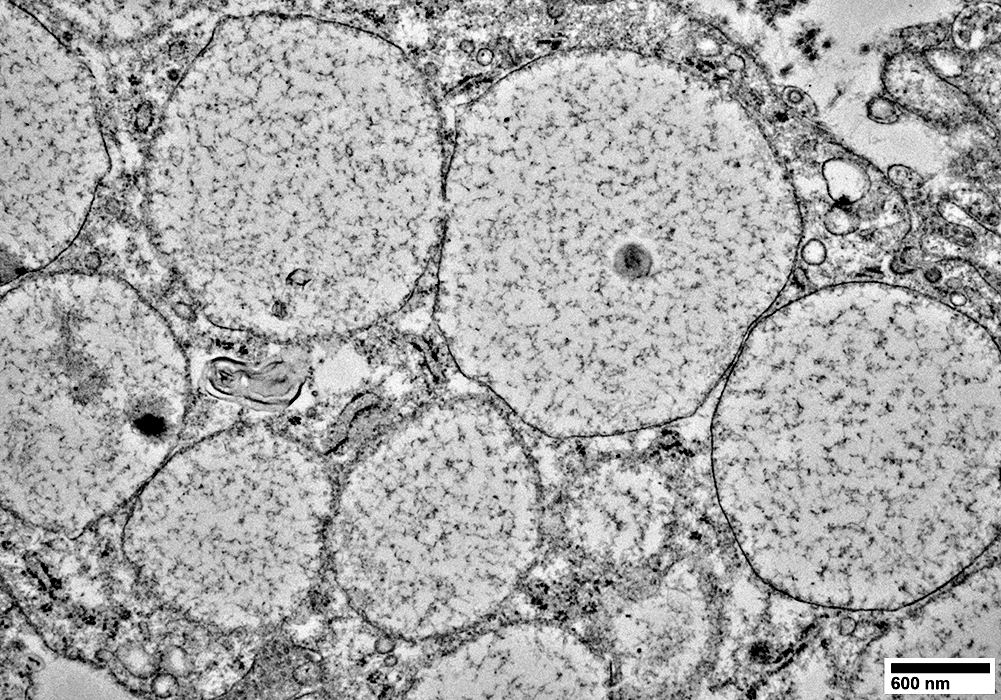
Cryptococci contain no mitochondria
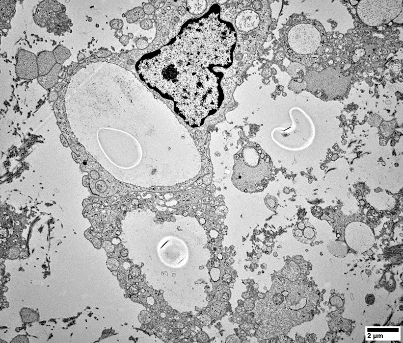
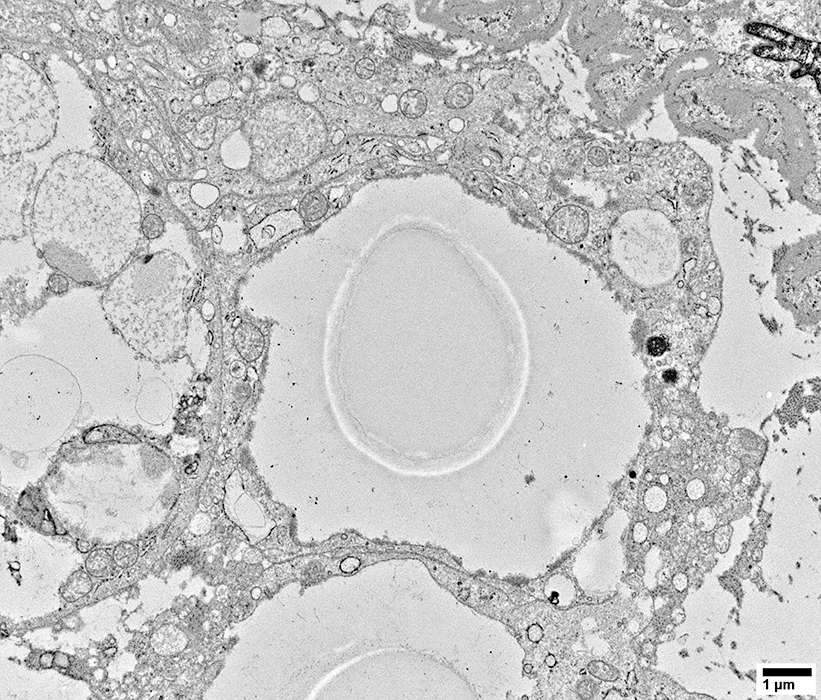
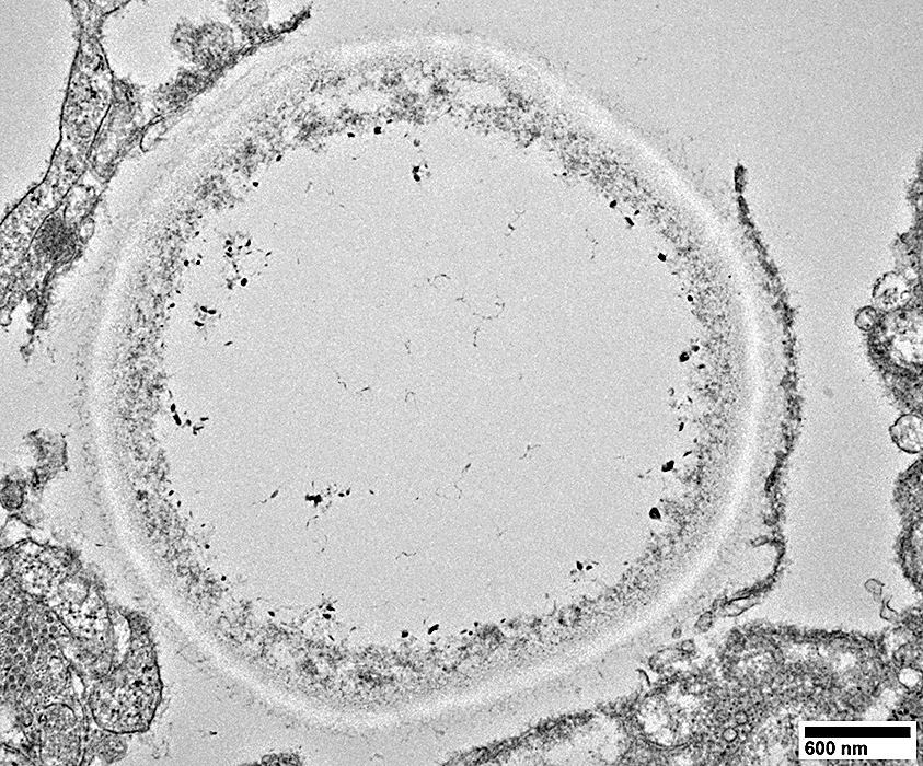
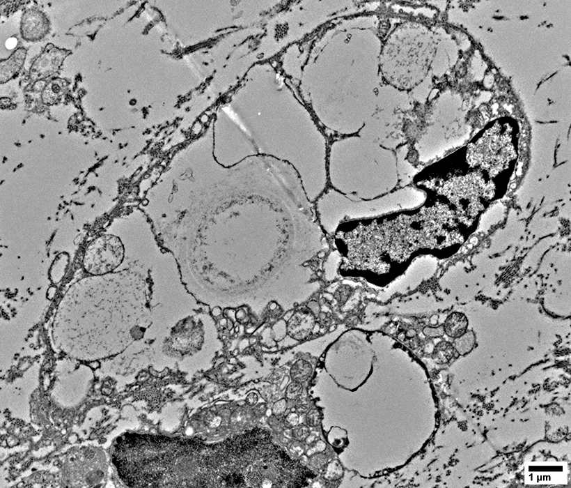
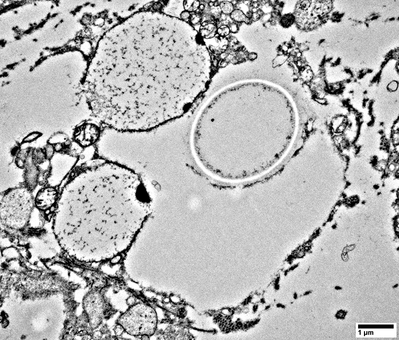
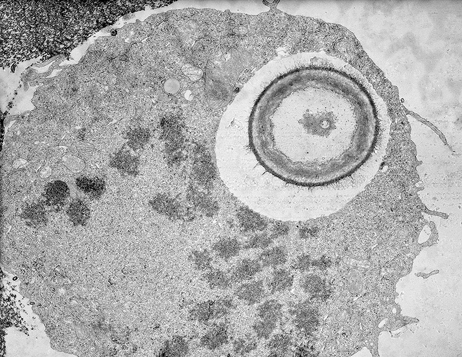
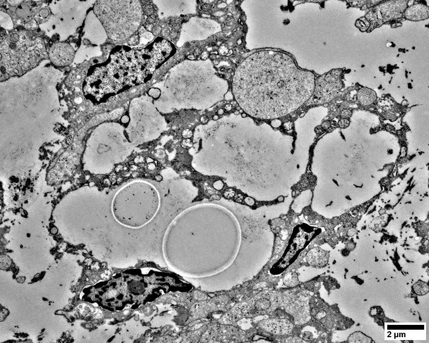
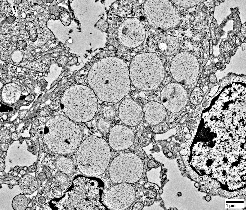
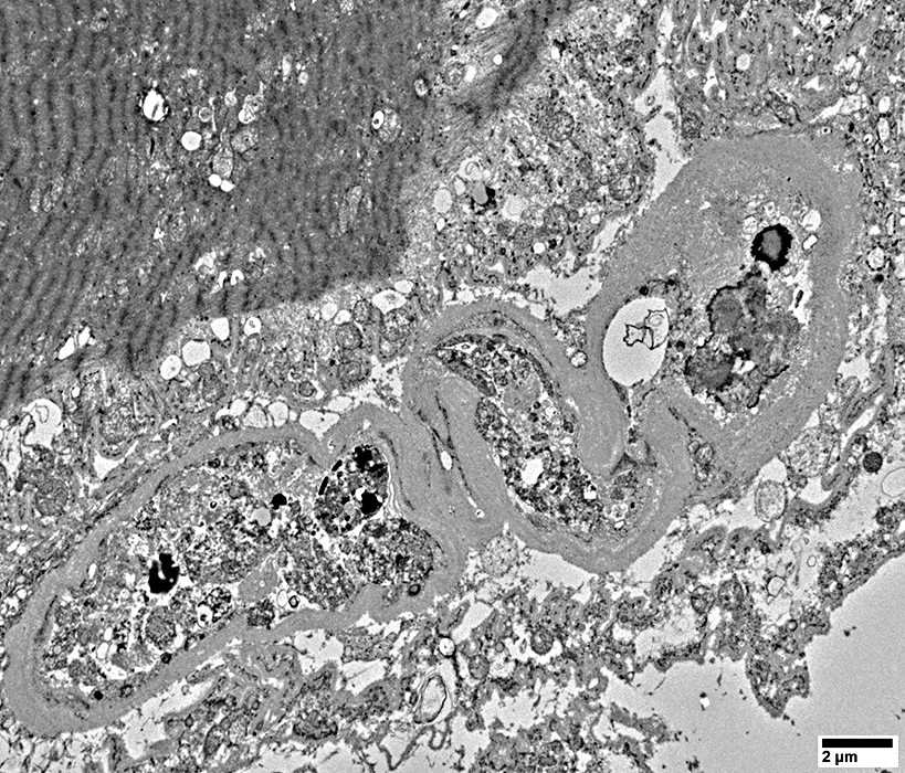
Cryptococci: Ultrastructure
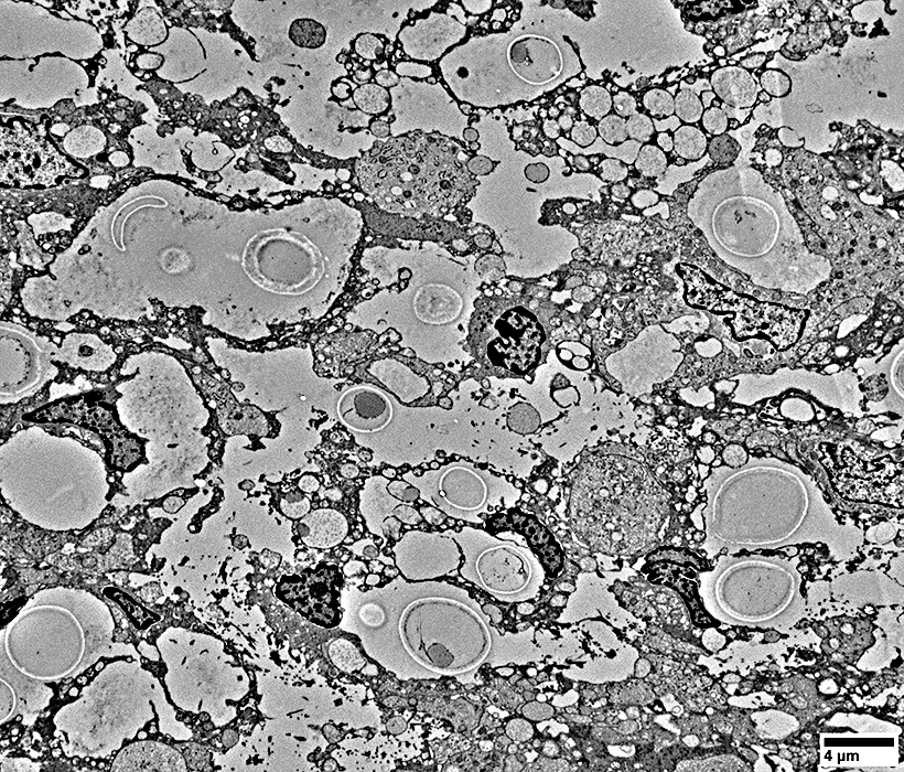 From: R Schmidt |
 From: R Schmidt |
 From: R Schmidt |
 From: R Schmidt |
 From: R Schmidt |
 From: R Schmidt |
 From: R Schmidt |
 From: R Schmidt |
 From: R Schmidt |
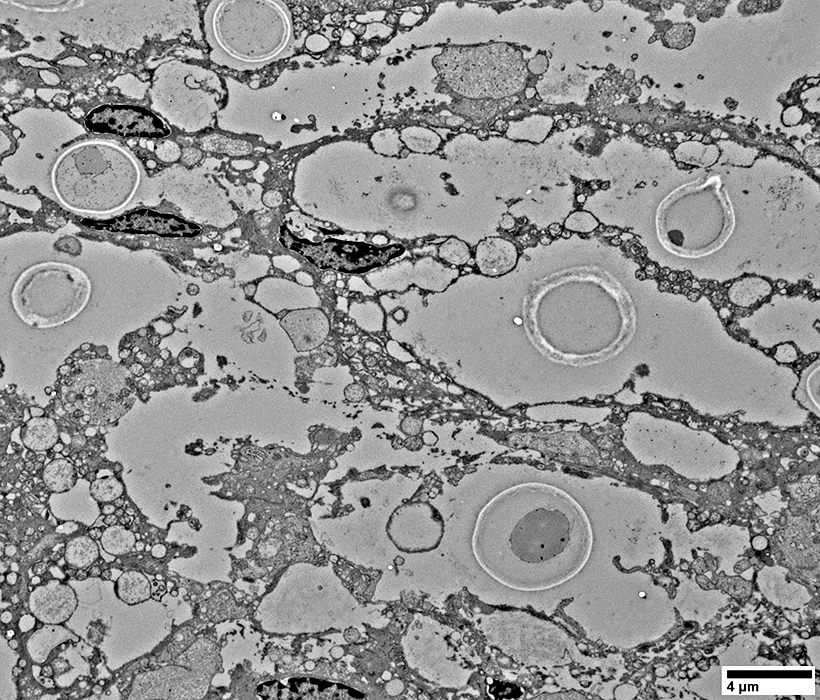
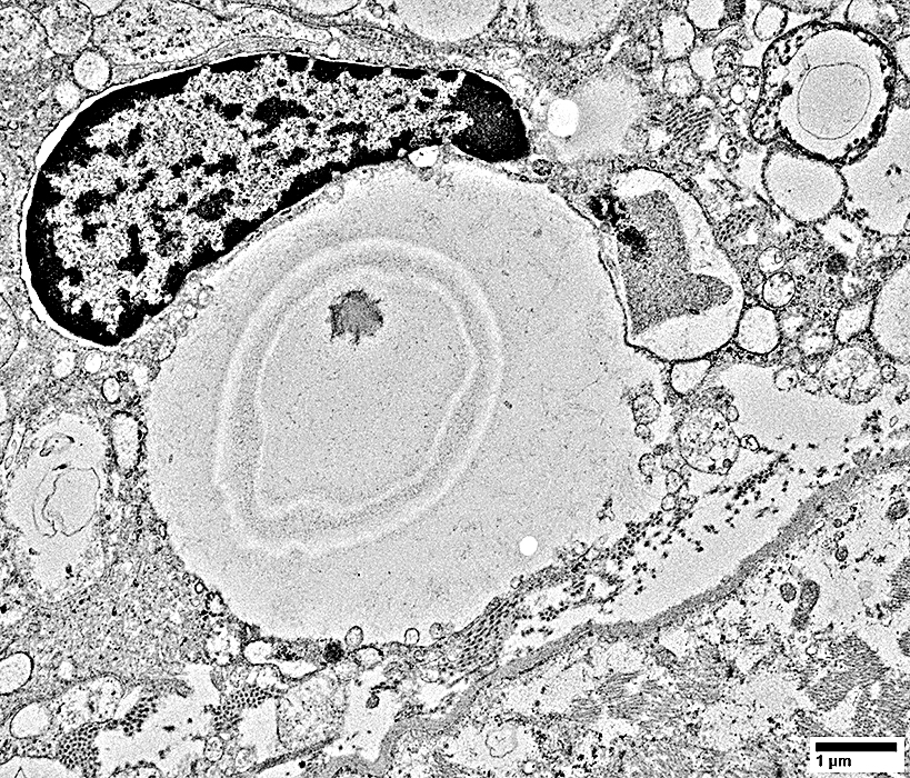
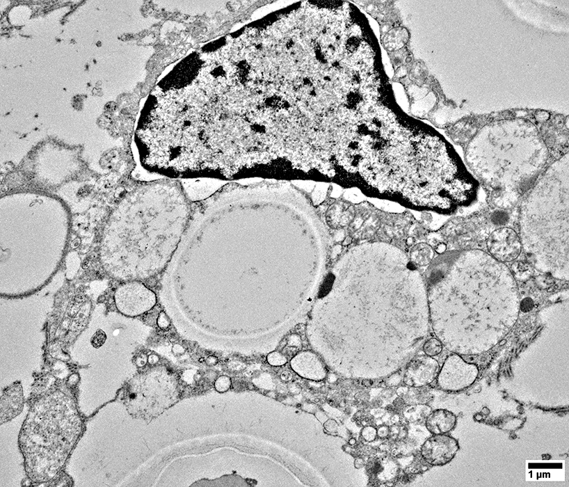
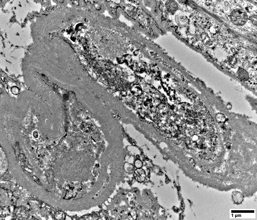
Cryptococcus within a Histiocyte
 From: Wikimedia DC/ Carolina Coelho |
 From: R Schmidt |
 From: R Schmidt |
 From: R Schmidt |
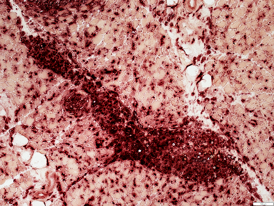
C5b-9 deposited on tissue within and surrounding a cryptococcal focus
 C5b-9 stain |
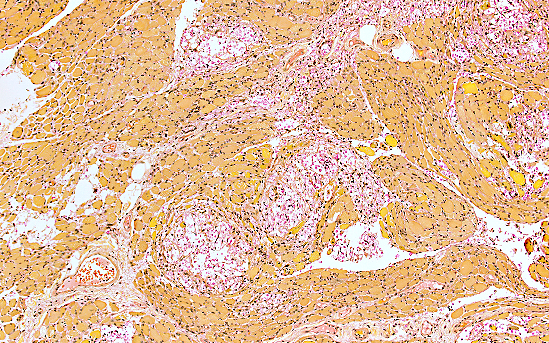
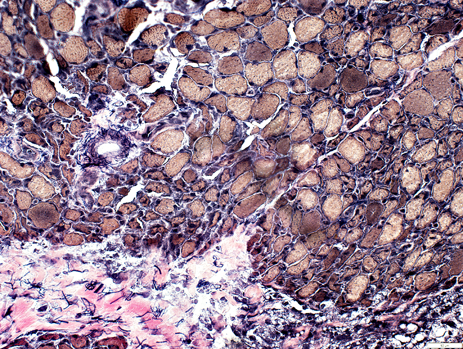
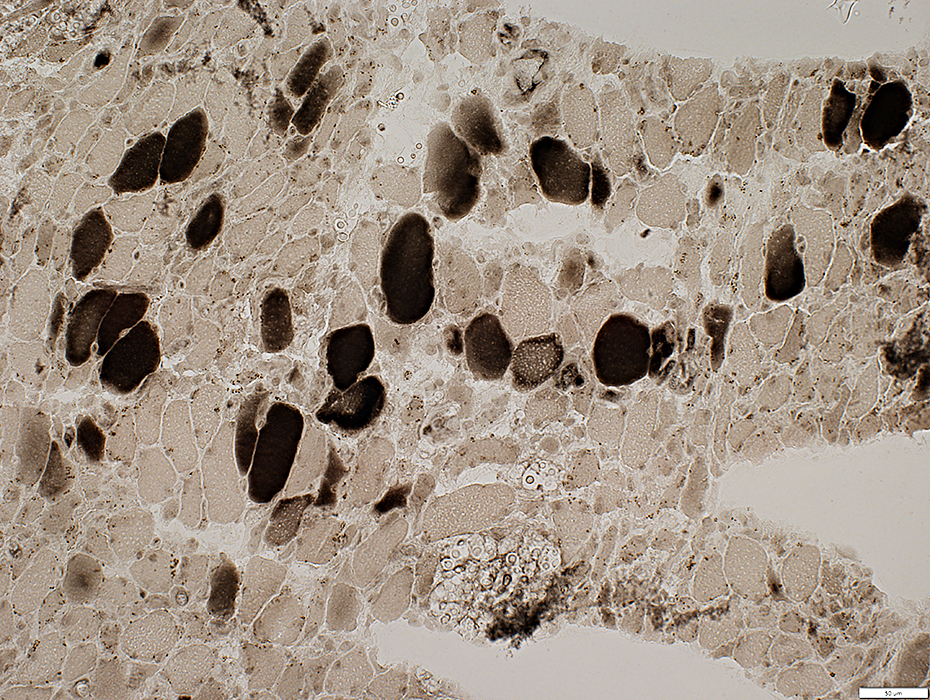
Cryptococcal myopathy: Muscle fibers
 Gomori trichrome stain |
Diffuse atrophy
Smallest muscle fibers are located at edge of fascicles near cryptococcal focus
 VvG stain |
 Gomori trichrome stain |
Diffuse atrophy
Fiber sizes: Varied
 VvG stain |
 VvG stain |
Muscle fibers
Diffuse atrophy
Fiber sizes: Varied
Smaller fibers at edge of fascicle near damaged perimysium
NADH stains small fibers dark (Below)
 NADH stain |
 COX stain |
Megaconial: Most mitochondria are large
Some COX- muscle fibers are present
 COX stain |
Type 2 fiber predominance
 ATPase pH 4.3 stain |
MHC Class I: Upregulated on surface of all muscle fibers
 MHC Class I stain |
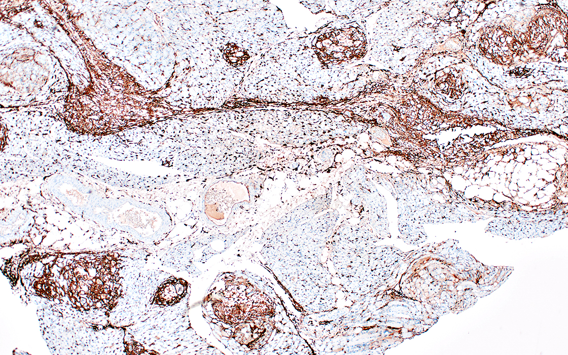
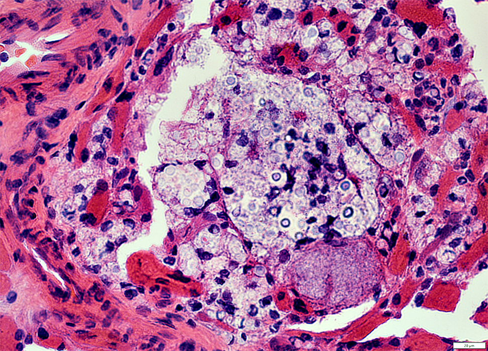
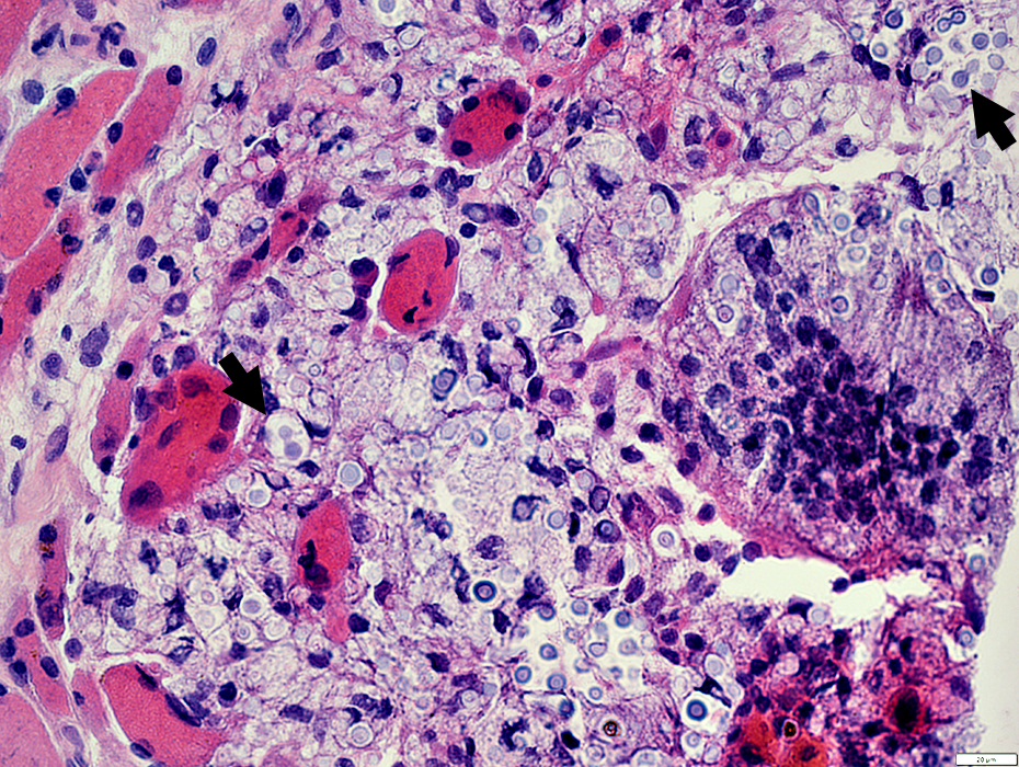
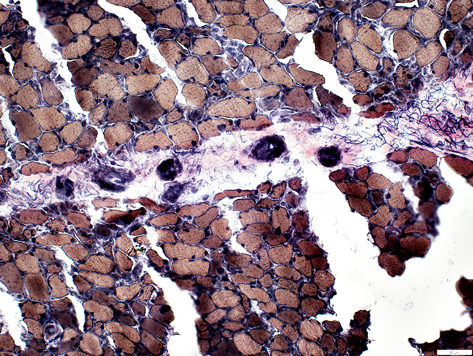
Cryptococcus: Perimysial connective tissue involvement
 H&E stain |
Some foci contain mostly cryptococcal organisms (Above)
A few foci contain cells and cryptococci (Below)
 H&E stain |
Histiocytes are present in
Perimysial cryptococcal foci
Endopmysium: Scattered
 Acid phosphatase stain |
 Alkaline phosphatase stain |
Perimysial connective tissue
Endomysial capillaries
 Alkaline phosphatase stain |
Capillary pathology
 UEA 1 stain |
Large & Abundant (Above)
C5b-9 staining (Below)
Also see: Endoneurial microvessel staining
 C5b-9 stain |
 From: R Schmidt |
 From: R Schmidt |
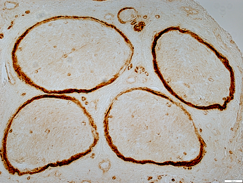
Cryptococcal infection: Associated Neuropathy
 H&E stain |
No cryptococcal infection
 Gomori trichrome stain |
 VvG stain |
 Acid phosphatase stain |
Few scattered in endoneurium
Some in epineurium, especially near vessels
 Acid phosphatase stain |
 Acid phosphatase stain |
C5b-9
Mild staining on endoneurial microvessels
Also see: Endomysial capillary staining
 C5b-9 stain |
Return to Cryptococcus
5/15/2023