Focal demyelination: Chronic
No, or little, remyelination
Probably at region of persistent temporal dispersion
|
Demyelinated axons Hypomyelinated axons Axon loss |
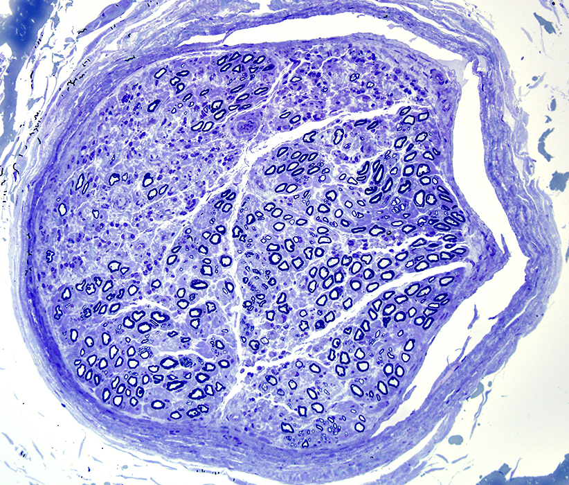 Toluidine blue stain |
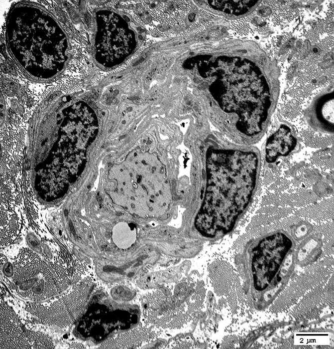
Focal endoneurial region with reduced numbers
Demyelinated axons are common in this area
Remaining myelinated axons often have thin myelin
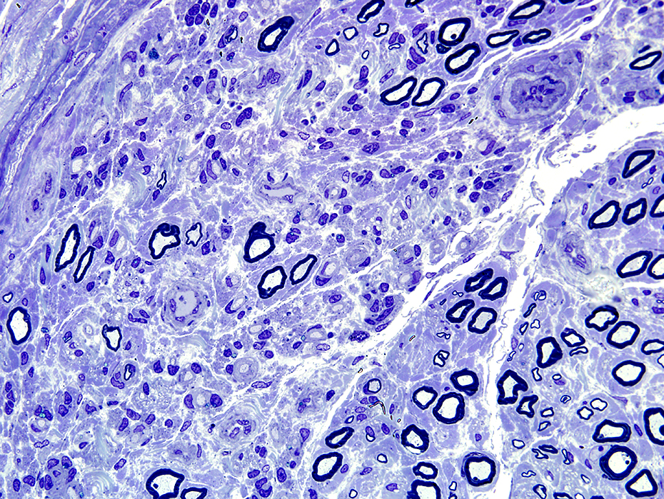 Toluidine blue stain |
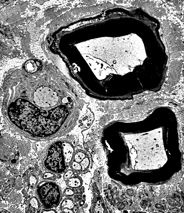
Region of nerve with chronic demyelination but little or no remyeliation
Many completely demyelinated axons
Schwann cells & their processes surrounding the demyelinated axons
Phagocytosis of myelin is complete: No histiocytes or myelin debris
This pathology is likely associated with a region of
chronic partial conduction block or temporal dispersion on electrodiagnostic testing
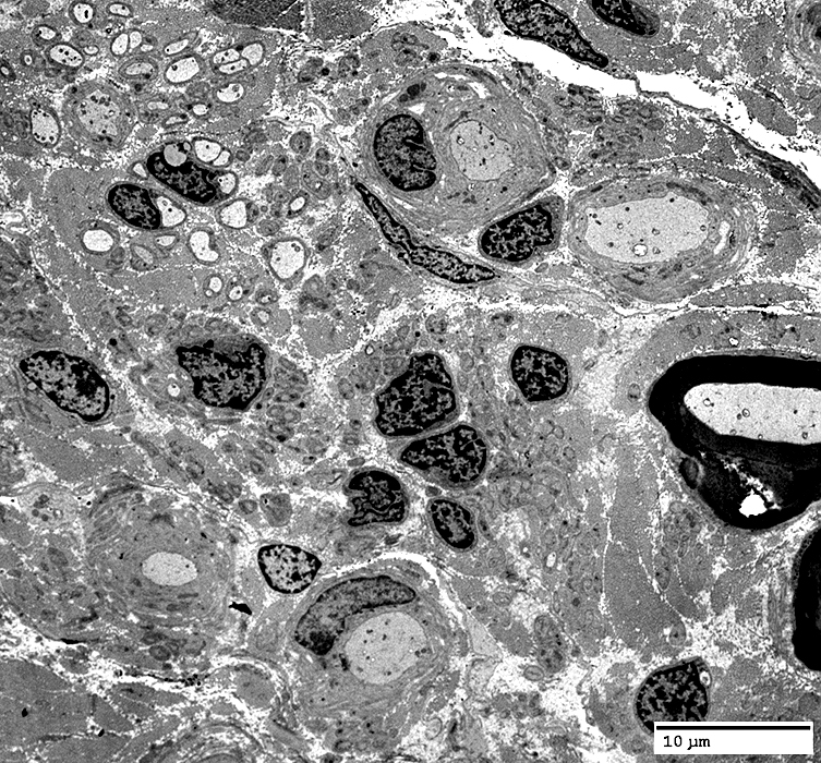 Electron micrographs: From Robert Schmidt MD |
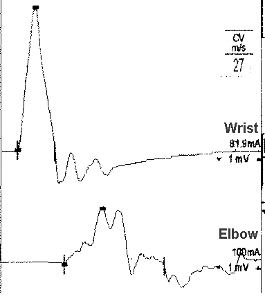 Nerve conduction studies (Median nerve) Temporal dispersion |
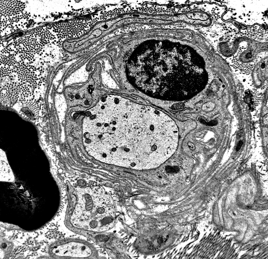
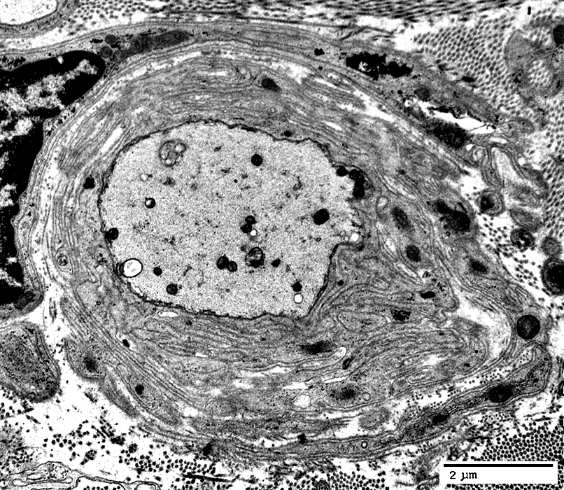
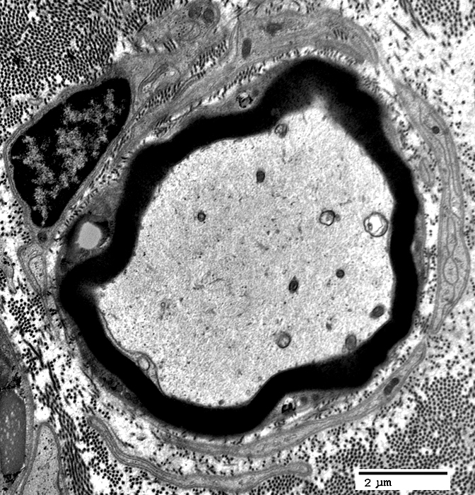
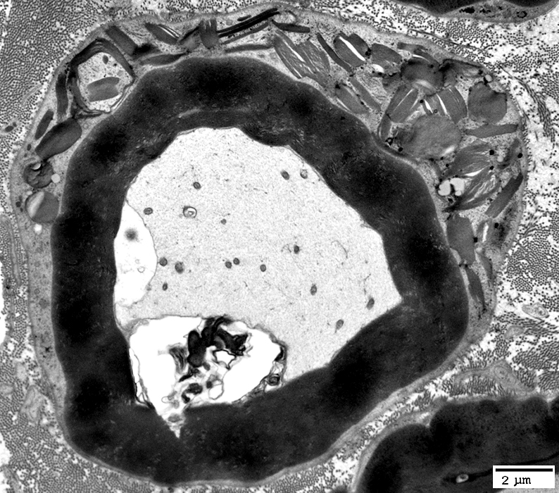
Axons
No surrounding myelin
Cytoplasm is dark & contains closely spaced neurofilaments.
Some myelinated axons may remain in the same area (Right)
Schwann cells & their processes
Surround the demyelinated axons
May be multiple Schwann cells around single axon
 Electron micrograph: From Robert Schmidt MD |
 |
 Electron micrographs: From Robert Schmidt MD |
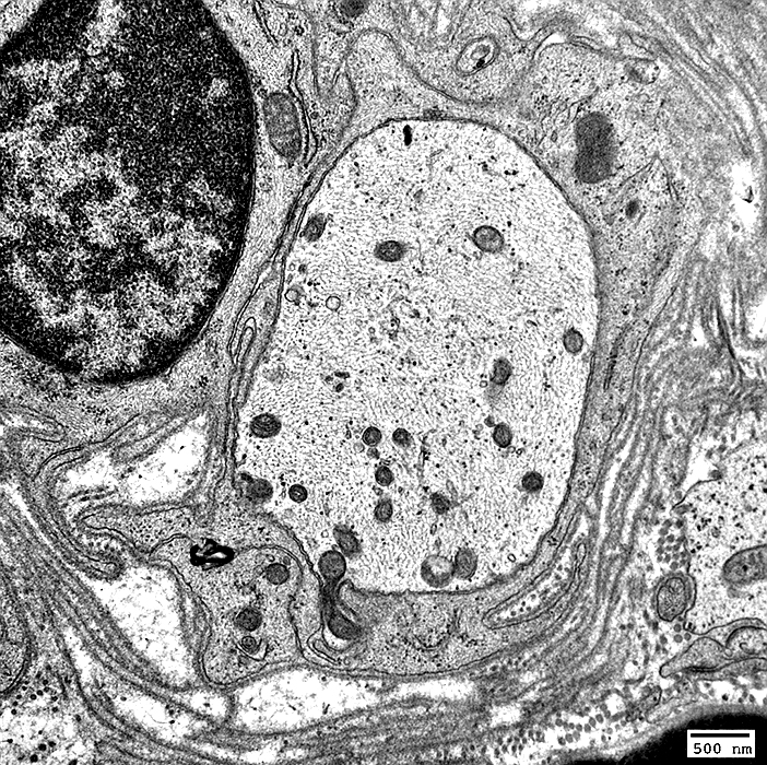
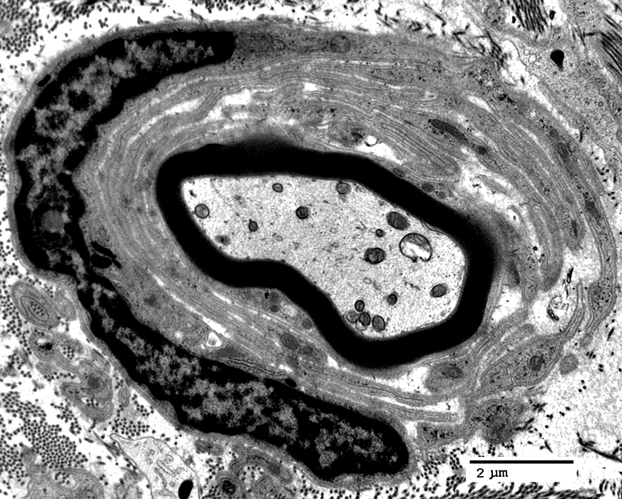
No myelination around axon
Schwann cell processes
Multiple
Surround axon
No onion bulbs
Lipofuscin debris (Arrow)
Usually only present in non-myelinating Schwann cells
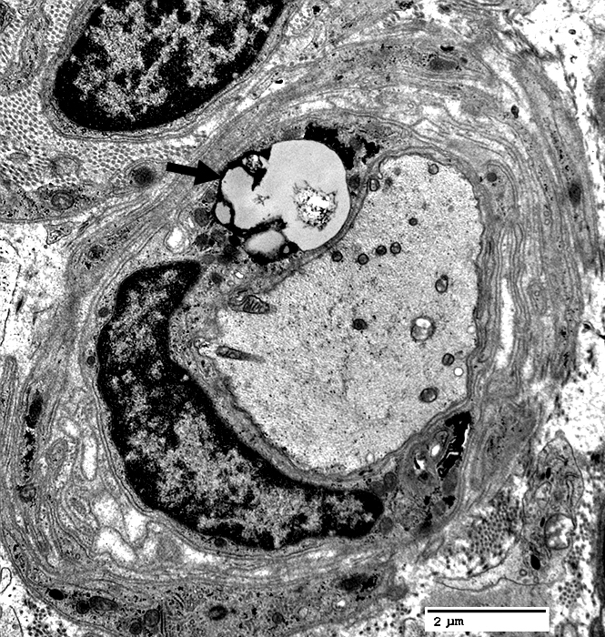 Electron micrographs: From Robert Schmidt MD |
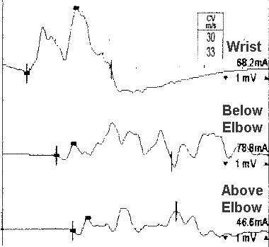 Nerve conduction studies (Ulnar nerve) Temporal dispersion NCV: Moderate slowing |
 Electron micrographs: From Robert Schmidt MD |
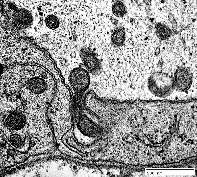
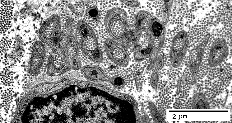
Surround demyelinated axon
Have associated basal lamina
 Electron micrographs: From Robert Schmidt MD |
 Electron micrographs: From Robert Schmidt MD |
 Electron micrographs: From Robert Schmidt MD |
Surrounded by few, or multiple, Schwann cell processes
 Electron micrographs: From Robert Schmidt MD |
Loss of Small Axons: Collagen Pockets
(Same nerve as above)
 Electron micrographs: From Robert Schmidt MD |
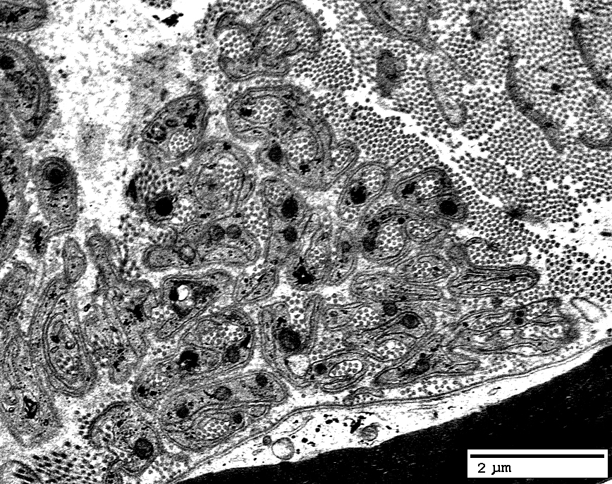 Electron micrographs: From Robert Schmidt MD |
Pi Granules
 Electron micrographs: From Robert Schmidt MD |
Return to Normal nerve biopsies
Return to Biopsy illustrations
Return to Neuromuscular Home Page
Return to Nerve biopsy
Return to Demyelinating neuropathies
Return to Chronic demyelination
4/7/2021