CALCIPHYLAXIS
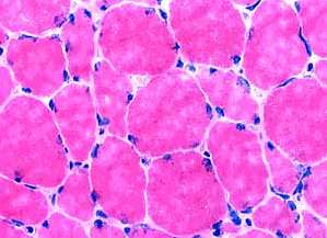
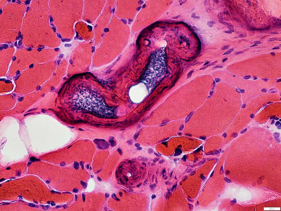 H & E stain |
 H & E stain |
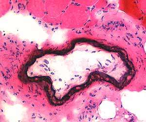
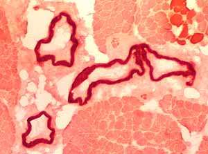
 Congo Red stain |
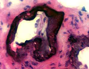
 Alizarin red stain |
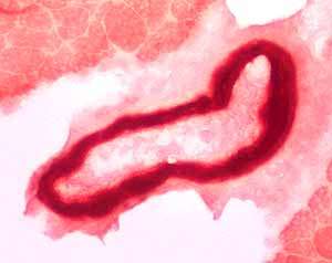
 Alizarin red stain |
Calcium deposited in capillaries
 Alizarin red stain |
Muscle pathology in calciphylaxis
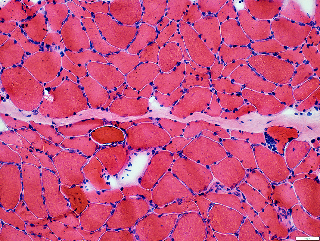 H & E stain |
Fiber sizes: Varied; Bimodal distribution
Myonuclei: Large; Irregular shapes
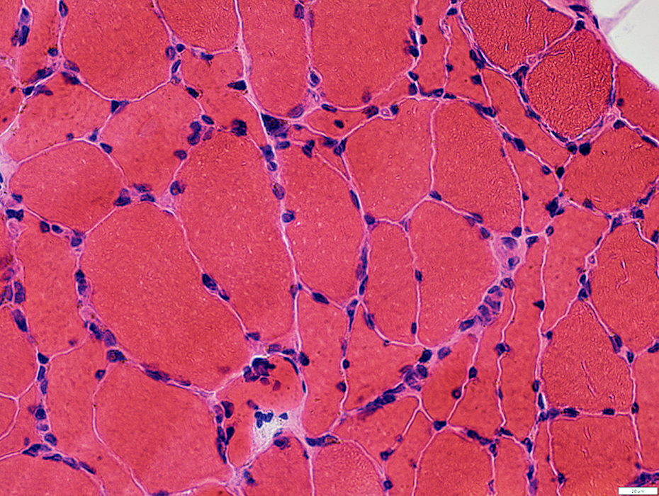 H & E stain |
Fiber sizes: Varied; Bimodal distribution
Small muscle fibers: Angular
Myonuclei: Large; Irregular shapes Endomysial capillaries: Some are large
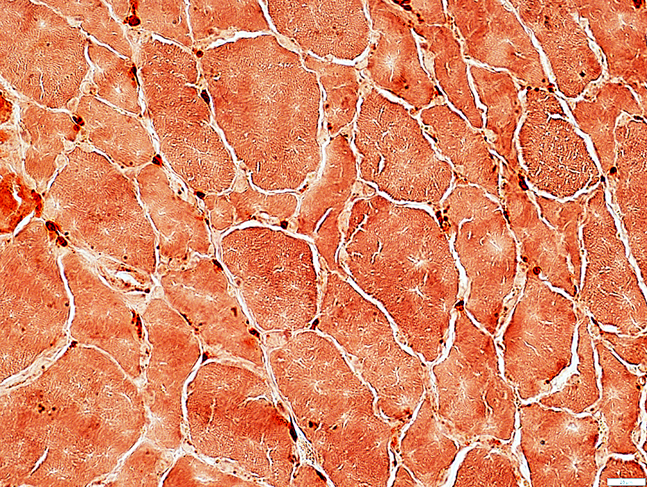
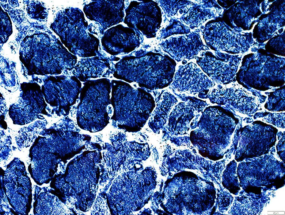
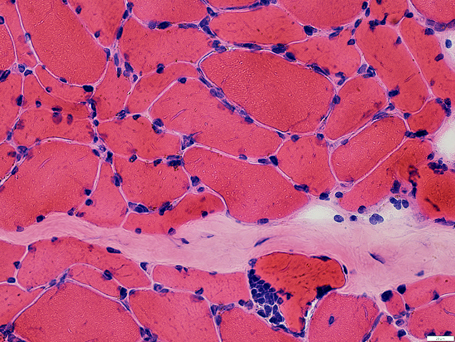 H & E stain |
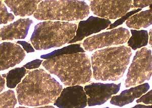
 Gomori Trichrome stain |
Fiber sizes: Varied; Bimodal distribution
Small muscle fibers: Angular
Endomysial capillaries: Some are large
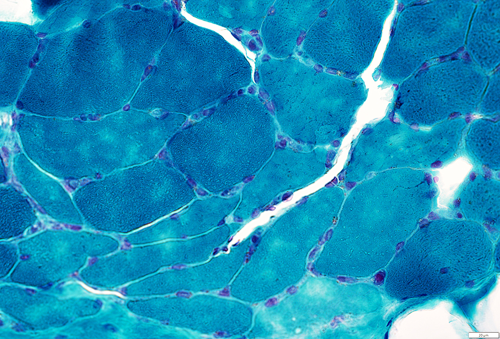
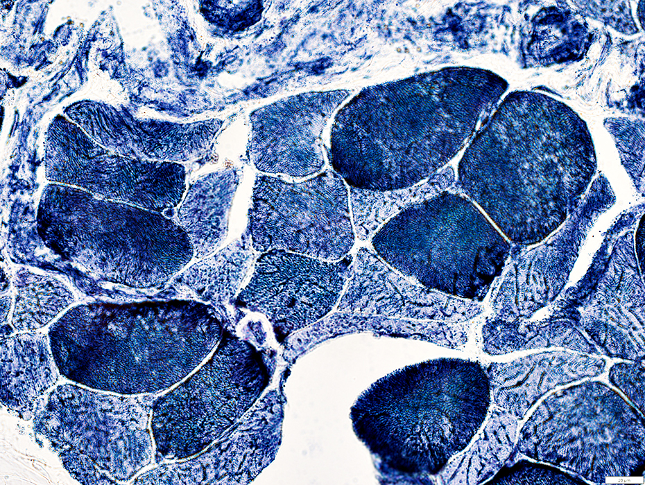 NADH stain |
Muscle fiber internal architecture: Irregular
Endomysial capillaries: Large (Below)
 NADH stain |
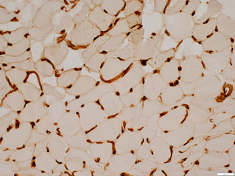
Endomysial capillaries: Pathology in calciphylaxis
Large
Misoriented (Circumferential around muscle fibers
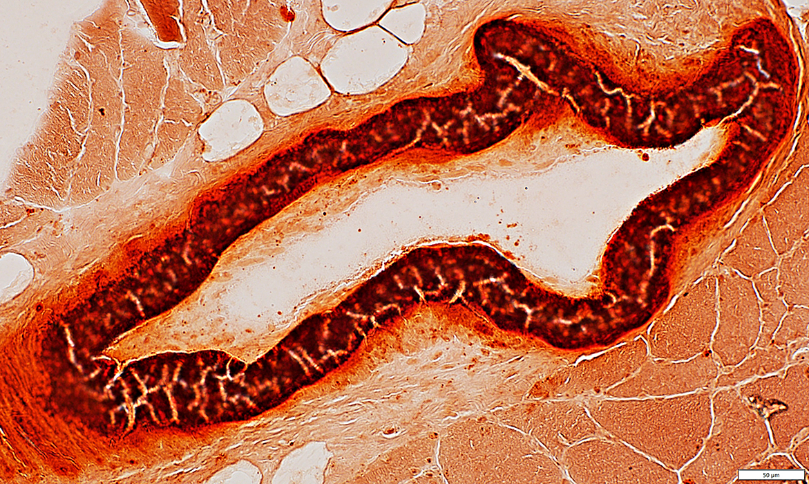
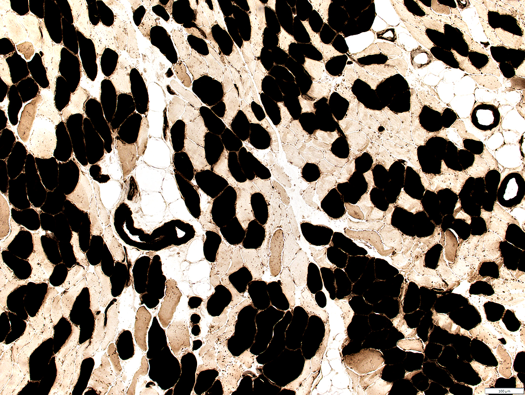
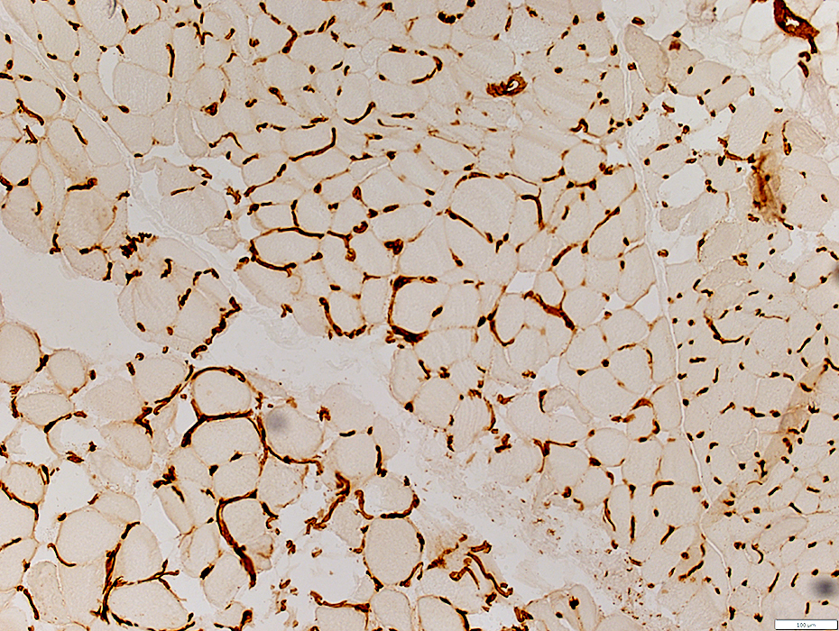 UEA I stain |
Endomysial capillaries: Large
 UEA I stain |
|
Muscle fiber sizes
Varied
Atrophy: Most fibers
Type 2 muscle fibers: Smaller than type 1
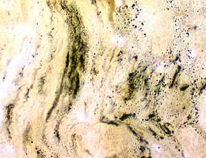
Perimysial connective tissue
Replaced by fat
 ATPase pH 4.3 stain |

|
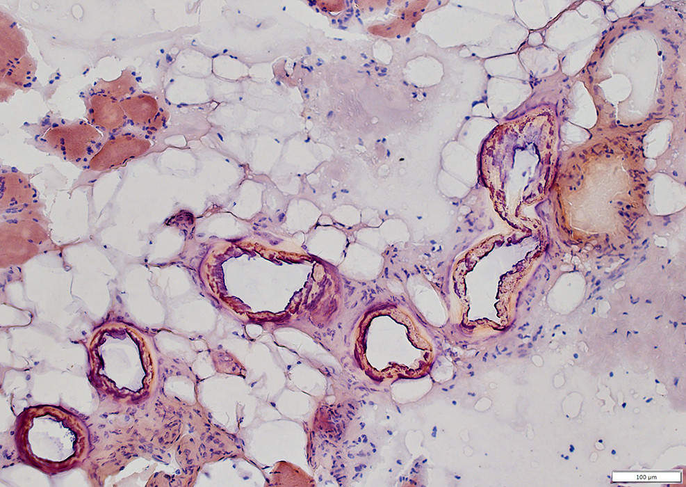
Subcutaneous tissue pathology in caliciphylaxis
|
Return to Calciphylaxis
Return to Neuromuscular Home Page
1/31/2022